. 1998 Apr;72(4):3051-9.
doi: 10.1128/JVI.72.4.3051-3059.1998.

Cleavage of the feline calicivirus capsid precursor is mediated by a virus-encoded proteinase

Affiliations

Cleavage of the feline calicivirus capsid precursor is mediated by a virus-encoded proteinase

S V Sosnovtsev et al. J Virol. 1998 Apr.

Abstract

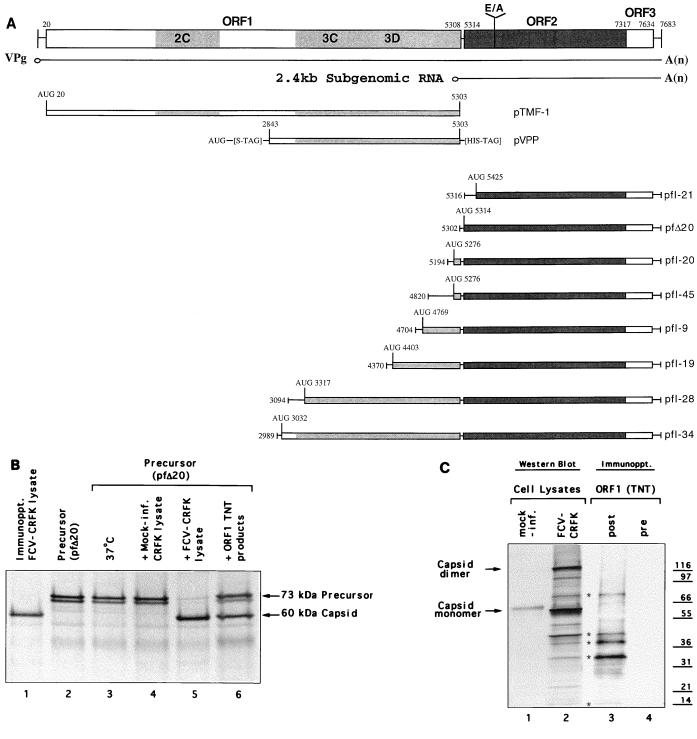
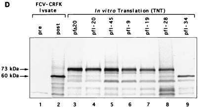
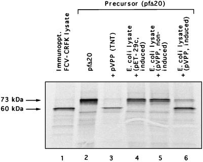
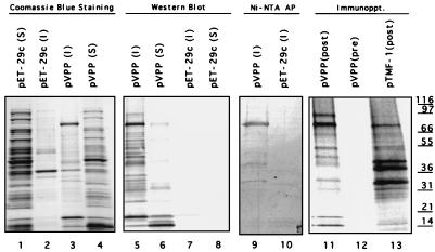
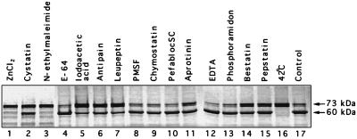
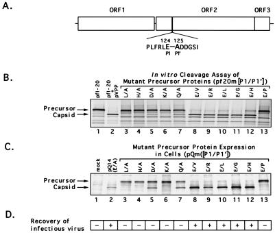
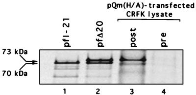
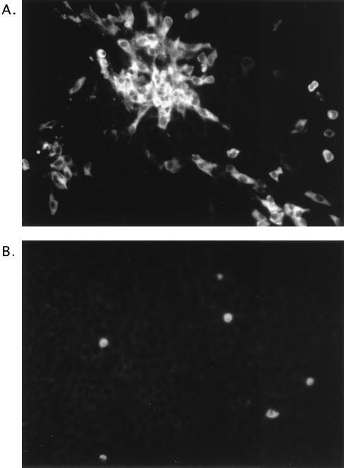
Feline calicivirus (FCV), a member of the Caliciviridae, produces its major structural protein as a precursor polyprotein from a subgenomic-sized mRNA. In this study, we show that the proteinase responsible for processing this precursor into the mature capsid protein is encoded by the viral genome at the 3'-terminal portion of open reading frame 1 (ORF1). Protein expression studies of either the entire or partial ORF1 indicate that the proteinase is active when expressed either in in vitro translation or in bacterial cells. Site-directed mutagenesis was used to characterize the proteinase Glu-Ala cleavage site in the capsid precursor, utilizing an in vitro cleavage assay in which mutant precursor proteins translated from cDNA clones were used as substrates for trans cleavage by the proteinase. In general, amino acid substitutions in the P1 position (Glu) of the cleavage site were less well tolerated by the proteinase than those in the P1' position (Ala). The precursor cleavage site mutations were introduced into an infectious cDNA clone of the FCV genome, and transfection of RNA derived from these clones into feline kidney cells showed that efficient cleavage of the capsid precursor by the virus-encoded proteinase is a critical determinant in the growth of the virus.


Figures

FIG. 1
FIG. 1
(A) Organization of the FCV genome (URB strain) and depiction of cDNA clones analyzed in this study. The corresponding genomic locations of the two major positive-sense RNA species (7.6 and 2.4 kb) found in infected cells and the location of the cleavage site (E124/A125) of the capsid precursor protein encoded in ORF2 are indicated. Clones pfI-21, pfI-20, pfI-45, pfI-9, pfI-19, pfI-28, and pfI-34 were selected from a cDNA library of the URB strain that was constructed by using the pSPORT plasmid (31) and contained nt 5316, 5194, 4820, 4704, 4370, 3094, and 2989, respectively, through a poly(A) tract from the URB genome, each under control of the T7 promoter. The location of the first in-frame AUG following the T7 promoter is indicated for each clone. Plasmids pTMF1, pVPP, and pfΔ20 were engineered as described in Materials and Methods. (B) Conditions under which in vitro cleavage of the FCV capsid precursor protein derived from plasmid pfΔ20 were observed. Lane 1, 60-kDa protein corresponding to the mature viral capsid protein immunoprecipitated (Immunoppt.) from an FCV-infected CRFK lysate with gp α-FCV; lane 2, in vitro TNT translation products synthesized from pfΔ20 without treatment. The translation products from pfΔ20 were treated as follows prior to analysis by SDS-PAGE: lane 3, incubation at 37°C for 3 h; lane 4, incubation (3 h, 37°C) with a nonradiolabeled CRFK lysate prepared from mock-infected cells; lane 5, incubation (3 h, 37°C) with a nonradiolabeled CRFK lysate prepared from FCV-infected cells; lane 6, incubation (3 h, 37°C) with nonradiolabeled translation products synthesized from pTMF-1. (C) Comparison of radiolabeled translation products derived from pTMF-1 (encoding the URB ORF1) with proteins produced in FCV-infected CRFK cells. Proteins produced in mock-infected (lane 1) or FCV-infected (lane 2) CRFK cells were analyzed in a Western blot reacted with cat FCV postinfection serum. The same infection serum was used to immunoprecipitate (Immunoppt.) radiolabeled TNT products derived from pTMF-1 (lane 3). Lane 4, immunoprecipitation analysis of radiolabeled TNT products derived from pTMF-1 with cat preinfection serum. The location of the mature capsid protein and its dimeric form in the FCV-infected CRFK cell lysate is shown. Asterisks denote proteins similar in size (indicated in kilodaltons) between ORF1 TNT products and the FCV-infected CRFK cell lysate. (D) Comparison of products produced by in vitro translation of cDNA clones encoding the ORF2 (with various lengths of upstream ORF1 sequence) with the mature 60-kDa capsid protein produced in virus-infected cells. Radiolabeled products (in all lanes) underwent immunoprecipitation with either preimmunization (lane 1) or postimmunization (lanes 2 to 9) gp α-FCV, as follows: lanes 1 and 2, FCV-infected CRFK lysate; lane 3, pfΔ20; lane 4, pfI-20; lane 5, pfI-45; lane 6, pfI-9; lane 7, pfI-19; lane 8, pfI-28; lane 9, pfI-34.
FIG. 1
FIG. 1
(A) Organization of the FCV genome (URB strain) and depiction of cDNA clones analyzed in this study. The corresponding genomic locations of the two major positive-sense RNA species (7.6 and 2.4 kb) found in infected cells and the location of the cleavage site (E124/A125) of the capsid precursor protein encoded in ORF2 are indicated. Clones pfI-21, pfI-20, pfI-45, pfI-9, pfI-19, pfI-28, and pfI-34 were selected from a cDNA library of the URB strain that was constructed by using the pSPORT plasmid (31) and contained nt 5316, 5194, 4820, 4704, 4370, 3094, and 2989, respectively, through a poly(A) tract from the URB genome, each under control of the T7 promoter. The location of the first in-frame AUG following the T7 promoter is indicated for each clone. Plasmids pTMF1, pVPP, and pfΔ20 were engineered as described in Materials and Methods. (B) Conditions under which in vitro cleavage of the FCV capsid precursor protein derived from plasmid pfΔ20 were observed. Lane 1, 60-kDa protein corresponding to the mature viral capsid protein immunoprecipitated (Immunoppt.) from an FCV-infected CRFK lysate with gp α-FCV; lane 2, in vitro TNT translation products synthesized from pfΔ20 without treatment. The translation products from pfΔ20 were treated as follows prior to analysis by SDS-PAGE: lane 3, incubation at 37°C for 3 h; lane 4, incubation (3 h, 37°C) with a nonradiolabeled CRFK lysate prepared from mock-infected cells; lane 5, incubation (3 h, 37°C) with a nonradiolabeled CRFK lysate prepared from FCV-infected cells; lane 6, incubation (3 h, 37°C) with nonradiolabeled translation products synthesized from pTMF-1. (C) Comparison of radiolabeled translation products derived from pTMF-1 (encoding the URB ORF1) with proteins produced in FCV-infected CRFK cells. Proteins produced in mock-infected (lane 1) or FCV-infected (lane 2) CRFK cells were analyzed in a Western blot reacted with cat FCV postinfection serum. The same infection serum was used to immunoprecipitate (Immunoppt.) radiolabeled TNT products derived from pTMF-1 (lane 3). Lane 4, immunoprecipitation analysis of radiolabeled TNT products derived from pTMF-1 with cat preinfection serum. The location of the mature capsid protein and its dimeric form in the FCV-infected CRFK cell lysate is shown. Asterisks denote proteins similar in size (indicated in kilodaltons) between ORF1 TNT products and the FCV-infected CRFK cell lysate. (D) Comparison of products produced by in vitro translation of cDNA clones encoding the ORF2 (with various lengths of upstream ORF1 sequence) with the mature 60-kDa capsid protein produced in virus-infected cells. Radiolabeled products (in all lanes) underwent immunoprecipitation with either preimmunization (lane 1) or postimmunization (lanes 2 to 9) gp α-FCV, as follows: lanes 1 and 2, FCV-infected CRFK lysate; lane 3, pfΔ20; lane 4, pfI-20; lane 5, pfI-45; lane 6, pfI-9; lane 7, pfI-19; lane 8, pfI-28; lane 9, pfI-34.
FIG. 2
FIG. 2
Analysis of the proteolytic activity of the proteins encoded in plasmid pVPP in the capsid precursor trans cleavage assay. Lane 1, immunoprecipitation (Immunoppt.) of radiolabeled FCV-infected CRFK cell lysate with gp α-FCV. Lane 2, pfΔ20 translation products, without treatment. The radiolabeled capsid precursor protein was incubated with the following nonradiolabeled preparations prior to analysis by SDS-PAGE: lane 3, in vitro TNT translation products synthesized from pVPP; lane 4, E. coli crude cell lysate prepared from IPTG-induced bacteria containing the pET-29c vector plasmid; lane 5, E. coli crude cell lysate from noninduced bacteria carrying plasmid pVPP; lane 6, E. coli crude cell lysate from IPTG-induced bacteria carrying plasmid pVPP.
FIG. 3
FIG. 3
Analysis of proteins encoded in plasmid pVPP. Bacteria carrying either plasmid pVPP or the vector plasmid pET-29c were induced with IPTG, and the soluble (S) or insoluble (I) bacterial products were prepared as described in the text. The bacterial products were subjected to SDS-PAGE and visualized with Coomassie blue stain: lane 1, pET-29c, soluble fraction; lane 2, pET-29c, insoluble fraction; lane 3, pVPP, insoluble fraction; lane 4, pVPP, soluble fraction. The same products were analyzed in a Western blot developed with cat postinfection serum: lane 5, pVPP, insoluble fraction; lane 6, pVPP, soluble fraction; lane 7, pET-29C, insoluble fraction; lane 8, pET-29c, soluble fraction. The insoluble fractions of pVPP or pET-29c were transferred to nitrocellose and probed with Ni-NTA–AP: lane 9, pVPP; lane 10, pET-29c. The radiolabeled TNT in vitro translation products derived from pVPP were immunoprecipitated (Immunoppt.) with either postinfection (lane 11) or preinfection (lane 12) cat serum. Lane 13, radiolabeled TNT products derived from pTMF-1 immunoprecipitated with cat postinfection serum.
FIG. 4
FIG. 4
Effects of selected protease inhibitors or temperature on the ability of the protease present in FCV-infected CRFK lysates to cleave in trans the capsid precursor protein translated from pfΔ20. In lanes 1 to 15, the indicated protease inhibitors were incubated with the FCV-infected CRFK lysate prior to incubation with the translated precursor protein under conditions described in the text. Lane 16, incubation of FCV-infected cell lysate at 42°C for 20 min prior to incubation with the capsid precursor; lane 17, precursor protein incubated with nontreated FCV-infected CRFK lysate.
FIG. 5
FIG. 5
Analysis of the effects of amino acid mutations introduced into the cleavage site of the FCV capsid precursor protein on the efficiency of cleavage by the viral proteinase as measured in the in vitro trans cleavage assay or by the ability of the virus to grow in cell culture. (A) Sequence of cleavage site between the precursor leader sequence and the mature capsid protein into which amino acid substitutions were introduced. (B) The radiolabeled capsid precursor proteins derived from in vitro translation of pfI-20 or engineered plasmids containing mutant cleavage sites [clones pf20m(P1/P1′)] were incubated with nonradiolabeled translation products derived from plasmid pVPP. The first lane contains the pfI-20 translation products without treatment. (C) Capped, synthetic RNA from individual full-length clones that contained the mutant cleavage sites [clones pQm(P1/P1′)] were transfected into CRFK cells, and proteins synthesized in cells were radiolabeled with [35S]methionine. The following cell lysates were analyzed by immunoprecipitation with gp α-FCV serum: lane 1, mock-infected CRFK cells; lane 2, CRFK cells transfected with RNA derived from pQ14; lanes 3 to 13, CRFK cells transfected with RNA derived from plasmids pQm(P1/P1′). (D) Effects of mutations in the P1 or P1′ position of the precursor cleavage site on the ability to recover viable virus from engineered full-length infectious clones. In a separate experiment, an aliquot of cell culture medium from cells transfected with RNA derived from each of the pQm(P1/P1′) clones was transferred to a fresh CRFK monolayer. Clones that did (+) and did not (−) yield viable progeny are indicated.
FIG. 6
FIG. 6
Comparison of FCV capsid precursor polyprotein expressed in vitro and in transfected cells. Lanes 1 and 2, radiolabeled products of in vitro translation of pfI-21 and pfΔ20, respectively; lanes 3, and 4, immunoprecipitation of radiolabeled precursor with gp α-FCV and gp preimmunization serum, respectively, from CRFK cells transfected with capped genomic RNA that encoded the H/A mutant cleavage site.
FIG. 7
FIG. 7
Immunofluorescence of CRFK cells transfected 26 h previously with full-length capped, synthetic RNA from pQm E/G (A) and pQm Q/A (B).

References

    1. Allaire M, Chernaia M M, Malcolm B A, James M N G. Picornaviral 3C cysteine proteinases have a fold similar to chymotrypsin-like serine proteinases. Nature. 1994;369:72–76. - PubMed
    1. Black D N, Burroughs J N, Harris T J R, Brown F. The structure and replication of calicivirus RNA. Nature. 1978;274:614–615. - PubMed
    1. Boniotti B M, Wirlblich C, Sibilia M, Meyers G, Thiel H-J, Rossi C. Identification and characterization of 3C-like protease from rabbit hemorrhagic disease virus, a calicivirus. J Virol. 1994;68:6487–6495. - PMC - PubMed
    1. Carter M J. Feline calicivirus protein synthesis investigated by Western blotting. Arch Virol. 1989;108:69–79. - PMC - PubMed
    1. Carter M J. Transcription of feline calicivirus RNA. Arch Virol. 1990;114:143–152. - PMC - PubMed

Associated data

LinkOut - more resources