. 1998 Apr;72(4):3146-54.
doi: 10.1128/JVI.72.4.3146-3154.1998.

Adenovirus E1B 55K represses p53 activation in vitro

Affiliations

Adenovirus E1B 55K represses p53 activation in vitro

M E Martin et al. J Virol. 1998 Apr.

Abstract

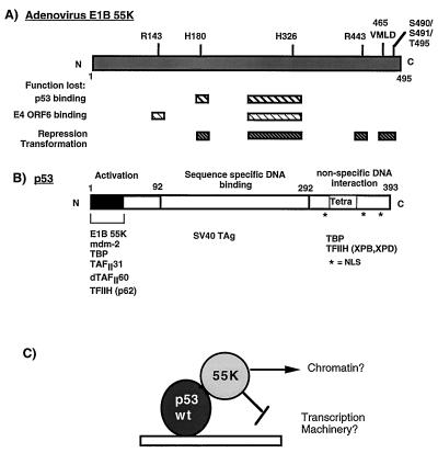
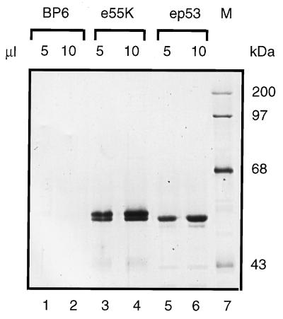
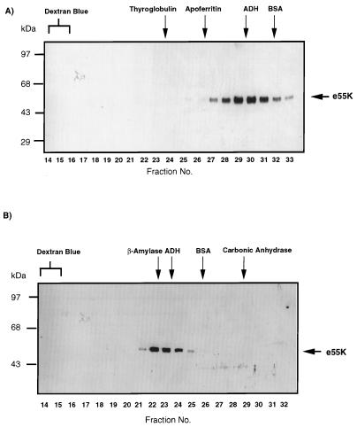
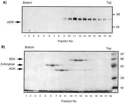
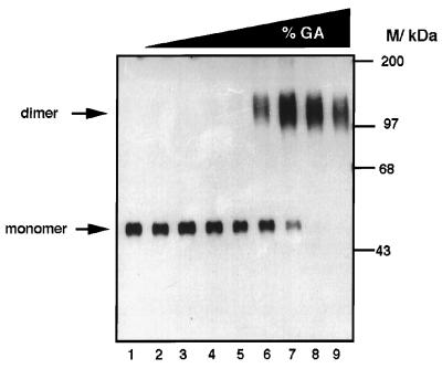
Adenovirus E1B 55K protein cooperates with E1A gene products to induce cell transformation. E1B 55K mediates its effects by binding to and inhibiting the transcriptional activation and growth-suppression functions of the tumor suppressor p53. Previous studies in vivo have suggested that E1B 55K has an active role in repressing p53 transcriptional activation and that this repression function is directed to specific promoters through E1B 55K's interaction with DNA-bound p53. Flag-tagged E1B 55K (e55K) was expressed with the baculovirus expression system and immunopurified. Gel filtration, velocity sedimentation centrifugation, and glutaraldehyde cross-linking indicated that e55K is a dimer with a nonglobular conformation. e55K bound directly to purified p53, causing an approximately 10-fold increase in p53 affinity for tandem binding sites. Using in vitro transcription assays reconstituted with purified p53, e55K, and HeLa cell nuclear extracts, we found that e55K specifically repressed p53 activation. These results demonstrate that as postulated from earlier transient expression experiments, E1B 55K is a specific repressor of transcription from a promoter with bound p53. Since HeLa nuclear extracts contain little detectable histone protein, E1B 55K probably represses transcription through direct or indirect interactions with the RNA polymerase II transcription machinery.


Figures

FIG. 1
FIG. 1
Schematic diagram illustrating the functional domains in Ad E1B 55K (A) and the tumor suppressor p53 (B). (A) Hatched boxes indicate regions in E1B 55K which when mutated result in loss of the described functions. Positions where amino acids have been mutated by linker insertion or site-directed mutagenesis (57, 67) are indicated above the E1B 55K molecule. (B) The activation, DNA binding, and nonspecific DNA binding domains of p53 are indicated. The tetramerization domain (Tetra) is located in the C terminus. Nuclear localization signals (NSL) are indicated by asterisks. Proteins which are known to interact with the defined regions are indicated at the bottom. (C) Model of how E1B 55K may affect the activity of p53. E1B 55K may mediate alterations in chromatin structure or may target the general transcriptional machinery. SV40 TAg, simian virus 40 T antigen.
FIG. 2
FIG. 2
Silver-stained SDS-PAGE (10% polyacrylamide) gels of purified Flag-tagged 55K (e55K), the control BP6 protein fraction purified under identical conditions to e55K from Sf9 cells infected with a baculovirus vector expressing β-galactosidase, and ep53 proteins. Lanes 1 to 6 contain BP6 (lanes 1 and 2 [5 and 10 μl, respectively]), e55K (lanes 3 and 4 [5 and 10 μl, respectively]), and ep53 (lanes 5 and 6 [5 and 10 μl, respectively]). M, molecular mass markers in kilodaltons (lane 7).
FIG. 3
FIG. 3
Gel filtration analysis of e55K through Superose 6 (A) and Superose 12 (B) columns. After chromatography, fractions were trichloroacetic acid precipitated and analyzed by SDS-PAGE and Western blotting with the anti-55K antibody 2A6. e55K was visualized by ECL. Fraction numbers are indicated at the bottom of each panel. The fractions in which the protein standards eluted are indicated at the top of each panel. Protein standards were thyroglobulin (a tetramer of 670 kDa [85Å]), apoferritin (a multimer of 440 kDa [61Å]), β-amylase (200 kDa), ADH (a tetramer of 150 kDa [46Å]), BSA (a monomer of 68 kDa [35Å]), and carbonic anhydrase (29 kDa).
FIG. 4
FIG. 4
Sucrose gradient analysis of e55K. e55K, BSA, β-amylase, and ADH were centrifuged together through a 5 to 20% sucrose gradient. Fractions were collected, trichloroacetic acid precipitated, and analyzed by SDS-PAGE, followed by Western blotting with anti-55K antibody 2A6 (A) and Coomassie blue staining (B). The top and bottom of the gradient are indicated. Molecular mass markers are indicated on the right in kilodaltons. Protein molecular mass standards from the bottom of the gradient are β-amylase (8.9S; 200 kDa; tetramer), ADH (7.4S; 150 kDa; tetramer), and BSA (4.3S; 68 kDa; monomer).
FIG. 5
FIG. 5
Glutaraldehyde cross-linking analysis of e55K. e55K was incubated with increasing concentrations of glutaraldehyde (%GA): lanes 1 to 9: 0, 10−4, 3 × 10⁻⁴, 10−3, 3 × 10⁻³, 10−2, 3 × 10⁻², 10−1, and 3 × 10⁻¹%, respectively. The positions of molecular mass markers (kilodaltons) are indicated. The positions of the monomeric and dimeric forms of e55K are indicated by arrows.
FIG. 6
FIG. 6
Highly purified e55K interacts with ep53 bound to DNA. (A) Two 32P-labeled DNA fragments were used in the DNA immunoprecipitation assay, one containing five Gal4 binding sites (lanes 1 and 2) and one containing five p53 binding sites (lanes 3 to 7). ep53 (150 ng) was added to binding reaction mixtures in lanes 2, 4, and 6. e55K (150 ng) was added to binding reaction mixtures in lanes 6 and 7. ep53 was immunoprecipitated with an anti-p53 antibody, Ab421, and e55K was immunoprecipitated with the anti-55K antibody 2A6. (B) The 32P-labeled DNA fragment containing five p53 binding sites was incubated with 150 ng of ep53 (lanes 2 and 3) and 150 ng of e55K (lane 3). Immunoprecipitation was with anti-p53 monoclonal antibody 421. For both panels A and B, immunoprecipitated DNA was resolved by PAGE and visualized by autoradiography.
FIG. 7
FIG. 7
E1B 55K increases the affinity of p53 for its DNA binding sites. DNase I footprinting of DNA-protein complexes. The positions of the p53 binding sites are indicated. (A) Lanes 1, 8, and 15 show the DNase I pattern in the absence of added proteins. Lanes 2 to 4 show decreasing concentrations of purified ep53 (150, 75, and 30 ng, respectively [10 ng/μl]). Lanes 5 to 7 show binding reactions with 3 μl (30 ng) of ep53 plus increasing amounts of e55K (10 ng/μl [3, 6, and 9 μl, respectively]). Lanes 9 to 11 show DNase I footprinting after incubation of the probe with 3, 6, and 9 μl, respectively, of e55K. Lanes 12 to 14 show binding reactions with 3 μl (30 ng) of ep53 plus 3, 6, and 9 μl of the control BP6 fraction. (B) Lanes 1 and 19 show the DNase I digestion pattern in the absence of added protein. Lanes 2 to 5 show footprinting with decreasing amounts of ep53 (10 ng/μl): 15, 10, 3, and 1 μl, respectively. Lanes 6 to 12 show footprinting with 3 μl of ep53 plus decreasing amounts of e55K (100, 30, 10, 3, 1, 0.3, and 0.1 ng, respectively). Lanes 13 to 18 show footprinting in the presence of 30 ng of e55K and decreasing amounts of ep53 (30, 10, 3, 1, 0.3, and 0.1 ng, respectively).
FIG. 8
FIG. 8
(A) In vitro transcription templates. p5BSE4CAT contains five p53 binding sites upstream of the Ad E4 TATA box and CAT gene. This template is responsive to activation by p53. The primer extensions of RNAs transcribed from this template are 160 nucleotides (160 n) in length. pG5E1BCAT contains five Gal4 binding sites upstream of the Ad E1B TATA box and CAT gene. This template is responsive to activation by Gal4-AH. Primer extensions of RNAs transcribed from this template are 110 nucleotides (110 n) in length. (B) e55K specifically represses p53-mediated activation at the transcriptional level. In vitro transcription reaction mixtures contained HeLa nuclear extract, template DNAs, and ep53 (20 ng [lanes 3 to 8 and 11 to 16]), Gal4-AH (10 ng [lanes 2, 4 to 8, and 10, and 12 to 16]) and increasing concentrations of e55K (10 ng/μl [lanes 5 to 8, 2, 4, 8, and 16 μl, respectively]) or increasing concentrations of the control BP6 protein fraction (lanes 13 to 16, 2, 4, 8, and 16 μl, respectively). Primer extension products (160 n and 110 n) indicate levels of transcription arising from pB5E4CAT and pG5E1BCAT, respectively and their positions are indicated by arrows. (C) In vitro transcription with templates p5BSCAT and pG5E1BCAT; HeLa nuclear extract; and 40 ng of ep53 (lanes 2 to 5); 10 ng of Gal4-AH; and 30, 70, and 140 ng of e55K (lanes 3, 4, and 5, respectively).

References

    1. Alland L, Muhle R, Hou H, Jr, Potes J, Chin L, Schreiber-Agus N, DePinho R A. Role for N-CoR and histone deacetylase in Sin3-mediated transcriptional repression. Nature. 1997;387:49–55. - PubMed
    1. Auble D T, Hansen K E, Mueller C G, Lane W S, Thorner J, Hahn S. Mot1, a global repressor of RNA polymerase II transcription, inhibits TBP binding to DNA by an ATP-dependent mechanism. Genes Dev. 1994;8:1920–1934. - PubMed
    1. Austin R J, Biggin M D. A domain of the even-skipped protein represses transcription by preventing TFIID binding to a promoter: repression by cooperative blocking. Mol Cell Biol. 1995;15:4683–4693. - PMC - PubMed
    1. Ayer D E, Kretzner L, Eisenman R N. Mad: a heterodimeric partner for Max that antagonizes Myc transcriptional activity. Cell. 1993;72:211–222. - PubMed
    1. Buckbinder L, Talbott R, Velasco-Miguel S, Takenaka I, Faha B, Seizinger B R, Kley N. Induction of the growth inhibitor IGF-binding protein 3 by p53. Nature. 1995;377:646–649. - PubMed

Publication types

MeSH terms