. 1998 Apr;72(4):3289-99.
doi: 10.1128/JVI.72.4.3289-3299.1998.

Rat parvovirus type 1: the prototype for a new rodent parvovirus serogroup

Affiliations

Rat parvovirus type 1: the prototype for a new rodent parvovirus serogroup

L J Ball-Goodrich et al. J Virol. 1998 Apr.

Abstract

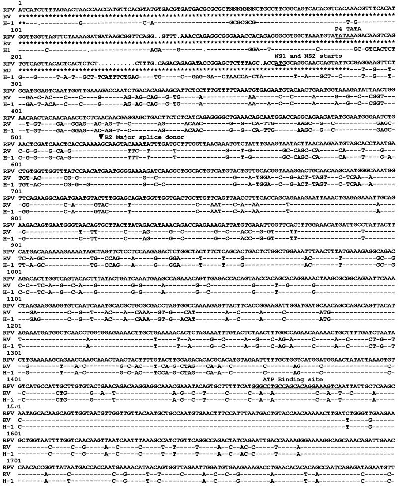
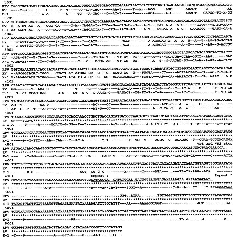
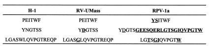
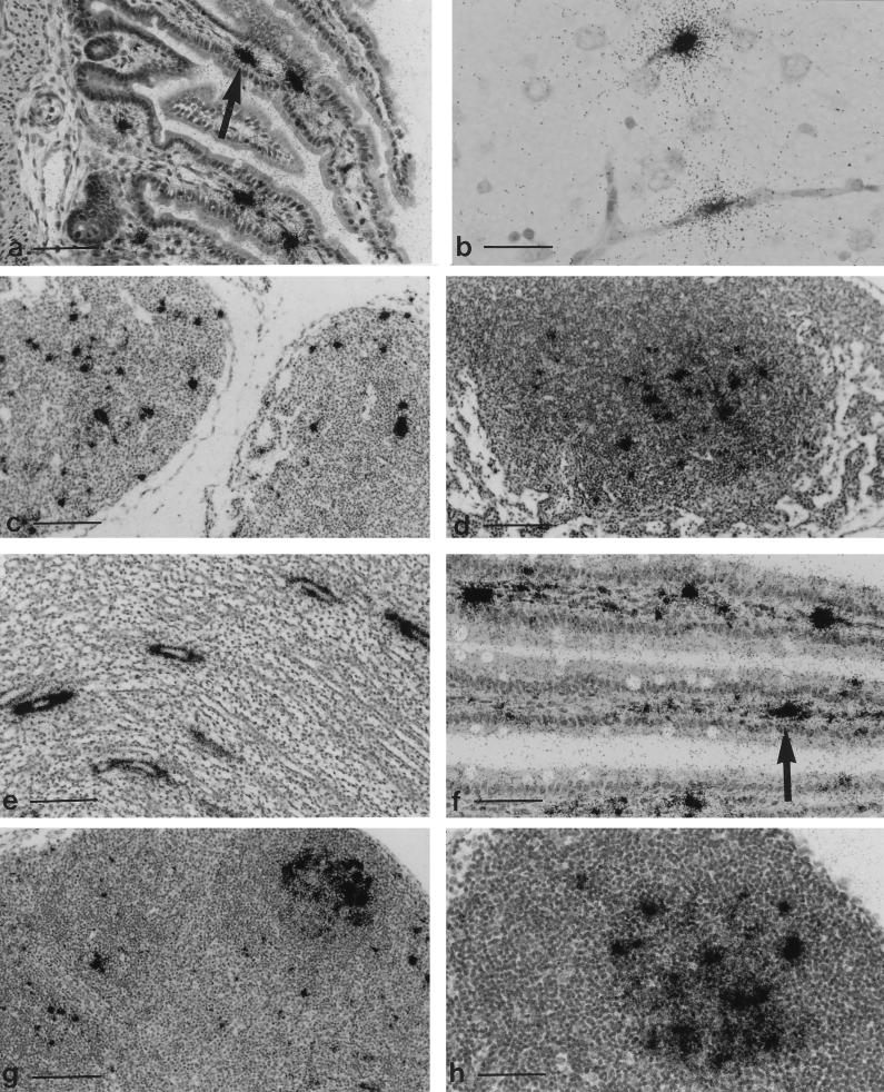
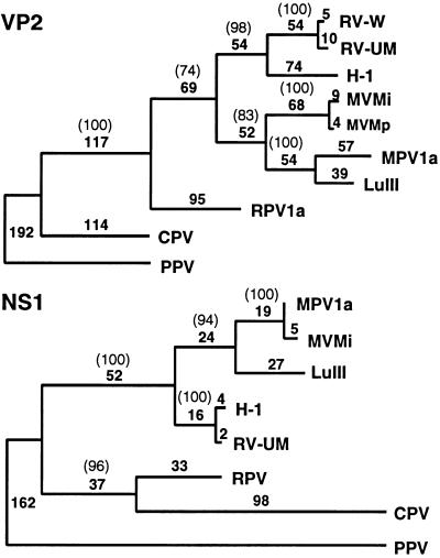
A newly recognized parvovirus of laboratory rats, designated rat parvovirus type 1a (RPV-1a), was found to be antigenically distinct. It was cloned, sequenced, and compared with the University of Massachusetts strain of rat virus (RV-UMass) and other autonomous parvoviruses. RPV-1a VP1 identity with these viruses never exceeded 69%, thus explaining its antigenic divergence. In addition, RPV-1a had reduced amino acid identity in NS coding regions (82%), reflecting phylogenetic divergence from other rodent parvoviruses. RPV-1a infection in rats had a predilection for endothelium and lymphoid tissues as previously reported for RV. Infectious RPV-1a was isolated 3 weeks after inoculation of infant rats, suggesting that it, like RV, may result in persistent infection. In contrast to RV, RPV-1a was enterotropic, a characteristic previously associated with parvovirus infections of mice rather than rats. RPV-1a also differed from RV in that infection was nonpathogenic for infant rats under conditions where RV infection causes high morbidity and mortality. Thus, RPV-1a is the prototype virus of an antigenically, genetically, and biologically distinct rodent parvovirus serogroup.


Figures

FIG. 1
FIG. 1
Sequence comparison of RPV-1a, RV-UMass, and H-1. The right terminus of H-1 virus is not included, as this region was not cloned or sequenced for RPV-1a or RV-UMass. Dashes indicate nucleotides identical to those of RPV-1a, dots indicate spaces inserted for maximal sequence alignment, and asterisks indicate nonsequenced regions. Regions conserved among parvoviruses are underlined, and MVM splice donors and acceptors are indicated with arrows. GCG programs Pileup and Pretty were used to generate the alignment.
FIG. 1
FIG. 1
Sequence comparison of RPV-1a, RV-UMass, and H-1. The right terminus of H-1 virus is not included, as this region was not cloned or sequenced for RPV-1a or RV-UMass. Dashes indicate nucleotides identical to those of RPV-1a, dots indicate spaces inserted for maximal sequence alignment, and asterisks indicate nonsequenced regions. Regions conserved among parvoviruses are underlined, and MVM splice donors and acceptors are indicated with arrows. GCG programs Pileup and Pretty were used to generate the alignment.
FIG. 2
FIG. 2
Carboxy termini of NS2 in H-1, RV-UMass, and RPV-1a.
FIG. 3
FIG. 3
(a to d) Tissues from rats inoculated o.n. with RPV-1a at 2 days of age and hybridized with a plus-sense 35S-labeled riboprobe to detect minus-sense DNA. (a) Small intestine at PID 7, with an arrow indicating signal in the lamina propria. Bar, 150 μm. (b) Brain at PID 7. Capillary endothelium is labeled, but there is no hemorrhage. Bar, 37 μm. (c) Mesenteric lymph node at PID 7. Bar, 150 μm. (d) Mesenteric lymph node at PID 20. Signal is localized over germinal center, a typical pattern for persistent lymphocytotropic infection with rodent parvoviruses. Bar, 150 μm. (e) Kidney from a rat inoculated with RPV-1a at 2 days of age, collected at PID 20, and hybridized with a 32P-labeled random-primed RPV probe. Signal is localized over collecting tubules. Bar, 150 μm. (f to h). Tissues from rats inoculated o.n. with RPV-1a at 3 weeks of age and hybridized with a plus-sense 35S-labeled riboprobe for minus-sense DNA. (f) Small intestine at PID 7, with an arrow indicating signal localized in the lamina propria. Bar, 75 μm. (g) Mesenteric lymph node at PID 7. Positive cells are in paracortical zones, and localization of signal over germinal centers has begun. Bar, 150 μm. (h) Mesenteric lymph node at PID 20 with signal over a germinal center. Bar, 75 μm.
FIG. 4
FIG. 4
Phylogeny-derived using parsimony analysis of 10 VP2 (A) and 8 NS1 (B) amino acid sequences. RV-W refers to the unpublished sequence of an RV strain kindly provided by Rene Wicker. The phylogeny was rooted by using PPV as an outgroup. The number of nucleotide changes in each horizontal branch is given, and the number in parentheses is the bootstrap support for this analysis that was obtained in 1,000 repetitions.

References

    1. Anouja F, Wattiez R, Mousset S, Caillet-Fauquet P. The cytotoxicity of the parvovirus minute virus of mice nonstructural protein NS1 is related to changes in the synthesis and phosphorylation of cell proteins. J Virol. 1997;71:4671–4678. - PMC - PubMed
    1. Astell C R, Gardiner E M, Tattersall P. DNA sequence of the lymphotropic variant of minute virus of mice, MVM(i), and comparison with the DNA sequence of the fibrotropic prototype strain. J Virol. 1986;57:656–659. - PMC - PubMed
    1. Astell C R, Smith M, Chow M B, Ward D C. Structure of the 3′ hairpin termini of four rodent parvovirus genomes: nucleotides sequence homology at origins of DNA replication. Cell. 1979;17:691–703. - PubMed
    1. Astell C R, Thomson M, Merchlinsky M, Ward D C. The complete DNA sequence of minute virus of mice, an autonomous parvovirus. Nucleic Acids Res. 1983;11:999–1018. - PMC - PubMed
    1. Ball-Goodrich, L. J. 1997. Unpublished results.

Publication types

Associated data

LinkOut - more resources