. 1998 Apr;72(4):3330-9.
doi: 10.1128/JVI.72.4.3330-3339.1998.

Herpes simplex virus gD and virions accumulate in endosomes by mannose 6-phosphate-dependent and -independent mechanisms

Affiliations

Herpes simplex virus gD and virions accumulate in endosomes by mannose 6-phosphate-dependent and -independent mechanisms

C R Brunetti et al. J Virol. 1998 Apr.

Abstract

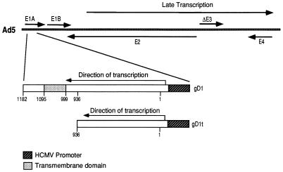
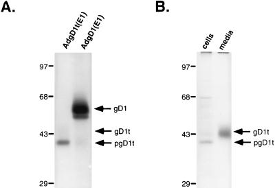
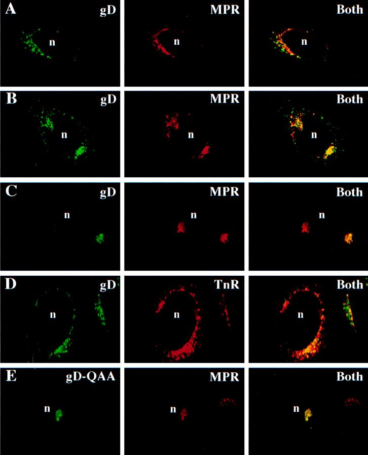
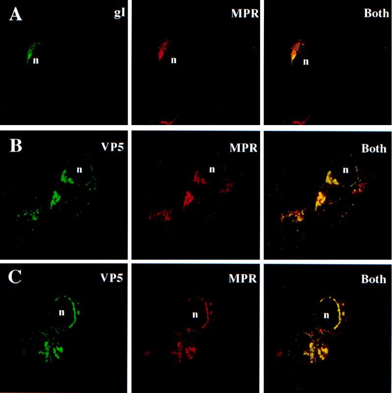
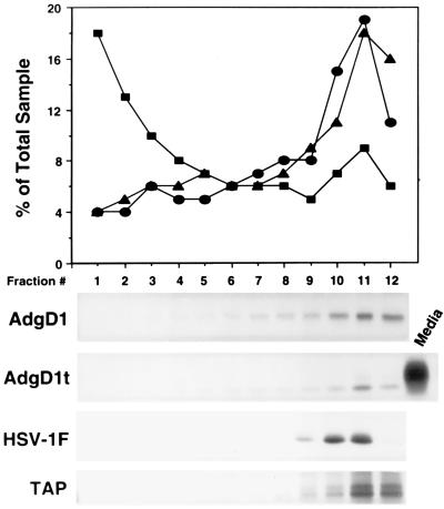
Herpes simplex virus (HSV) glycoprotein D (gD) is modified with mannose 6-phosphate (M6P) and binds to M6P receptors (MPRs). MPRs are involved in the well-characterized pathway by which lysosomal enzymes are directed to lysosomes via a network of endosomal membranes. Based on the impaired ability of HSV to form plaques under conditions in which glycoproteins could not interact with MPRs, we proposed that MPRs may function during HSV egress or cell-to-cell spread (C. R. Brunetti, R. L. Burke, B. Hoflack, T. Ludwig, K. S. Dingwell, and D. C. Johnson, J. Virol. 69:3517-3528, 1995). To further analyze M6P modification and intracellular trafficking of gD in the absence of other HSV proteins, adenovirus (Ad) vectors were used to express soluble and membrane-anchored forms of gD. Both membrane-bound and soluble gD were modified with M6P residues and were localized to endosomes that contained the 275-kDa MPR or the transferrin receptor. Similar results were observed in HSV-infected cells. Cell fractionation experiments showed that gD was not present in lysosomes. However, a mutant form of gD and another HSV glycoprotein, gI, that were not modified with M6P were also found in endosomes in HSV-infected cells. Moreover, a substantial fraction of the HSV nucleocapsid protein VP6 was found in endosomes, consistent with accumulation of virions in an endosomal compartment. Therefore, it appears that HSV glycoproteins and virions are directed to endosomes, by M6P-dependent as well as by M6P-independent mechanisms, either as part of the virus egress pathway or by endocytosis from the cell surface.


Figures

FIG. 1
FIG. 1
Features of recombinant Ad vectors expressing gD1 or gD1t. Sequences encoding a full-length, membrane-anchored gD (gD1; residues 1 to 394; nucleotides 1 to 1182) or a truncated version of gD missing the transmembrane and cytosolic domains (gD1t; residues 1 to 312; nucleotides 1 to 936) were inserted into the E1 region of the Ad5 genome. The gD genes were coupled to the human cytomegalovirus (HCMV) immediate-early promoter in the right-to-left orientation, opposite to the direction of E1 transcription. The Ad vectors, AdgD1(E1) and AdgD1t(E1), are nonreplicating Ad propagated on 293 cells that supply E1 proteins.
FIG. 2
FIG. 2
Expression of gD by recombinant Ad vectors. (A) Human R970 cells were infected with AdgD1(E1) or AdgD1t(E1) at 10 PFU/cell, and after 44 h the cells were labelled for 2 h with [35S]cysteine and [35S]methionine. Detergent extracts of the cells were made, and gD was immunoprecipitated from cellular extracts with a mixture of anti-gD MAbs DL6 and LP2. (B) Human R970 cells were infected as for panel A, and then the cells were labelled for 30 min with [35S]cysteine and [35S]methionine. The medium was removed, medium containing excess unlabelled cysteine and methionine was added, and the cells were incubated for an additional 2 h. The gD present in the cell culture supernatant or in detergent extracts of the cells was immunoprecipitated with a mixture of anti-gD MAbs DL6 and LP2, and the proteins were separated on SDS-polyacrylamide gels.
FIG. 3
FIG. 3
HSV gD accumulates in endosomes. R970 cells grown on glass coverslips were infected with AdgD1(E1) (A) or AdgD1t(E1) (B) at 100 PFU/cell for 44 h or with wild-type HSV-1 (C and D) or HSV (QAA) (E) at 1 PFU/cell for 8 h. The cells were fixed with paraformaldehyde and permeabilized with 0.2% Triton X-100. The following primary antibodies were applied to the cells: affinity-purified rabbit anti-275-kDa MPR antibodies and, simultaneously, anti-gD MAb DL6 (A, B, C, and E) and rabbit anti-gD polyclonal antibodies and a mouse anti-transferrin receptor (TnR) MAb (D). The cells were washed, and secondary antibodies, goat anti-rabbit Texas red (red signal) and goat anti-mouse FITC (green signal), were applied to the cells. In panel D the red signal produced by rabbit anti-gD antibodies was switched to a green signal and the green signal from the mouse anti-transferrin receptor MAb was switched to green for consistency. The cells were washed, and the coverslips were mounted on glass slides and viewed with a Zeiss confocal microscope. “Both” indicates images in which both signals (green for gD and red for MPR or TnR) were superimposed. n, cell nucleus.
FIG. 4
FIG. 4
HSV gI and VP5 capsid protein colocalize with 275-kDa MPR. R970 cells were infected with HSV-1, and 12 to 16 h later, the cells were fixed and subsequently permeabilized with 0.2% Triton X-100. (A) Cells incubated simultaneously with affinity-purified rabbit anti-275-kDa MPR and anti-gI MAb 3104. (B and C) Cells incubated with rabbit anti-275-kDa MPR antibodies for 60 min, washed, and incubated with Texas red-conjugated goat anti-rabbit IgG. The cells were washed and subsequently incubated with rabbit anti-VP5 antibodies that had been directly conjugated with FITC. The cells were washed again and viewed with a confocal microscope. n, cell nucleus.
FIG. 5
FIG. 5
Subcellular fractionation of AdgD1t(E1)-, AdgD1(E1)-, and HSV-1 (F)-infected cells. Human MRC-5 fibroblasts were infected with AdgD1(E1) or AdgD1t(E1) at 20 PFU/cell for approximately 40 h or with HSV-1 (F) for 8 h. Cells were labelled for 30 min with [35S]cysteine and [35S]methionine followed by incubation for 2 h in unlabelled medium or with Na 125I and lactoperoxidase or were left unlabelled (for enzyme assays). The cells were disrupted with a Dounce homogenizer, nuclei were removed by centrifugation, and cellular membranes were applied to an 18% Percoll gradient, which was centrifuged for 30 min. The gradients were fractionated from the bottom, and fractions from unlabelled cells were assayed for galactosyltransferase activity (circles) (Golgi marker) and β-hexosaminidase activity (squares) (lysosomal marker). Fractions from cells labelled with 125I (triangles) (plasma membrane marker) were analyzed by using a liquid scintillation counter. Fractions from cysteine- or methionine-labelled cells were immunoprecipitated with gD-specific MAbs LP2 and DL6 or with rabbit polyclonal serum specific for TAP (ER marker).

References

    1. Addison C, Rixon F J, Palfreyman J W, O’Hara M, Preston V G. Characterization of a herpes simplex virus mutant which has a temperature sensitive defect in penetration into cells and assembly of capsids. Virology. 1984;138:246–259. - PubMed
    1. Alconada A, Bauer U, Hoflack B. A tyrosine-based motif and a casein kinase II phosphorylation site regulate the intracellular trafficking of the varicella zoster virus glycoprotein I, a protein localized in the trans-Golgi network. EMBO J. 1996;15:6096–6110. - PMC - PubMed
    1. Banfield B W, Leduc Y, Esford L, Visalli R J, Brandt C R, Tufaro F. Evidence for an interaction of herpes simplex virus with chondroitin sulfate proteoglycans during infection. Virology. 1995;208:531–539. - PubMed
    1. Baranski T J, Koelsch G, Hartsuck J A, Kornfeld S. Mapping and molecular modeling of a recognition domain for lysosomal enzyme targeting. J Biol Chem. 1991;266:23365–23372. - PubMed
    1. Bett A J, Haddara W, Prevec L, Graham F L. An efficient and flexible system for construction of adenovirus vectors with insertions or deletions in early regions 1 and 3. Proc Natl Acad Sci USA. 1994;91:8802–8806. - PMC - PubMed

Publication types

MeSH terms

Substances

LinkOut - more resources