

doi: 10.1128/JVI.72.4.3370-3376.1998.
A cytopathogenic, apoptosis-inducing variant of hepatitis A virus
Affiliations
- PMID: 9525664
- PMCID: PMC109820
- DOI: 10.1128/JVI.72.4.3370-3376.1998
Item in Clipboard
A cytopathogenic, apoptosis-inducing variant of hepatitis A virus
J Virol.
1998 Apr.
Abstract
A cytopathogenic variant of hepatitis A virus (HAV(cyt/HB1.1)) was isolated from persistently infected BS-C-1 cells by serial passages in FRhK-4 cells. This virus shows a rapid replication pattern and high final titers are obtained, which are main characteristics of cytopathogenic HAVs. Sequencing of the nontranslated regions and the coding regions for 2ABC and 3AB revealed that mutations are distributed all over these regions and that certain mutated sites correspond to those in other cytopathogenic HAV variants. Investigating the mechanisms causing the cytopathic effect in FRhK-4 cells infected with this variant, we found that an apoptotic reaction takes place.
Figures
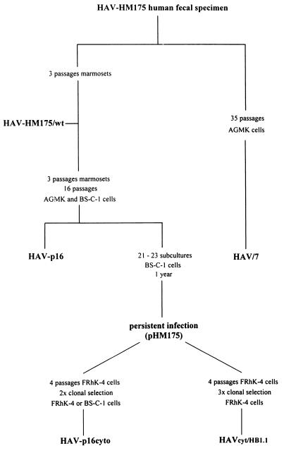
Passage history of variants of HAV strain HM175. The cytopathogenic variant HAVcyt/HB1.1 was isolated in our laboratory. HAV/7 (7), which was recovered after transfection of FRhK-4 cells with the RNA of the infectious cDNA clone pHAV/7 (7), was used as a reference in our experiments characterizing HAVcyt/HB1.1. Phenotypic and genotypic features of HAVcyt/HB1.1 were compared with those of the p16cyto viruses HM175/18f, -43c, and -24a (20), which were obtained independently from HAVcyt/HB1.1 from the same persistent infection (8).
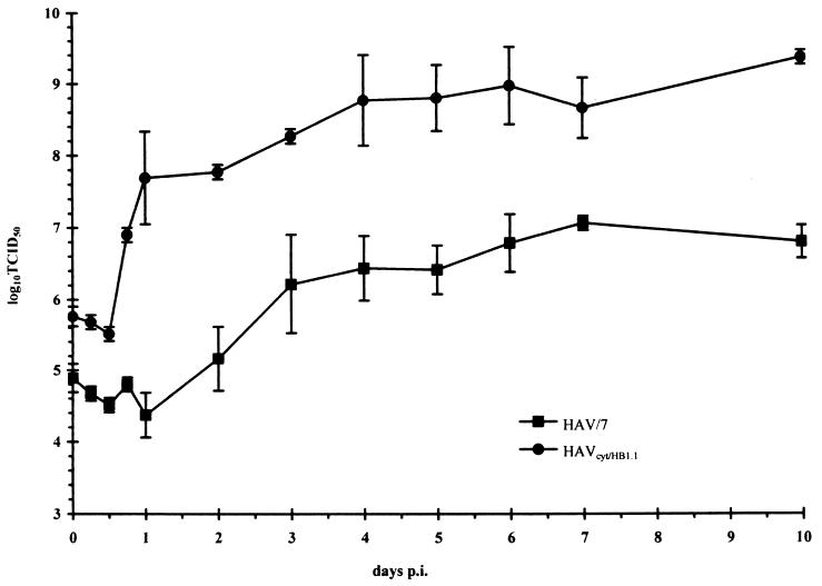
Replication kinetics under one-step growth curve conditions for HAVcyt/HB1.1 in FRhK-4 cells in comparison with HAV/7. The kinetics show the total titers in the course of 10 days. The cells were infected with an MOI of 5, and at the times indicated the 50% tissue culture infective dose (TCID50) titer was determined in FRhK-4 cells 2 weeks after inoculation by indirect immunofluorescence. Each data point is an average obtained from two separate experiments. Error bars indicate standard deviations of the means.
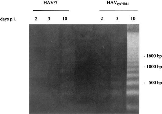
DNA fragmentation analysis of FRhK-4 cells infected with HAVcyt/HB1.1. FRhK-4 cells were infected with HAVcyt/HB1.1 or with HAV/7 at an MOI of 4. Cytoplasmic extracts from 2 × 10⁶ cells were prepared at 2, 3, and 10 days p.i., and DNA was extracted with phenol-chloroform. After treatment with RNase A, the samples were analyzed on a 1.6% agarose gel. Two days after infection, no DNA fragments indicative for apoptosis could be seen. From 3 days p.i. onward, increasing amounts of DNA fragments could be detected in HAVcyt/HB1.1-infected cells. In HAV/7-infected cells, no fragmentation occurred.
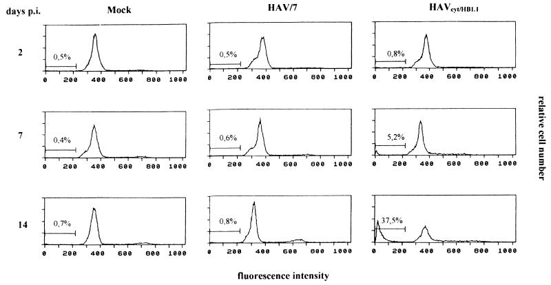
Flow cytometric DNA fluorescence profiles of HAVcyt/HB1.1-infected FRhK-4 cells in comparison with mock- and HAV/7-infected cells. At the days indicated, the cells were trypsinized and fixed with 70% ethanol. For nuclear staining, the cells were incubated with 50 μg of propidium iodide per ml for 30 min at room temperature. Apoptotic nuclei were identified as a subdiploid peak with a DNA content of less than 2 N. The percentages of cells with a DNA content of less than 2 N are indicated (see also Table 2).
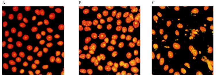
Nuclear morphology in FRhK-4 cells infected with HAVcyt/HB1.1. FRhK-4 cells were infected with HAVcyt/HB1.1 or HAV/7. At 10 days p.i., the cells were prepared for nuclear staining with propidium iodide (orange) and for HAV antigen detection by indirect immunofluorescence (green) with the HAV-neutralizing monoclonal antibody 7E7. Nuclear changes, which are indicative for apoptosis, could not be detected in uninfected (A) and HAV/7-infected (B) cells but were detected in cells infected with HAVcyt/HB1.1 (C). In apoptotic cells, the nuclei are irregularly formed, show protuberances on the surface, and disintegrate into densely stained nuclear apoptotic bodies. The characteristic segregation of chromatin in apoptotic nuclear fragments and nuclear budding are evident. In HAVcyt/HB1.1-infected cells (C), an accumulation of viral antigen was detected.
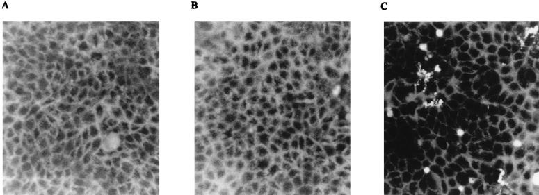
Generation of ROI in HAVcyt/HB1.1-infected FRhK-4 cells. Cells were incubated at day 3 p.i. with 2′,7′-dichlorofluorescein diacetate, a fluorescent probe for intracellular ROI, for 1 h and analyzed by fluorescence microscopy. ROI could not be detected in noninfected cells (A) or in HAV/7-infected cells (B) but were detected in cells infected with HAVcyt/HB1.1 (C).
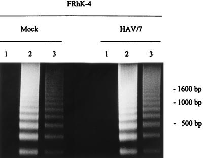
DNA fragmentation analysis of FRhK-4 cells and cells persistently infected with HAV/7 after treatment with actinomycin D and cycloheximide. FRhK-4 cells as well as cells persistently infected with HAV/7 were incubated with actinomycin D (1 μg/ml) (lanes 2) and cycloheximide (50 μg/ml) (lanes 3). Both inhibitors of macromolecular synthesis induced an apoptotic reaction in FRhK-4 cells and HAV/7-infected cells, which was revealed by DNA fragmentation after 1 day of incubation. No fragmentation occurred in cells not treated with the inhibitors (lanes 1).
References
-
- Ameisen J C, Capron A. Cell dysfunction and depletion in AIDS: the programmed cell death hypothesis. Immunol Today. 1991;12:102–105. - PubMed
-
- Anderson D A. Cytopathology, plaque assay, and heat inactivation of hepatitis A virus strain HM 175. J Med Virol. 1987;22:35–44. - PubMed
-
- Busciglio J, Yankner B A. Apoptosis and increased generation of oxygen species in Down’s syndrome neurons in vitro. Nature. 1995;378:776–779. - PubMed
-
- Buttke T M, Sandstrom P A. Oxidative stress as a mediator of apoptosis. Immunol Today. 1994;15:7–10. - PubMed
Publication types
MeSH terms
Substances
LinkOut - more resources
Full Text Sources
