. 1998 Apr;72(4):3459-63.
doi: 10.1128/JVI.72.4.3459-3463.1998.

Cleavage of human immunodeficiency virus type 1 proteinase from the N-terminally adjacent p6* protein is essential for efficient Gag polyprotein processing and viral infectivity

Affiliations

Cleavage of human immunodeficiency virus type 1 proteinase from the N-terminally adjacent p6* protein is essential for efficient Gag polyprotein processing and viral infectivity

U Tessmer et al. J Virol. 1998 Apr.

Abstract

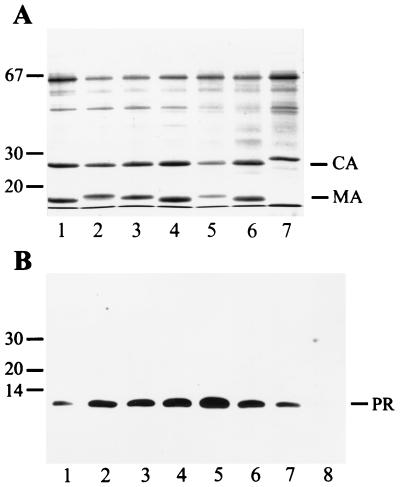
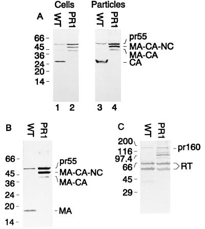
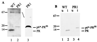
Maturation of infectious human immunodeficiency virus (HIV) particles requires proteolytic cleavage of the structural polyproteins by the viral proteinase (PR), which is itself encoded as part of the Gag-Pol polyprotein. Expression of truncated PR-containing sequences in heterologous systems has mostly led to the autocatalytic release of an 11-kDa species of PR which is capable of processing all known cleavage sites on the viral precursor proteins. Relatively little is known about cleavages within the nascent virus particle, on the other hand, and controversial results concerning the active PR species inside the virion and the relative activities of extended PR species have been reported. Here, we report that HIV type 1 (HIV-1) particles of four different strains obtained from different cell lines contain an 11-kDa PR, with no extended PR proteins detectable. Furthermore, mutation of the N-terminal PR cleavage site leading to production of an N-terminally extended 17-kDa PR species caused a severe defect in Gag polyprotein processing and a complete loss of viral infectivity. We conclude that N-terminal release of PR from the HIV-1 polyprotein is essential for viral replication and suggest that extended versions of PR may have a transient function in the proteolytic cascade.


Figures

FIG. 1
FIG. 1
Analysis of particle-associated Gag and PR proteins from several HIV-1 and HIV-2 strains. (A) Particles collected by sedimentation through a cushion of sucrose were separated on SDS-polyacrylamide gels, and proteins were detected by Coomassie blue staining. Lanes 1 to 4 show particles from MT4 cells, and lanes 5 to 7 show particles from C8166 cells. HIV-1 strains NL4-3 (lane 1), IIIB (lane 2), HXB2 (lanes 3 and 5), and SF2 (lanes 4 and 6) and HIV-2 strain CBL20 (lane 7) were analyzed in parallel. Molecular mass markers (in kilodaltons) are indicated on the left, and the positions of HIV CA and MA proteins are marked on the right. The 65-kDa protein is bovine serum albumin. (B) Immunoblot detection of particle-associated PR species. Proteins were separated on SDS-polyacrylamide gels and transferred to nitrocellulose membranes (Schleicher & Schuell) by electroblotting. Membranes were blocked with 10% low-fat dry milk and reacted with rabbit polyclonal antiserum against HIV-1 PR diluted 1:2,000 in buffer containing 5% low-fat dry milk and 0.01% Tween 20. Incubation was performed overnight at room temperature with shaking. Peroxidase-conjugated antirabbit antiserum (Jackson Immunochemicals Inc.) was used as the secondary antibody at a dilution of 1:10,000 and was incubated for 90 min at room temperature. Immune complexes were visualized with the ECL system (Amersham) according to the manufacturer’s instructions. In lane 1, 50 ng of purified PR was loaded; lanes 2 to 8 contain particle extracts from HIV-1 strains NL4-3 (lane 2), IIIB (lane 3), HXB2 (lanes 4 and 6), and SF2 (lanes 5 and 7) and HIV-2 strain CBL20 (lane 8), as described above. Note that 10 ng of purified PR was analyzed in the same experiment and was detected on a longer exposure.
FIG. 2
FIG. 2
Western blot analysis of gag and pol gene products after transient transfection. COS-7 cells transfected with pNL4-3 (WT) or pNL43-PR1 (PR1) and corresponding media were harvested 72 h after transfection. Lysates of transfected cells (A, lanes 1 and 2) and particles sedimented through a sucrose cushion (A, lanes 3 and 4, B, and C) were resolved on SDS-polyacrylamide gels. Western blots were stained with rabbit polyclonal antiserum to CA (A; dilution, 1:500) (16), with sheep polyclonal antiserum to MA (B; dilution, 1:200), or with rabbit polyclonal antiserum to RT (C; dilution, 1:200) (16). Alkaline phosphatase-conjugated antirabbit or antisheep antisera (Jackson Immunochemicals Inc.) were used as the secondary antibody, and immune complexes were visualized by color reaction. Molecular mass standards (in kilodaltons) are shown on the left, and relevant HIV-specific proteins are indicated on the right.
FIG. 3
FIG. 3
Western blot analysis of PR species in wild-type (WT) and PR1-derived virus particles following E. coli expression. (A) Particles from WT (lane 1) or PR1-transfected (lanes 2 and 3) COS-7 cells were collected from the culture media and resolved on SDS-polyacrylamide gels. Western blots were stained with rabbit polyclonal antiserum to PR and were visualized by ECL. In lane 3, particles collected from three transfections were combined to produce an increased signal and were analyzed on a different gel system, which gives higher resolution in the low-molecular-mass region of the gel (25). (B) E. coli BL21 DE3 cells were transformed with plasmids pHIV-proPII, containing either the WT sequence or the PR1 mutation. Bacteria were grown and induced as described previously (11), and samples of uninduced (u) and induced (i) bacteria were separated on SDS-polyacrylamide gels as indicated above each lane. The blot was reacted with rabbit polyclonal antiserum against PR, and alkaline phosphatase-conjugated antirabbit serum and immune complexes were detected by color reaction. P6*-PRm designates the fusion protein containing the Phe-to-Ile cleavage site mutation.

References

    1. Adachi A, Gendelman H E, Koenig S, Folks T, Willey R, Rabson A, Martin M A. Production of acquired immunodeficiency syndrome-associated retrovirus in human and nonhuman cells transfected with an infectious molecular clone. J Virol. 1986;59:284–291. - PMC - PubMed
    1. Almog N, Roller R, Arad G, Passi-Even L, Wainberg M A, Kotler M. A p6pol-protease fusion protein is present in mature particles of human immunodeficiency virus type 1. J Virol. 1996;70:7228–7232. - PMC - PubMed
    1. Beissinger M, Paulus C, Bayer P, Wolf H, Rösch P, Wagner R. Sequence-specific resonance assignments of the 1H-NMR spectra and structural characterization in solution of the HIV-1 transframe protein p6*. Eur J Biochem. 1996;237:383–392. - PubMed
    1. Chen C, Okayama H. High-efficiency transformation of mammalian cells by plasmid DNA. Mol Cell Biol. 1987;7:2745–2752. - PMC - PubMed
    1. Co E, Koelsch G, Lin Y, Ido E, Hartsuck J A, Tang J. Proteolytic processing mechanisms of a miniprecursor of the aspartic proteinase of human immunodeficiency virus type 1. Biochemistry. 1994;33:1248–1254. - PubMed

Publication types