. 1998 Apr 15;18(8):2808-21.
doi: 10.1523/JNEUROSCI.18-08-02808.1998.

Functional evidence that BDNF is an anterograde neuronal trophic factor in the CNS

Affiliations

Functional evidence that BDNF is an anterograde neuronal trophic factor in the CNS

J P Fawcett et al. J Neurosci. .

Abstract

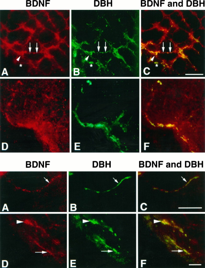
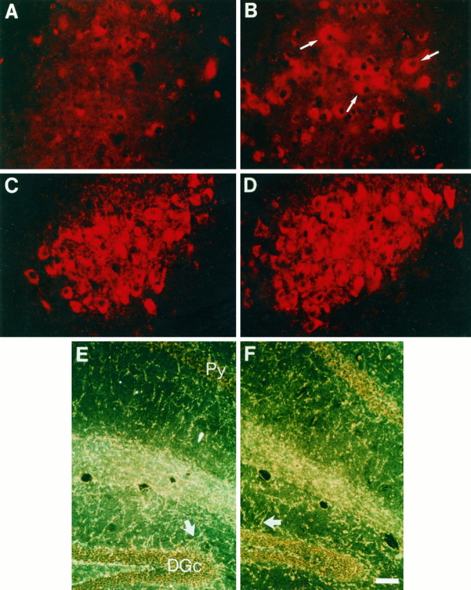
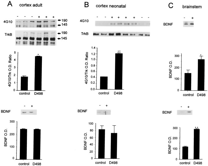
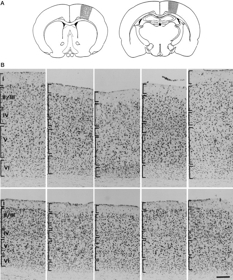
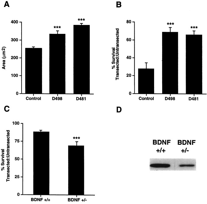
In this report, we have tested the hypothesis that brain-derived neurotrophic factor (BDNF) is an anterograde neurotrophic factor in the CNS and have focused on central noradrenergic neurons that synthesize BDNF. Double-label immunocytochemistry for BDNF and dopamine-beta-hydroxylase (DBH), a marker for noradrenergic neurons, demonstrated that BDNF is partially localized to noradrenergic nerve fibers and terminals in the adult rat brain. To test the functional importance of this anterograde BDNF, we analyzed transgenic mice carrying a DBH-BDNF minigene. Increased synthesis of BDNF in noradrenergic neurons of DBH-BDNF mice caused elevated TrkB tyrosine kinase activation throughout postnatal life in the neocortex, a noradrenergic target region. This afferently regulated increase in TrkB receptor activity led to long-lasting alterations in cortical morphology. To determine whether noradrenergic neuron-expressed BDNF also anterogradely regulated neuronal survival, we examined a second noradrenergic target, neonatal facial motoneurons. One week after axotomy, 72% of facial motoneurons were lost in control animals, whereas only 30-35% were lost in DBH-BDNF transgenic mice. Altogether, these results indicate that BDNF is anterogradely transported to fibers and terminals of noradrenergic neurons, that anterogradely secreted BDNF causes activation of TrkB in target regions, and that this secretion has functional consequences for target neuron survival and differentiation. This presynaptic secretion of BDNF may provide a cellular mechanism for modulating neural circuitry, in either the developing or mature nervous system.


Figures

Fig. 1.
Fig. 1.
Colocalization of BDNF and dopamine-β-hydroxylase (DBH) in fibers and nerve terminals in the adult rat brain. Fluorescence photomicrographs of the rat brainstem double-labeled with antibodies to BDNF (A, D, visualized using a CY3-labeled secondary antibody) and DBH (B, E, visualized using a CY2-labeled secondary antibody). C and F are photographic double exposures; in these panels, yellow indicates regions of double labeling. A–C, The region of noradrenergic innervation rostral to the ventrolateral reticular region. Noradrenergic neuron cell bodies are DBH-positive (asterisk) but are largely negative for BDNF, with some cells exhibiting faint BDNF immunoreactivity (large arrowhead). However, the processes of these noradrenergic neurons are largely positive for both BDNF and DBH (arrows). D–F, The ventrolateral periolivary region is immunoreactive for both the BDNF (D) and DBH antibody (E), with punctate staining that may represent terminals or cut fibers. Space bar, 50 μm.
Fig. 3.
Fig. 3.
A, B, BDNF expression is increased in the locus coeruleus of DBH–BDNF mice. Fluorescence photomicrographs at the level of the locus coeruleus in control (A) and line D481 DBH–BDNF (B) animals that were immunostained with an antibody specific to BDNF. Note that in control locus coeruleus, the level of immunostaining is not above background, whereas in the DBH–BDNF locus coeruleus, there are many BDNF-immunoreactive cell bodies (arrows). C–F, The level and pattern of dopamine-β-hydroxylase immunostaining is not altered in DBH–BDNF mice. C, D, Fluorescence micrographs at the level of the locus coeruleus in control (C) and line D481 DBH–BDNF (D) animals that were immunostained with an antibody specific to dopamine-β-hydroxylase, which recognizes noradrenergic and adrenergic neurons and fibers. The pattern and level of DBH immunostaining are similar in both cases.E, F, Dark-field micrographs of coronal sections of the hippocampus from control (E) and line D481 DBH–BDNF (F) brains that have been immunostained with anti-DBH and visualized with peroxidase. Both photographs derive from a similar level of the hippocampus, and the dentate granule cell (DGc) and pyramidal cell (Py) layers are marked. Note that in dark field the DBH-positive nerve fibers appear bright silver–yellow (arrows), and there are no apparent differences in the pattern or density of DBH-immunoreactive fibers in transgenic versus control animals. Scale bar, 100 μm.
Fig. 4.
Fig. 4.
A, B, Endogenous levels of TrkB autophosphorylation are increased in the cortex of DBH–BDNF mice, whereas BDNF levels are unchanged. Top, Cortical lysates from individual adult (A) and 1-week-old (B) control (−) and line D498 DBH–BDNF (+) animals were immunoprecipitated with anti-panTrk, and then analyzed by Western blots with antiphosphotyrosine (4G10). To ensure that the observed increases reflected an increase in the activation of TrkB, the blots were reprobed with anti-TrkBout (TrkB). Image analysis quantitation was used to normalize the level of autophosphorylation of the 145 kDa TrkB band relative to levels of TrkB protein. The normalized data (shown in the graphs, with ann of at least 3 individual animals in each case) were analyzed statistically for significance using a Student’st test. *p < 0.05; **p < 0.005. The size of the two TrkB isoforms is indicated by 145 and 190. Bottom, Western blot analysis of BDNF protein in the cortex of individual adult (A) and 1-week-old (B) control (−) and line D498 DBH–BDNF (+) mice. Thegraphs represent image analysis quantification of the data obtained on the same Western blot of three individual control and transgenic animals, with the optical density (O. D.) being arbitrary numbers. C, BDNF levels are increased in the brainstem of DBH–BDNF mice. Western blot analysis of BDNF protein in the brainstem of individual 1-week-old (top) and adult (bottom) control (−) versus line D498 DBH–BDNF (+) mice. The graphsrepresent image analysis quantification of the data obtained on the same Western blot of three individual control and transgenic animals, with the optical density (O. D.) being arbitrary numbers. Statistical analysis of these data demonstrates that BDNF is significantly increased in the brainstem of adult and neonatal DBH–BDNF animals. *p < 0.05; **p < 0.005.
Fig. 5.
Fig. 5.
Overexpression of BDNF in noradrenergic neurons leads to decreased neuronal numbers in the anterior neocortex.A, Schematic drawing showing the rostral and caudal levels that were analyzed for morphology of the neocortex.B, Photomicrographs of coronal Nissl-stained sections of the neocortex of control and DBH–BDNF transgenic mice.Rightmost and leftmost panels are from control animals, whereas the three innermost panels are all from line D498 DBH–BDNF animals. The top setrepresents the rostral level of the neocortex indicated inA, and the bottom set represents the caudal level. Brackets define approximate boundaries of the different cortical layers indicated in the leftmost panel. Scale bar, 70 μm.
Fig. 6.
Fig. 6.
A, Facial motorneurons are hypertrophied in neonatal DBH–BDNF transgenic mice. The average cross-sectional area of P12 facial motoneurons was determined by image analysis of cresyl violet-stained coronal sections of the appropriate brainstem level of DBH–BDNF mice of lines D498 and D481 versus their control littermates. In both lines of DBH–BDNF mice, facial motoneurons were significantly hypertrophied. ***p < 0.005. B, Neonatal facial motoneurons are rescued from axotomy-induced death in DBH–BDNF transgenic animals. The facial nerves of 5-d-old control and DBH–BDNF animals were unilaterally transected, and 7 d later, serial 16 μm sections were collected throughout the entirety of the facial nuclei. The number of facial motoneurons was then determined in the contralateral, control versus ipsilateral, transected facial nuclei by image analysis quantification of the number of facial motoneurons in every fifth section. The results of this analysis are presented as a ratio of the number of neurons in the transected versus untransected nuclei. The mean number of facial motoneurons within each group is presented within the text. Note that in control littermates, only 28% of facial motoneurons remain 1 week after a facial nerve transection, whereas in lines D498 and D481, 69 and 66% of facial motoneurons remain, respectively. ***p < 0.005;n = at least three animals in each group.C, Survival of transected facial motoneurons is reduced in adult BDNF+/− mice compared with their BDNF+/+ littermates. Methods similar to those described in B were used, with the exception that unilateral facial nerve transections were performed on 3-month-old BDNF+/− versus BDNF+/+ animals. The results of this analysis are presented as a ratio of the number of neurons in the transected versus untransected nuclei. Note that in BDNF+/+ littermates, 87% of motoneurons remain 1 week after a facial nerve transection, whereas in BDNF+/− mice, only 71% of facial motoneurons remain. ***p < 0.05; n = 3 animals in each group. D, BDNF levels are decreased in the BDNF+/− brain. Western blots of equal amounts of protein from the cortex of BDNF+/− versus BDNF+/+ mice were probed with an antibody to BDNF. A similar decrease was observed in other regions of the BDNF+/− brain (data not shown).

References

    1. Altar CA, Cai N, Bliven T, Juhasz M, Conner JM, Acheson AL, Lindsay R, Wiegand SJ. Anterograde transport of brain-derived neurotrophic factor and its role in the brain. Nature. 1997;389:856–860. - PubMed
    1. Altman J, Bayer SA. Vertical compartmentation and cellular transformations in the germinal matrices of the embryonic rat cerebral cortex. Exp Neurol. 1990;107:23–35. - PubMed
    1. Barde Y-A, Edgar D, Thoenen H. Purification of a new neurotrophic factor from mammalian brain. EMBO J. 1982;1:549–553. - PMC - PubMed
    1. Bayer SA, Altman J. Development of layer I and the subplate in the rat neocortex. Exp Neurol. 1990;107:48–62. - PubMed
    1. Bayer SA, Altman J. Neurogenesis and neuronal migration. In: Paxinos G, editor. The rat nervous system. Academic; San Diego: 1994. pp. 1041–1078.

Publication types

MeSH terms

Substances

LinkOut - more resources