. 1998 Apr 15;18(8):2891-906.
doi: 10.1523/JNEUROSCI.18-08-02891.1998.

Contributions of the optic tectum and the retina as sources of brain-derived neurotrophic factor for retinal ganglion cells in the chick embryo

Affiliations

Contributions of the optic tectum and the retina as sources of brain-derived neurotrophic factor for retinal ganglion cells in the chick embryo

K H Herzog et al. J Neurosci. .

Abstract

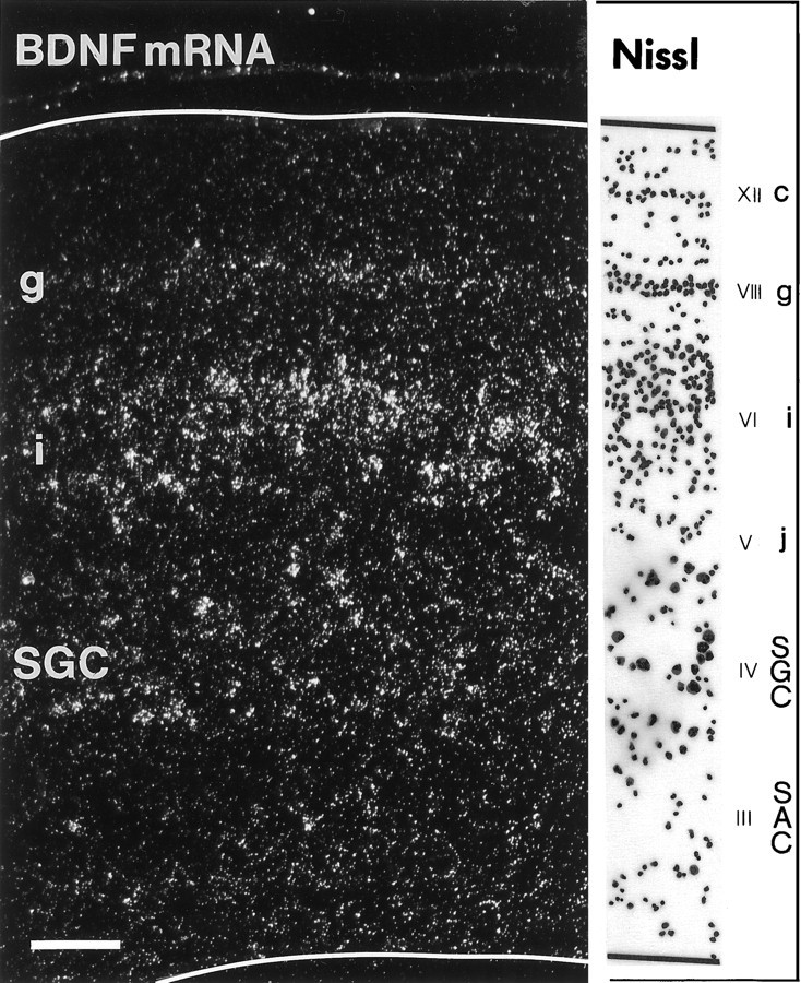
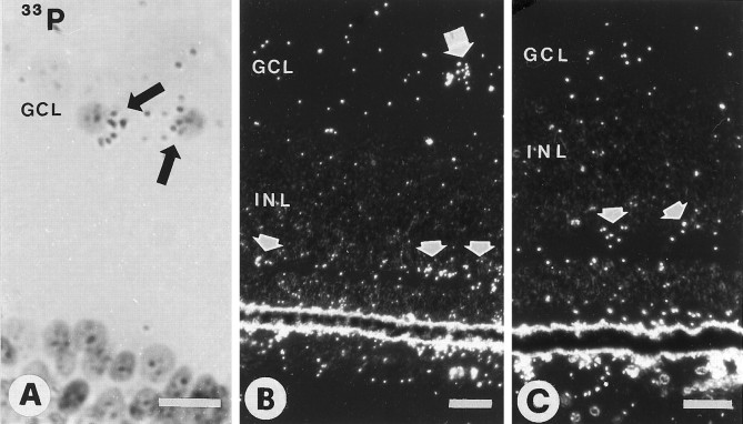
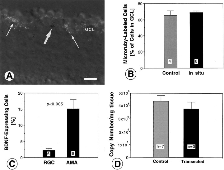
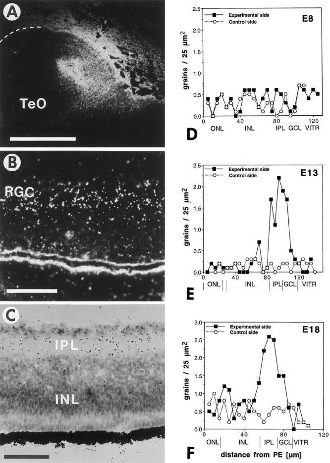
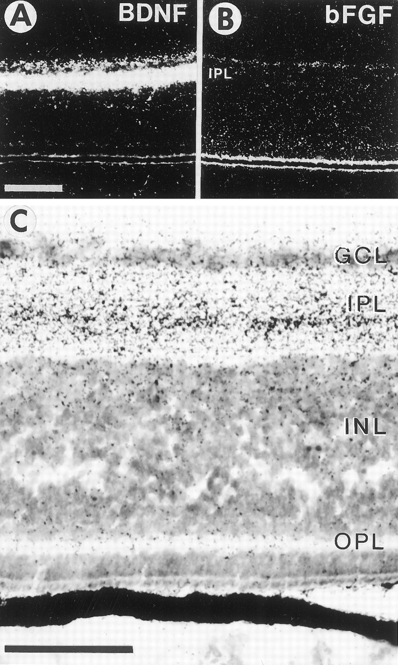
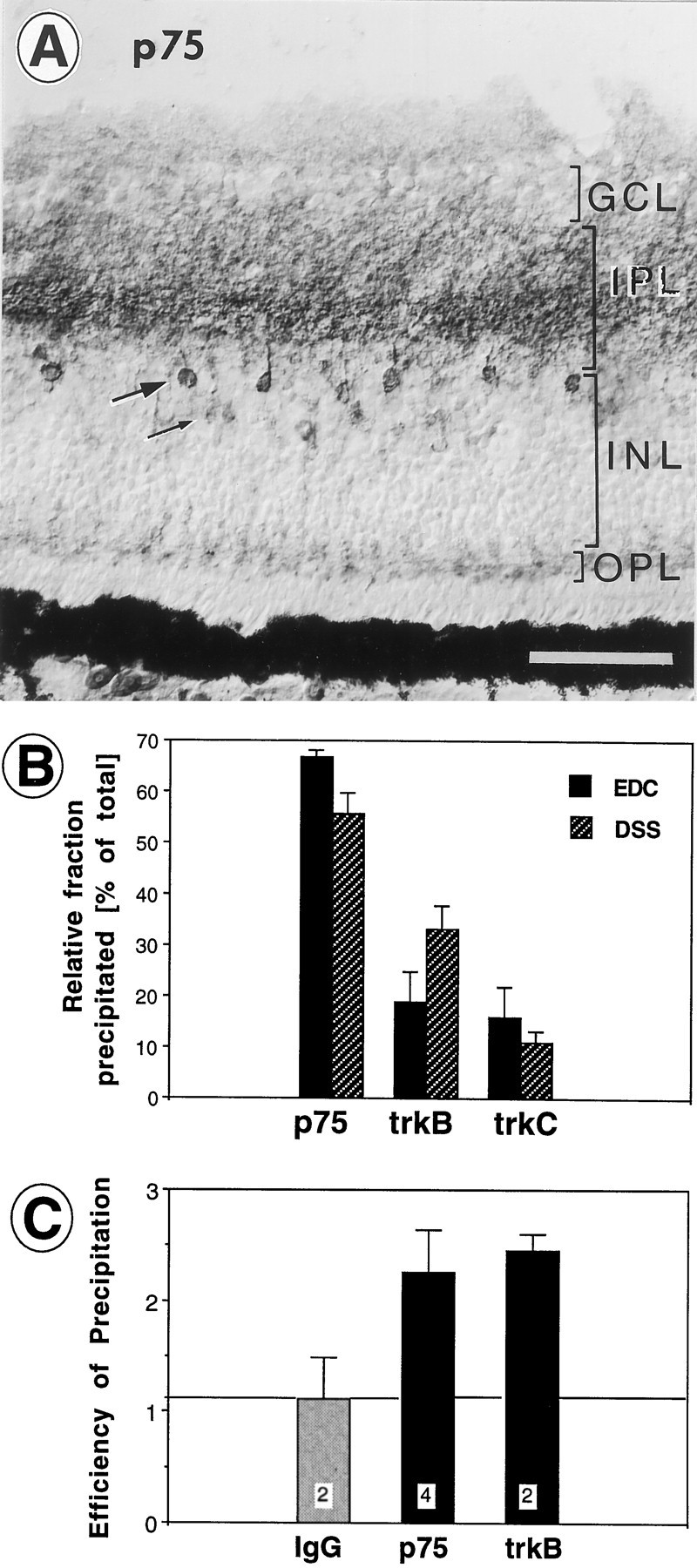
Retinal ganglion cells (RGC) are supported by brain-derived neurotrophic factor (BDNF), but it is not known if BDNF acts as a target-derived factor or as an afferent or autocrine trophic factor. Here we demonstrate that BDNF mRNA is expressed in the retinorecipient layer of the chick optic tectum as well as in the inner nuclear layer and ganglion cell layer of the retina. Amacrine cells rather than RGC were the main source of BDNF mRNA in the ganglion cell layer, as determined by in situ hybridization that was combined with retrograde labeling of RGC and destruction of RGC by optic stalk transection, followed by quantitative RT-PCR. Cells in the ganglion cell layer as well as the retinorecipient layers of the optic tectum were BDNF-immunolabeled. After injections into the tectum, radio-iodinated BDNF was transported to the retina where autoradiographic label accumulated in the inner plexiform and ganglion cell layers. After intraocular injection, iodinated BDNF accumulated in these same retinal layers and correlated with the distribution of p75 neurotrophin receptor protein. The majority of cross-linked receptor-bound BDNF in the retina immunoprecipitated with p75 antibodies. No difference in the intensity of BDNF immunolabel was observed in the experimental retina or tectum after optic stalk transection, indicating that most of the BDNF in the RGC was not derived from the optic tectum. These data indicate that a substantial fraction of the BDNF in the ganglion cell layer is derived from local sources, afferents within the retina, rather than from the optic tectum via retrograde transport.


Figures

Fig. 1.
Fig. 1.
Expression of BDNF mRNA in the optic tectum of a chick embryo at 15.5 d of incubation (E15.5). Dark-field view of a transverse section through the central region of the tectum hybridized with a 35S-labeled riboprobe for chicken BDNF. BDNF mRNA-expressing layers and the corresponding Nissl staining of the same section (camera lucida tracing) are indicated on theright [embryonic layers III–VI,VIII, and XII, according to LaVail and Cowan (1971)]. Note that the expression of BDNF is most intense in the deeper half of layer SGFSi. SGFS sublayers c,g, i, and j are indicated.SGC, Stratum griseum centrale; SAC, stratum album centrale. Scale bar, 100 μm.
Fig. 2.
Fig. 2.
BDNF mRNA expression in neurons of tectal layer SGFSi at E15.5. Note that silver grains (35S-labeled) frequently accumulate over the apical half of the soma (arrowheads) from which the apical dendrite extends into the retinorecipient layers. The pial surface of the tectum is toward the top of the micrograph. Scale bar, 10 μm.
Fig. 3.
Fig. 3.
Retinae of E15 chick embryos hybridized with33P-labeled riboprobes for chicken BDNF. A, Bright-field view of two labeled cells in the ganglion cell layer (GCL, arrows). B, Dark-field image of a labeled cell in the GCL (larger arrow) and several labeled cells in the outer half of the inner nuclear layer (INL, smaller arrows).C, Dark-field image of E15 retina after optic stalk transection at E4. Label in the outer half of the INL is maintained (arrows). Scale bars: 10 μm in A; 20 μm in B, C.
Fig. 4.
Fig. 4.
Identification and quantification of retinal ganglion cells (RGC) and amacrine cells with BDNF expression in the ganglion cell layer (GCL) of E15 chick embryos (A–C) and quantification of BDNF mRNA in the retina after degeneration of RGC induced by optic stalk transection (D). A, Section through the retina retrogradely labeled with Microruby (white grains) and hybridized for BDNF mRNA (dark silver grains). Note that many RGC are retrogradely labeled (thin arrows), but most of the BDNF-expressing cells (large arrow) are not fluorescent. Not all Microruby grains are in the focus plane. Scale bar, 20 μm. B, Comparison of the percentage of Microruby-labeled cells (RGC) in the GCL before and after hybridization. Note that there is no apparent quenching of the fluorescent signal. The number of visual fields (with ∼70 cells each) analyzed is indicated within white squares. Error bars indicate SEM. C, Percentages of BDNF-expressing cells for the RGC population (identified by labeling with Microruby) and for the amacrine cells (AMA; identified by the lack of fluorescent label). Note that <3% of RGC express BDNF, as compared with ≈15% of BDNF-expressing amacrine cells in the GCL. The number of visual fields analyzed (with a total of 392 cells) is indicated withinwhite squares. Error bars indicate SEM.D, BDNF mRNA was measured at age E17 by quantitative RT-PCR in normal retinae (Control) and compared with retinae lacking most RGC (Transected). BDNF mRNA levels were not reduced significantly in retinae after the destruction of their RGC. Error bars indicate SEM. Similar data were obtained for retinae from age E14.
Fig. 5.
Fig. 5.
Specificity of the BDNF antiserum in tissue sections. A, The BDNF antiserum specifically recognizes cells in the ganglion cell layer (GCL) of an E8.5 retina. B, No label was detected in theGCL after preincubation and precipitation of the serum with the B6 peptide coupled to Sepharose beads. Scale bar, 50 μm.
Fig. 6.
Fig. 6.
BDNF immunoreactivity in the retina.A, The ganglion cell layer (GCL) of an E7 retina is weakly labeled. B, At E8.5 newly formed cells adjacent to the ora serrata are BDNF-positive. These cells presumably represent retinal ganglion cells (RGC). C, In an E.8.5 retina BDNF is not visibly reduced in the GCL after optic stalk transection (X) at E4 (compare with Fig.5A). D, At E11 many cells in the normal GCL are strongly labeled. E, After optic stalk transection (X) many RGC have degenerated at E14. Some large cells display strong BDNF immunoreactivity.F, An E14 retina labeled with the BDNF antiserum at higher magnification displays heterogeneity of the label within the GCL. Scale bars: 25 μm in A–C, E; 50 μm in D; 10 μm in F.
Fig. 7.
Fig. 7.
BDNF immunocytochemistry in the optic tectum of a 16-d-old chick embryo. The left panel shows a section through the normal tectum (ipsilateral to the manipulated eye); theright panel shows a section through the experimental tectum (contralateral to the injection of exogenous BDNF in the eye). The two panels are from the same tissue section. Note that many more neurons in the i sublayer of the stratum griseum et fibrosum superficiale (SGFSi) are BDNF-labeled after injection in the contralateral eye. Label represents endogenous BDNF (not anterogradely transported exogenous BDNF), because it was not apparent in this tectal layer after the injection of radiolabeled BDNF in the eye. The induction of endogenous BDNF in layer SGFSi was not a specific effect of the exogenous BDNF in the eye, because intraocular injection of vehicle elicited the same effect (data not shown). Immunolabel of neurons in the stratum griseum centrale (SGC) or stratum opticum (SO) was not visibly altered by manipulations of the eye. Scale bar, 100 μm.
Fig. 8.
Fig. 8.
Retrograde transport of exogenous125I-labeled BDNF from the tectum to the retina.A, Dark-field view of an injection site in the tectum (TeO) of an 18-d-old (E18) chick embryo. The surface of the tectum is indicated with a dashed line. Dorsal is to the top, and lateral is to the left. Scale bar, 1 mm. B, Dark-field view of the contralateral retina of the same animal showing increased grain density over the inner nuclear layer and the ganglion cell layer, which comprises mostly retinal ganglion cells (RGC). Scale bar, 50 μm.C, Bright-field view of the contralateral retina of an E13 embryo showing increased grain densities (125I-BDNF) in the ganglion cell layer and inner plexiform layer (IPL) after injection into the tectum. The outer nuclear layer (ONL) is not labeled. D–F, Grain density profiles of I125-BDNF label in the retinae after unilateral injections into the tectum of E8 (D), E13 (E), and E18 (F) embryos. The filled squares represent averages from 10 traces through the contralateral retina; the open circlesrepresent averages from 10 traces through the ipsilateral (control) retina (background). The retinal layers and the distances from the pigment epithelium (PE) are indicated at thebottom. ONL, Outer nuclear layer;INL, inner nuclear layer; IPL, inner plexiform layer; GCL, ganglion cell layer;VITR, vitreous body. Note that iodinated BDNF accumulates in the retina at E13 and E18, but not at E8, and that most of the radioactivity accumulates in the IPL layer; a smaller amount is present in the GCL, which contains the retinal ganglion cell bodies.
Fig. 9.
Fig. 9.
Accumulation of radio-iodinated BDNF (A, C) and basic fibroblast growth factor (bFGF, B) in the retina after intraocular injection. A, Dark-field view of an autoradiographic section through the E16 retina after the injection of ∼10 ng of iodinated BDNF in the eye. Scale bar, 100 μm. B, Dark-field view of an autoradiographic section through the E16 retina after the injection of ∼10 ng of bFGF. Note that bFGF does not accumulate significantly in any retinal layer. C, Bright-field view at a higher magnification of the same retina shown inA. Note that BDNF accumulates differentially in the retinal layers, with the highest grain densities in the central part of the inner plexiform layer (IPL), with moderate densities in the ganglion cell layer (GCL) and the inner border of the inner nuclear layer (INL), and with low densities in the outer plexiform layer (OPL). Scale bar, 50 μm.
Fig. 10.
Fig. 10.
A, Distribution of p75 receptor immunoreactivity in the central retina of a 16-d-old chick embryo. Note the heavy labeling in the central parts of the inner plexiform layer (IPL) and heavily labeled cell bodies and processes of presumptive amacrine cells in the inner margin of the inner nuclear layer (INL, large arrow). Faintly labeled cell bodies are located in the center of the INL (thin arrow). Light labeling is present in the ganglion cell layer (GCL) and in the outer plexiform layer (OPL). Endogenous p75 is abundant in layers in which exogenous BDNF accumulates (see Fig. 9C). Scale bar, 50 μm. B, Relative contributions of p75, trkB, and trkC to the fraction of BDNF that was bound to receptors and could be immunoprecipitated with antibodies after cross-linking with EDC or DSS. Averages of four to seven independent experiments. Note that the majority of receptor-bound BDNF binds to p75. Error bars indicate SEM. C, Relative efficiencies of the antibodies for immunoprecipitation of p75 and trkB. Samples of the immunoprecipitates (pellets) and supernatants were Western-blotted (6% SDS gel) and visualized with secondary horseradish peroxidase chemoluminescence or 125I-labeled p75 or trkB antibodies. The resulting bands at 75 kDa (p75) and 140 kDa (trkB), respectively, were quantified by densitometry, and the ratios of density values (pellet/supernatant) were compared. Irrelevant antiserum (IgG) did not immunoprecipitate the antigen, whereas the p75 and trkB antibodies showed approximately equal efficiencies for immunoprecipitation (average ratios of 2.2. and 2.5). Error bars indicate SEM. The number of independent experiments is indicated within white squares.

Similar articles

Cited by

References

    1. Acheson A, Conover JC, Fandl JP, DeChiara TM, Russell M, Thadani A, Squinto SP, Yancopoulos GD, Lindsay RM. A BDNF autocrine loop in adult sensory neurons prevents cell death. Nature. 1995;374:450–453. - PubMed
    1. AryPires R, Nakatani M, Rehen SK, Linden R. Developmentally regulated release of intraretinal neurotrophic factors in vitro. Int J Dev Neurosci. 1997;15:239–255. - PubMed
    1. Auerbach R, Kubal L, Folkman J. A simple procedure for the long-term cultivation of chick embryos. Dev Biol. 1974;41:391–394. - PubMed
    1. Ballarin M, Ernfors N, Persson H. Hippocampal damage and kainic acid injection induce a rapid increase in mRNA for BDNF and NGF in the rat brain. Exp Neurol. 1991;114:35–43. - PubMed
    1. Barbacid M. The Trk family of neurotrophin receptors. J Neurobiol. 1994;25:1386–1403. - PubMed

Publication types

MeSH terms

Substances