. 1998 Apr 15;18(8):2914-22.
doi: 10.1523/JNEUROSCI.18-08-02914.1998.

A Ca2+-independent receptor for alpha-latrotoxin, CIRL, mediates effects on secretion via multiple mechanisms

Affiliations

A Ca2+-independent receptor for alpha-latrotoxin, CIRL, mediates effects on secretion via multiple mechanisms

M A Bittner et al. J Neurosci. .

Abstract

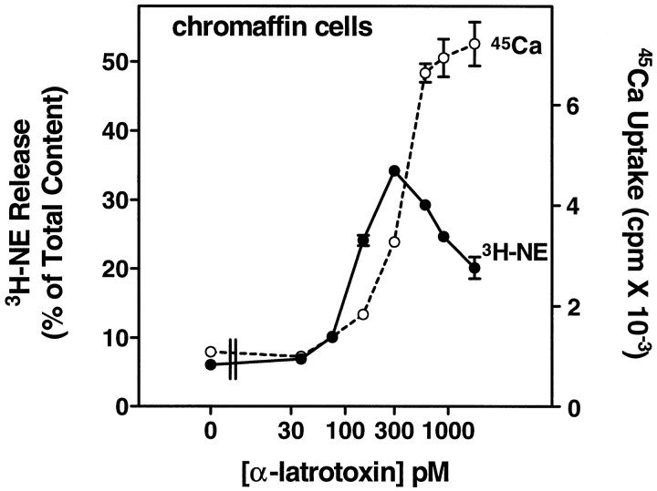
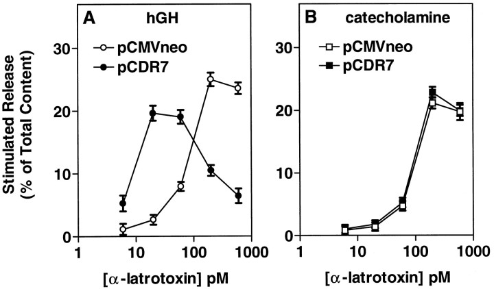
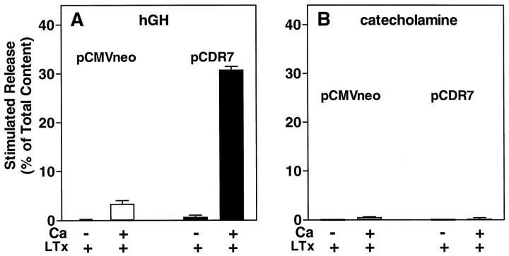
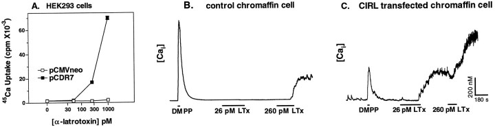
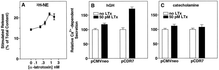
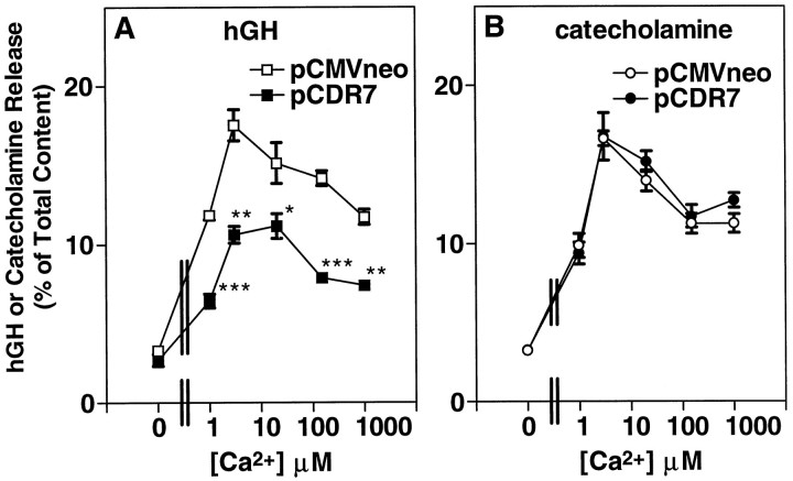
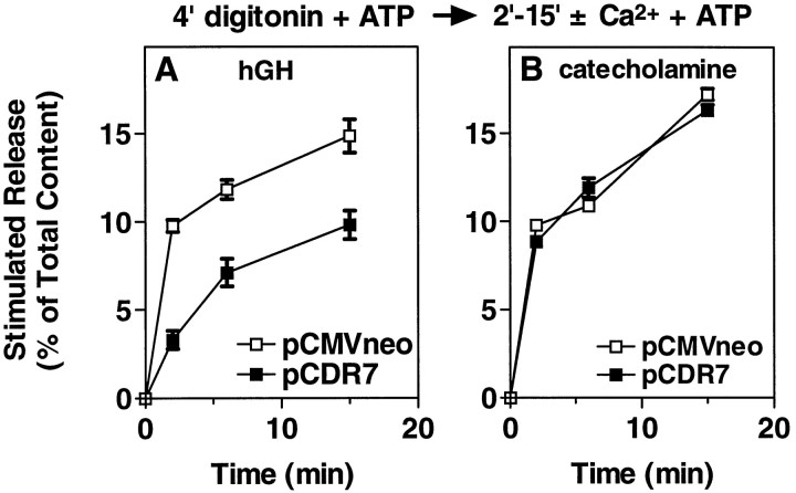
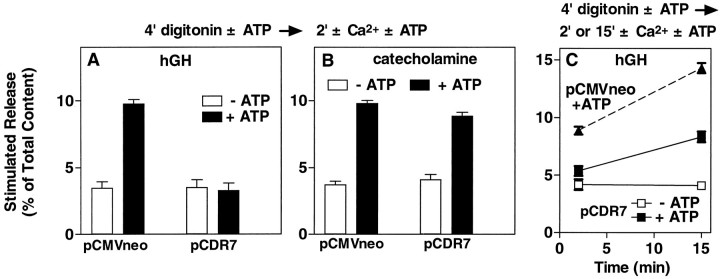
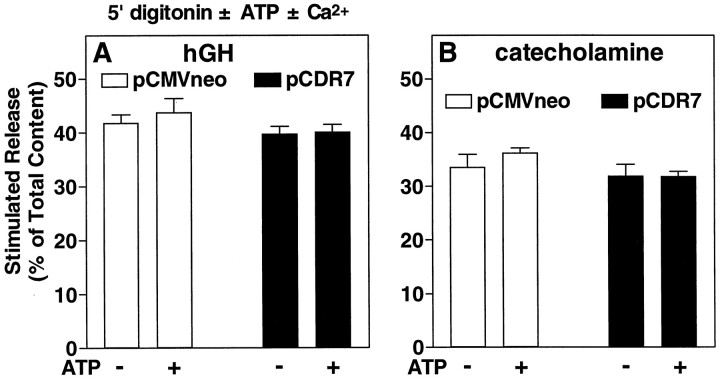
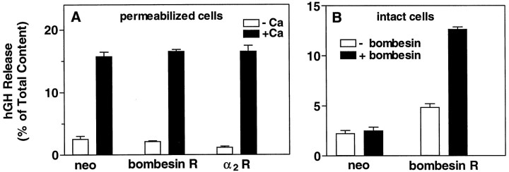
alpha-Latrotoxin (alpha-Ltx), a component of black widow spider venom, stimulates secretion from nerve terminals and from PC12 cells. In this study we examine the effects of expression of a newly cloned Ca2+-independent receptor for alpha-Ltx (CIRL) on secretion from bovine chromaffin cells. We first characterized the effect of alpha-Ltx on secretion from untransfected cells. alpha-Ltx, by binding in a Ca2+-independent manner to an endogenous receptor, causes subsequent Ca2+-dependent secretion from intact cells. The stimulation of secretion is correlated with Ca2+ influx caused by the toxin. In permeabilized cells in which the Ca2+ concentration is regulated by buffer, alpha-Ltx also enhances Ca2+-dependent secretion, indicating a direct role of the endogenous receptor in the secretory pathway. Expression of CIRL increased the sensitivity of intact and permeabilized cells to the effects of alpha-Ltx, demonstrating that this protein is functional in coupling to secretion. Importantly, in the absence of alpha-Ltx, the expression of CIRL specifically inhibited the ATP-dependent component of secretion in permeabilized cells without affecting the ATP-independent secretion. This suggests that this receptor modulates the normal function of the regulated secretory pathway and that alpha-Ltx may act by reversing the inhibitory effects of the receptor.


Figures

Fig. 1.
Fig. 1.
α-Latrotoxin stimulates catecholamine secretion and Ca2+ uptake in cultured chromaffin cells. Monolayer cultures of bovine adrenal chromaffin cells were labeled with3H-norepinephrine (3H-NE), rinsed, and incubated with the indicated concentrations of α-Ltx in physiological saline without Ca2+ or Mg2+ and with 0.1 mm EGTA for 4 min. The toxin was removed, and the cells were incubated for 6 min in PSS containing 2.2 mmCa2+ and 0.5 mm Mg2+. The incubation solution was removed, the cells were lysed with 1% Triton X-100, and the amount of 3H-NE was determined by liquid scintillation spectrometry; n = 3 wells/group. Parallel cultures that had not been labeled with3H-NE also were incubated with α-Ltx, followed by an incubation in PSS containing 2.2 mmCa2+, 0.5 mm Mg2+, and 3 μCi/ml 45Ca2+. After 4 min, the cells were rinsed immediately three times in PSS, and the amount of45Ca2+ in the cells was determined by liquid scintillation spectrometry; n = 4 wells/group.
Fig. 2.
Fig. 2.
Expression of CIRL increases the sensitivity of intact chromaffin cells to stimulation by α-Ltx. Chromaffin cells were transfected with plasmids for hGH (pXGH5) and either pCDR7, which encodes a newly cloned protein that binds α-Ltx (CIRL), or pCMVneo (a control) by calcium phosphate precipitates, as described. After 4 d the cells were incubated with the indicated concentrations of α-Ltx in PSS without Ca2+ or Mg2+ and with 0.1 mm EGTA. After 4 min, the toxin was removed, and the cells were incubated for an additional 5 min in PSS containing 2.2 mm Ca and 0.5 mm Mg. The amounts of hGH (A) and catecholamine (B) released into the medium and the amounts remaining in the cells were determined as described; n = 4 wells/group.
Fig. 3.
Fig. 3.
Secretion stimulated by α-latrotoxin requires extracellular Ca2+. Chromaffin cells were transfected with plasmids for hGH (pXGH5) and either pCDR7 or pCMVneo, as in Figure 2. After 4 d the cells were incubated for 10 min with or without 26 pm α-Ltx in either PSS without Ca2+ or Mg2+ and with 0.2 mm EGTA (indicated as −Ca), or PSS containing 2.2 mm Ca2+ and 0.5 mmMg2+ (indicated as +Ca). The amounts of hGH (A) and catecholamine (B) released into the medium and the amounts remaining in the cells were determined as described; n = 4 wells/group.
Fig. 4.
Fig. 4.
CIRL supports both Ca2+ entry and the sustained elevation of intracellular Ca2+elicited by α-latrotoxin. A, HEK293 cells were transfected with plasmids for green fluorescent protein (GFP) and either pCDR7 (CIRL) or pCMVneo by Lipofectamine, as described. After 2 d the cells were incubated with the indicated concentrations of α-Ltx in physiological saline without Ca2+ or Mg2+ and with 0.2 mm EGTA for 4 min. The incubation with toxin was followed by a 6 min incubation in PSS containing 2.2 mm Ca2+, 0.5 mm Mg2+, and 1 μCi/ml45Ca2+. The cells were rinsed immediately three times with PSS without45Ca2+, and the amount of45Ca2+ in the cells was determined by liquid scintillation spectrometry; n = 5 wells/group. B, C, Chromaffin cells were transfected with plasmids for GFP and either pCDR7 or pCMVneo, with GFP serving as a marker for the transfected cells. Cultures were loaded with 1 μm fura-2 AM and subsequently were perfused with a 10 μm concentration of a nicotinic agonist (dimethylphenylpiperazinium, DMPP), followed by the indicated concentrations of α-Ltx in PSS. The duration of each agonist application is indicated by a horizontal bar. Intracellular Ca2+ levels were obtained as described in Materials and Methods. Following are the mean resting Ca2+ levels: −CIRL, 56 ± 11 nm,n = 3; +CIRL, 45 ± 7 nm,n = 4.
Fig. 5.
Fig. 5.
Expression of CIRL increases the sensitivity of permeabilized chromaffin cells to the secretion-enhancing effects of α-Ltx. A, Monolayer cultures of nontransfected chromaffin cells were labeled with 3H-norepinephrine (3H-NE), rinsed, and incubated with the indicated concentrations of α-Ltx in physiological saline without Ca2+ or Mg2+ and with 0.2 mm EGTA for 4 min. The toxin was removed, and the cells were permeabilized for 6 min in KGEP buffer containing 20 μm digitonin and 2 mm MgATP, followed by incubation with or without 30 μm Ca2+in KGEP with 2 mm MgATP for 16 min. The incubation solution was removed, the cells were lysed with 1% Triton X-100, and the amount of 3H-NE was determined by liquid scintillation spectrometry. B, C, Chromaffin cells were transfected with plasmids for hGH (pXGH5) and either pCDR7 or pCMVneo, as in Figure 2. After 4–6 d the cells were incubated with the indicated concentrations of α-Ltx in PSS without Ca2+ or Mg2+ and with 0.1 mm EGTA. After 4 min the toxin was removed, and the cells were permeabilized with 20 μm digitonin in KGEP buffer without Ca2+ for 4 min, followed by incubation with or without 30 μm Ca2+ in KGEP for 15 min. MgATP (2 mm) was included in both incubations. The amounts of hGH (B) and catecholamine (C) released into the medium and the amounts remaining in the cells were determined as described. For each condition (± CIRL), release in the presence of α-Ltx was normalized to release in the absence of toxin. Actual values for Ca2+-dependent release of hGH in the absence of α-Ltx were 21.38 ± 0.80% (pCMVneo) and 15.75 ± 1.78% (pCDR7;p < 0.05 vs pCMVneo). Values for Ca2+-dependent release of catecholamine were 17.15 ± 0.59% (pCMVneo) and 17.49 ± 0.72% (pCDR7); n = 4 wells/group.
Fig. 6.
Fig. 6.
Expression of CIRL reduces secretion at all Ca2+ concentrations. Chromaffin cells were transfected with plasmids for hGH (pXGH5) and either pCDR7 or pCMVneo, as in Figure 2. After 4 d the cells were permeabilized with 20 μm digitonin in KGENP buffer (potassium glutamate, EGTA, nitrilotriacetic acid, and PIPES-containing solution; see Materials and Methods) without Ca2+ for 4 min, followed by incubation with the indicated Ca2+concentrations in KGENP for 15 min. MgATP (2 mm) was included in both incubations. The amounts of hGH (A) and catecholamine (B) released into the medium and the amounts remaining in the cells were determined as described; n = 4 wells/group. *p < 0.05 versus pCMVneo; **p< 0.001 versus pCMVneo; ***p < 0.0001 versus pCMVneo.
Fig. 7.
Fig. 7.
Time course of secretion in permeabilized chromaffin cells with or without overexpressed CIRL. Chromaffin cells were transfected with plasmids for hGH (pXGH5) and either pCDR7 or pCMVneo, as in Figure 2. After 4 d the cells were permeabilized with 20 μm digitonin in KGEP buffer without Ca2+ and with 2 mm MgATP for 4 min, followed by incubation with or without 30 μmCa2+ in KGEP containing 2 mm MgATP for the indicated times. The amounts of hGH (A) and catecholamine (B) released into the medium and the amounts remaining in the cells were determined as described;n = 4 wells/group.
Fig. 8.
Fig. 8.
Effect of overexpressed CIRL on ATP-dependent and ATP-independent secretion. Chromaffin cells were transfected with plasmids for hGH (pXGH5) and either pCDR7 or pCMVneo, as in Figure 2. After 4 d the cells were permeabilized with 20 μm digitonin in KGEP buffer without Ca2+ and with or without 2 mm MgATP for 4 min, followed by incubation with or without 30 μmCa2+ in the continuing presence or absence of 2 mm MgATP for 2 min (A, B) or for 2 and 15 min (C, a separate experiment). The amounts of hGH (A, C) and catecholamine (B) released into the medium and the amounts remaining in the cells were determined as described;n = 4 wells/group.
Fig. 9.
Fig. 9.
Effect of overexpressed CIRL on secretion stimulated in the presence of digitonin with and without exogenous ATP. Chromaffin cells were transfected with plasmids for hGH (pXGH5) and either pCDR7 or pCMVneo, as in Figure 2. After 6 d the cells were permeabilized with 20 μm digitonin in KGEP buffer with or without 30 μm Ca2+ and with or without 2 mm MgATP for 5 min. The amounts of hGH (A) and catecholamine (B) released into the medium and the amounts remaining in the cells were determined as described; n = 4 wells/group.
Fig. 10.
Fig. 10.
Effect of overexpressed G-protein-coupled receptors on secretion from digitonin-permeabilized chromaffin cells. Chromaffin cells were transfected with plasmids for hGH (pXGH5) and the α2 adrenergic receptor, the bombesin receptor, or pCMVneo, as in Figure 2. After 4 d the cells were permeabilized (A) with 20 μm digitonin in KGEP buffer without Ca2+ and with 2 mm MgATP for 4 min, followed by incubation with or without 30 μmCa2+ in KGEP containing 2 mm MgATP for 2 min. B, Intact cells transfected with either pCMVneo or bombesin receptor were challenged for 12 min with 20 nmbombesin in PSS containing 2.2 mm Ca2+and 0.5 mm Mg2+. The amounts of hGH and catecholamine (data not shown) released into the medium and the amounts remaining in the cells were determined as described;n = 4 wells/group.

References

    1. Barnett DW, Liu J, Misler S. Single-cell measurements of quantal secretion induced by α-latrotoxin from rat adrenal chromaffin cells: dependence on extracellular Ca2+. Pflügers Arch. 1996;432:1039–1046. - PubMed
    1. Battey JF, Way JM, Corjay MH, Shapira H, Kusano K, Harkins R, Wu JM, Slattery T, Mann E, Feldman RI. Molecular cloning of the bombesin/gastrin-releasing peptide receptor from Swiss 3T3 cells. Proc Natl Acad Sci USA. 1991;88:395–399. - PMC - PubMed
    1. Bittner MA, Holz RW. A temperature-sensitive step in exocytosis. J Biol Chem. 1992a;267:16226–16229. - PubMed
    1. Bittner MA, Holz RW. Kinetic analysis of secretion from permeabilized adrenal chromaffin cells reveals distinct components. J Biol Chem. 1992b;267:16219–16225. - PubMed
    1. Bittner MA, Bennett MK, Holz RW. Evidence that syntaxin 1A is involved in storage in the secretory pathway. J Biol Chem. 1996;271:11214–11221. - PubMed

Publication types

MeSH terms

LinkOut - more resources