. 1998 Apr 15;18(8):2954-61.
doi: 10.1523/JNEUROSCI.18-08-02954.1998.

Evidence for a tetrameric structure of recombinant NMDA receptors

Affiliations

Evidence for a tetrameric structure of recombinant NMDA receptors

B Laube et al. J Neurosci. .

Abstract

The amino acids L-glutamate and glycine are essential agonists of the excitatory NMDA receptor, a subtype of the ionotropic glutamate receptor family. The native NMDA receptor is composed of two types of homologous membrane-spanning subunits, NR1 and NR2. Here, the numbers of glycine-binding NR1 and glutamate-binding NR2 subunits in the NMDA receptor hetero-oligomer were determined by coexpressing the wild-type (wt) NR1 with the low-affinity mutant NR1(Q387K), and the wt NR2B with the low-affinity mutant NR2BE387A, subunits in Xenopus oocytes. In both cases, analysis of the resulting dose-response curves revealed three independent components of glycine and glutamate sensitivity. These correspond to the respective wild-type and mutant affinities and an additional intermediate hybrid affinity, indicating the existence of three discrete receptor populations. Binomial analysis of these data indicates the presence of two glycine and two glutamate binding subunits in the functional receptor. In addition, we analyzed the inhibitory effects of the negative dominant NR1(R505K) and NR2BR493K mutants on maximal inducible whole-cell currents of wt NR1/NR2B receptors. The inhibition profiles obtained on expression of increasing amounts of these mutant proteins again were fitted best by assuming an incorporation of two NR1 and two NR2 subunits into the receptor hetero-oligomer. Our data are consistent with NMDA receptors being tetrameric proteins that are composed of four homologous subunits.


Figures
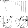
Fig. 1.
Fig. 1.
Membrane currents in oocytes expressing heteromeric NMDA receptors generated by coinjection of NR1, NR1Q387K, and NR2B subunits. A, Glycine-activated whole-cell currents in the presence of 100 μml-glutamate obtained at a holding potential of −70 mV. Oocytes injected with the wt NR1 and NR1Q387K cRNAs at a ratio of 1:1 were superfused with the glycine concentrations indicated above the application bars. Calibration: 200 nA, 15 sec. B, Glycine dose–response curves obtained from oocytes injected with NR1 and NR1Q387K (•) cRNAs at ratio of 1:1. Note the triphasic dose–response seen on coexpression of the wt and mutant NR1 subunits. Corresponding EC50 values for the high-affinity (HA), intermediate-affinity (IMA), and low-affinity (LA) components are 0.56 μm, 48 μm and 4.8 mm, with fractional contributions of 27, 44, and 29% of the maximal current, respectively. C, Glycine dose–response curves recorded from oocytes injected with the NR1 and NR1Q387KcRNAs at a ratios of 5:1 (▪) or 1:5 (▴), respectively. In both cases, clearly biphasic dose–response curves were obtained. The corresponding EC50 values of the HA- and IMA-, or IMA- and LA- components are 0.9 and 50 μm, or 84 μmand 7 mm, with fractional current contributions of 79 and 21%, or 26 and 74%, respectively, of the maximal current. In bothB and C, the solid linesrepresent least-squares fits of the data to the modified Hill Equation2. The dotted lines correspond to the dose–response curves of the pure NR1/NR2B and NR1Q387K/NR2B HA- and LA-components. Data from individual oocytes are shown; all measurements were repeated on at least three different oocytes with similar results. The mean (± SD) of the EC50 values and the Hill coefficients calculated from these experiments are given in Table 1. The subunit compositions predicted for tetrameric receptor complexes are indicated schematically (○ represents NR1 wt, • NR1Q387K, and ◍ NR2B).
Fig. 2.
Fig. 2.
Glycine affinities of the different NMDA receptor populations generated by coexpressing the wt NR1 and NR1Q387K subunits with NR2B. The EC50values of the HA-, IMA-, and LA-components obtained on coinjection of different ratios of the NR1 and NR1Q387K cRNAs as described in Figure 1 are plotted as a function of the relative fraction of the wt NR1 cRNA injected (the number of experiments is given in Table 1). The dashed lines represent the mean of the EC50 values determined at different cRNA ratios; error bars represent SD. Values in parentheses represent the relative fraction of the current contribution of each component. Note that the EC50 values of glycine for the different current components are independent of the relative ratios of cRNAs injected.
Fig. 3.
Fig. 3.
Membrane currents in oocytes coexpressing NR1, wt NR2B, and mutant NR2BE387A subunits. Glutamate dose–response curves obtained from oocytes injected with the NR2B and NR2BE387A cRNAs at ratios of (•) 1:1, (▪) 5:1, and (▴) 1:5 are shown. Note the clearly biphasic shape of the glutamate dose–response curves obtained at the 5:1 and 1:5 cRNA injection ratios. The corresponding EC50 values of the HA- and IMA-, or IMA- and LA-current components are 1.3 and 42 μm, or 22 and 360 μm, with fractional contributions of 68 and 32%, or 26 and 74%, respectively, of the maximal current. The solid lines represent least-squares fits of the modified Hill equation (Eq. 2) assuming the presence of three different channel species. The dashed linescorrespond to the dose–response curves of the pure NR2B and NR2BE387A high- and low-affinity components. Data from individual oocytes are shown; all measurements were repeated on at least three different oocytes with similar results. The mean (± SD) of the EC50 values and the Hill coefficients calculated from these experiments are summarized in Table 2. The subunit compositions predicted for the respective tetrameric NMDA receptor complexes are indicated schematically, with ○ representing NR2B wt, • NR2BQ387K, and ◍ NR1.
Fig. 4.
Fig. 4.
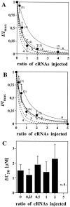
Incorporation of negative dominant NMDA receptor subunit mutants. A, Inactivation of NMDA receptor current on coexpression of the NR1 and NR1R505Ksubunits together with the NR2B protein. Membrane currents were determined in oocytes coinjected with different ratios of the NR1 and NR1R505K cRNAs; relative inducible whole-cell currents are plotted against increasing NR1R505K/NR1 cRNA ratios. The solid line represents the least-squares fit for the inactivation curve (calculated copy number of 2.2;r2 = 0.94). The dashed lines represent theoretical inactivation curves, assuming the presence of two (2), three (3), or four (4) NR1 subunits, and full inactivation on incorporation of a single NR1R505K polypeptide; these could be fitted to the experimental data with correlation coefficients ofr2 = 0.91, 0.75 and 0.58, respectively. The dotted line (a) corresponds to the expected inactivation curve for an NMDA receptor containing three copies of the NR1 subunit, assumingthat incorporation of a single copy of the negative dominant subunit would not impair channel function; the calculated correlation coefficient for our data isr2 = 0.64. B, Inactivation of NMDA receptor current on coexpression of the NR2B and NR2BR493K subunits together with the NR1 protein. Membrane currents were measured and analyzed as described inA. The least-squares fit for the inactivation curve gave a calculated copy number of 1.7 (r2 = 0.97). The respective correlation coefficients for the theoretical inactivation curves obtained from assuming the presence of two (2), three (3), or four (4) copies of the NR2 subunit arer2 = 0.93, 0.74, and 0.53, respectively. The inactivation curve predicted for three NR2 subunits with a putative single “silent” copy (a) fitted with a correlation coefficient of r2 = 0.74 only. Agonist concentrations used were 100 μm glutamate and 10 μm glycine. All data were normalized to the responses obtained on coinjecting the NR1 and the NR2B cRNAs alone and represent the mean ± SD (values in brackets represent the number of experiments). C, EC50 values obtained from the glutamate dose–response relations determined on coexpression of the NR2B and NR2BR493K subunits at the indicated cRNA ratios. Note that incorporation of the NR2BR493K mutant into the NMDA receptor has no significant effect on apparent glutamate affinity. n.d., Not determined because of a low current–response.

References

    1. Amin J, Weiss DS. GABAA receptor needs two homologous domains of the β-subunit for activation by GABA but not by pentobarbital. Nature. 1993;366:565–569. - PubMed
    1. Béhé P, Stern P, Wyllie D, Nasser M, Schoepfer R, Colquhoun D. Determination of NMDA NR1 subunit copy number in recombinant NMDA receptors. Proc R Soc Lond B Biol Sci. 1995;262:205–213. - PubMed
    1. Benveniste M, Mayer ML. Kinetic analysis of antagonist action at N-methyl-d-aspartic acid receptors. Two binding sites each for glutamate and glycine. Biophys J. 1991;59:560–573. - PMC - PubMed
    1. Betz H. Homology and analogy in transmembrane channel design: lessons from synaptic membrane proteins. Biochemistry. 1990;29:3591–3599. - PubMed
    1. Blackstone CD, Moss SJ, Martin LJ, Huganir R. Biochemical characterization and localization of a non-NMDA receptor in rat brain. J Neurochem. 1992;58:1118–1126. - PubMed

Publication types

MeSH terms

LinkOut - more resources