. 1998 Apr 15;18(8):2974-81.
doi: 10.1523/JNEUROSCI.18-08-02974.1998.

Evidence that increased hippocampal expression of the cytokine interleukin-1 beta is a common trigger for age- and stress-induced impairments in long-term potentiation

Affiliations

Evidence that increased hippocampal expression of the cytokine interleukin-1 beta is a common trigger for age- and stress-induced impairments in long-term potentiation

C A Murray et al. J Neurosci. .

Abstract

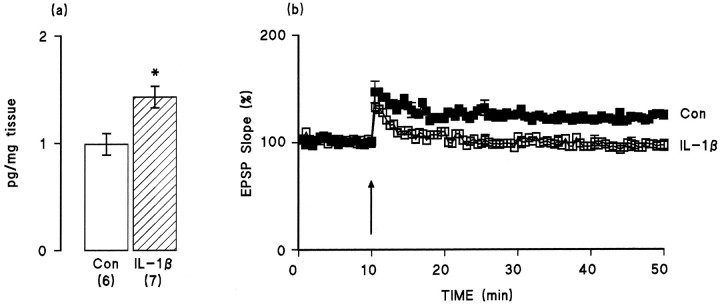
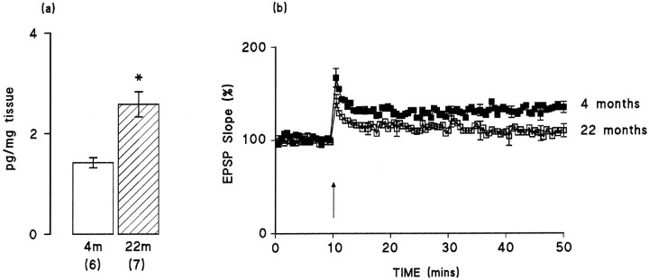
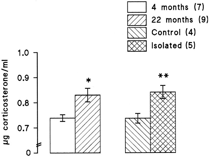
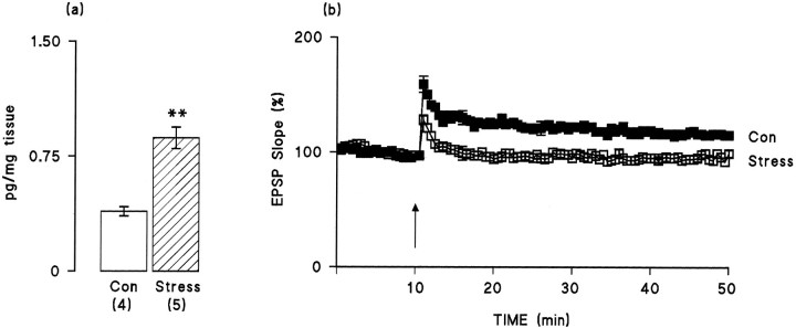
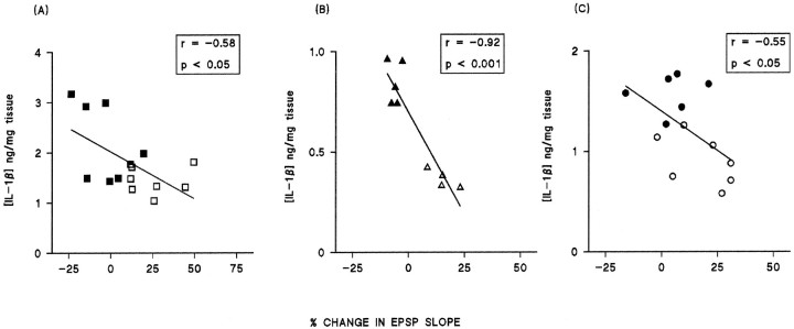
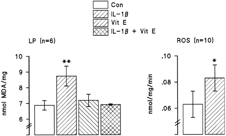
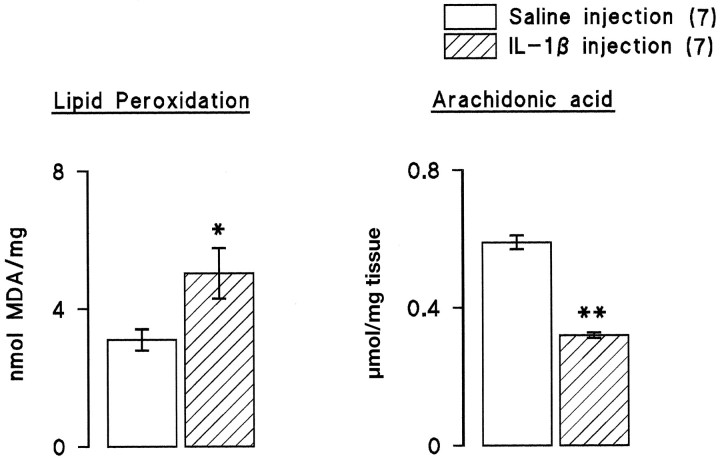
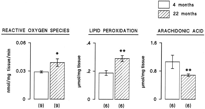
Several cytokines and their receptors are identified in brain; one of these is the proinflammatory cytokine interleukin-1beta that is synthesized and released from neurons and glia in response to stress or insult. Among the actions of interleukin-1beta is its ability to inhibit long-term potentiation in the hippocampus in vitro, an action that mimics one of the consequences of stress and age. It has been shown that the concentration of interleukin-1beta in brain tissue is increased in neurodegenerative conditions, and recent evidence from our laboratory has indicated an increase in the concentration of interleukin-1beta in the hippocampus of aged rats. These observations led us to consider that the underlying common cause of impaired long-term potentiation in aged and stressed rats might be increased endogenous interleukin-1beta concentration in hippocampus. The data presented here indicate that there was an inverse relationship between concentration of interleukin-1beta in the dentate gyrus and long-term potentiation in perforant path-->granule cell synapses in aged rats, stressed rats, and rats pretreated with interleukin-1beta. The evidence suggested that the cytokine induces formation of reactive oxygen species that triggers lipid peroxidation in vivo, as well as in vitro, and that these changes lead to depletion of membrane arachidonic acid that correlates with impaired long-term potentiation. We propose that three theories of aging, the glucocorticoid theory, the membrane theory, and the free radical theory, constitute three facets of age with one underlying trigger: an increase in the endogenous concentration of interleukin-1beta in hippocampus.


Figures

Fig. 1.
Fig. 1.
IL-1β concentration in dentate gyrus is increased and LTP is compromised after intraventricular injection of IL-1β. a, IL-1β concentration (picograms per milligrams of tissue) in dentate gyrus was significantly increased (p < 0.01, Student’s ttest; hatched bars) after intraventricular injection of IL-1β (10 μl, 3.5 ng/ml) 30 min before recording commenced compared with saline injection (open bars). Values are mean ± SEM. The number of observations is given inparentheses. b, Induction of LTP was not significantly affected by pretreatment with IL-1β, but EPSP slope decreased to baseline levels ∼20 min after tetanic stimulation (arrow). Values are given as mean percent of change in the slope of the EPSP over a 50 min period (n = 7 in both groups). Error bars indicating SE are included at 5 min intervals; in some instances these are so small as to be obscured by the symbols.
Fig. 2.
Fig. 2.
IL-1β concentration in dentate gyrus is increased and LTP is compromised in 22-month-old compared with 4-month-old rats. a, IL-1β concentration (picograms per milligram of tissue) in dentate gyrus was significantly increased (p < 0.01, Student’s ttest; hatched bars) in aged rats compared with young rats (open bars). Values are mean ± SEM, and the number of observations is given in parentheses.b, The mean percent of change in EPSP slope immediately after the tetanus was similar in both groups of rats, but EPSP slope in the last 5 min of the experiment was significantly reduced in the aged rats (n = 10) compared with the young rats (n = 7; p < 0.01, Student’st test). Error bars indicating SE are included at 5 min intervals; in some instances these are so small as to be obscured by the symbols.
Fig. 3.
Fig. 3.
Circulating corticosteroid concentrations were elevated in mildly stressed rats and in aged rats. There was a significant increase in circulating corticosteroids in 22-month-old compared with 4-month-old rats (p < 0.05, Student’s t test) and similarly in rats that had been housed in isolation, i.e., mildly stressed rats, compared with the control rats that had been housed as a group (p < 0.01, Student’s ttest). Analysis was made in samples obtained from rats at the end of electrophysiological recording in all cases. The number of observations is given in parentheses.
Fig. 4.
Fig. 4.
IL-1β concentration in dentate gyrus is increased and LTP is compromised in rats that were mildly stressed by isolation-housing compared with control rats. a, IL-1β concentration (picograms per milligram of tissue) was significantly increased (p < 0.01, Student’st test; hatched bars) in mildly stressed rats compared with control rats (open bars). Values are mean ± SEM, and the number of observations is given inparentheses. b, Induction of LTP was significantly reduced in the mildly stressed rats compared with the control rats (p < 0.05, Student’st test), and the mean percent of EPSP slope returned to baseline levels in ∼10 min in the stressed rats (n = 6), whereas it remained elevated for the duration of the experiment in the control rats (n = 4). Values are given as mean percent of change in the slope of the EPSP over a 50 min period. Error bars indicating SE are included at 5 min intervals; in some instances these are so small as to be obscured by the symbols.
Fig. 5.
Fig. 5.
Maintenance of LTP is inversely proportional to IL-1β concentration in hippocampus. Regression analysis on data from aged rats compared with young (a), individually housed rats compared with group-housed (b), and IL-1β-pretreated rats compared with saline-pretreated (c) revealed that there was a statistically significant correlation between IL-1β concentration and the mean percent of change in EPSP slope in the last 5 min of the experiment. Regression (r) and p values are indicated.
Fig. 6.
Fig. 6.
IL-1β induces lipid peroxidation and reactive oxygen species production in hippocampal tissue in vitro. IL-1β (3.5 ng/ml) significantly increased lipid peroxidation in hippocampal homogenate (p < 0.001) and this was inhibited by vitamin E (200 μm). IL-1β also significantly increased formation of reactive oxygen species (p < 0.01, Student’st test). The number of observations is given inparentheses.
Fig. 7.
Fig. 7.
Intraventricular injection of IL-1β increases lipid peroxidation and decreases membrane arachidonic acid concentration in hippocampus. Untetanized dentate gyri from the saline-injected and IL-1β-injected rats were dissected at the end of the period of electrophysiological recording, stored as described in Materials and Methods, and analyzed for lipid peroxidation and membrane arachidonic acid concentration. There was a significant increase in lipid peroxidation (p < 0.05, Student’st test), and this was accompanied by a significant decrease in membrane arachidonic acid concentration (p < 0.001, Student’s ttest). The number of observations is given inparentheses.
Fig. 8.
Fig. 8.
Aging was associated with increased reactive oxygen species formation and lipid peroxidation, coupled with a decrease in membrane arachidonic acid. Untetanized dentate gyri from 4- and 22-month-old rats were dissected at the end of the period of electrophysiological recording, stored as described in Materials and Methods, and analyzed for reactive oxygen species production, lipid peroxidation, and membrane arachidonic acid concentration. Reactive oxygen species production and lipid peroxidation were significantly increased in 22-month-old compared with 4-month-old rats (p < 0.05 and p < 0.01, respectively, Student’s t test). Mean arachidonic acid concentration was significantly decreased in 22-month-old compared with 4-month-old rats (p < 0.01, Student’st test). The number of observations is given inparentheses.

References

    1. Ban E, Milon G, Prudhomme N, Fillion G, Haour F. Receptors for interleukin-1 (α and β) in mouse brain: mapping and neuronal localization in hippocampus. Neuroscience. 1991;43:21–30. - PubMed
    1. Barnes CA. Memory deficits associated with senescence: a neurophysiological and behavioural study in the rat. J Comp Physiol Psychol. 1979;93:74–104. - PubMed
    1. Bellinger FP, Madamba S, Siggins GR. Interleukin-1β inhibits synaptic strength and long-term potentiation in the rat CA1 hippocampus. Brain Res. 1993;628:227–234. - PubMed
    1. Bodnoff SR, Humphreys AG, Lehman JC, Diamond DM, Rose GM, Meaney M. Enduring effects of chronic corticosterone treatment on spatial learning, synaptic plasticity, and hippocampal neuropathology in young and mid-aged rats. J Neurosci. 1995;15:61–69. - PMC - PubMed
    1. Bradford MM. A rapid and sensitive method for the quantitation of microgram quantities of proteins utilizing the principle of protein dye binding. Anal Biochem. 1976;72:248–254. - PubMed

Publication types