. 1998 Apr 15;18(8):3088-97.
doi: 10.1523/JNEUROSCI.18-08-03088.1998.

Overtraining does not mitigate contextual fear conditioning deficits produced by neurotoxic lesions of the basolateral amygdala

Affiliations

Overtraining does not mitigate contextual fear conditioning deficits produced by neurotoxic lesions of the basolateral amygdala

S Maren. J Neurosci. .

Abstract

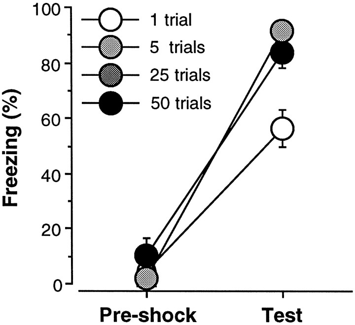
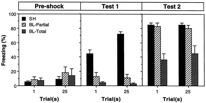
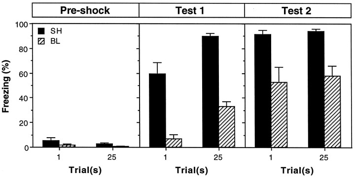
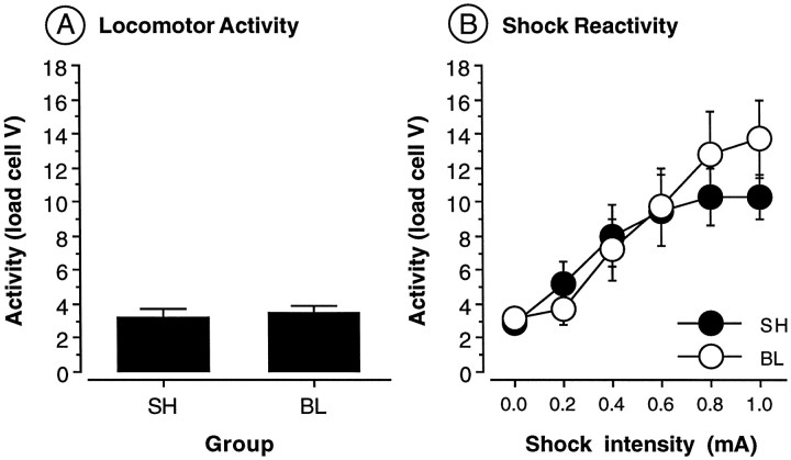
The influence of overtraining on the magnitude of fear-conditioning deficits produced by neurotoxic lesions of the basolateral amygdala (BLA) was examined. Either 1 d before or 1 week after the administration of neurotoxic BLA lesions, rats received either 1 or 25 conditioning trials consisting of the delivery of unsignaled foot shock in a novel observation chamber; freezing served as the measure of conditional fear. In this conditioning paradigm, asymptotic performance is reached in five conditioning trials, and 25 conditioning trials constitutes an overtraining procedure. The results revealed that overtraining does not affect the magnitude of the contextual freezing deficits produced by post-training BLA lesions. Similarly, overtraining did not influence the level of reacquisition obtained by rats with post-training BLA lesions after 10 reacquisition trials. A similar pattern of results was observed in rats with pretraining BLA lesions. Neurotoxic BLA lesions did not alter either motor activity or shock reactivity. These results indicate that overtraining does not limit the important role of the BLA in the acquisition and expression of contextual fear conditioning.


Figures

Fig. 1.
Fig. 1.
Number of conditioning trials required for overtraining. Mean ± SEM percentage of freezing during the 3 min period before foot shock on the conditioning day (Pre-shock) and during the 4 min context extinction test conducted 24 hr after conditioning (test). Rats received 1, 5, 25, or 50 conditioning trials consisting of the delivery of unsignaled foot shock. Asymptotic freezing was reached with five conditioning trials, and additional training did not increase the levels of freezing beyond the level obtained with five trials.
Fig. 2.
Fig. 2.
Photomicrographs showing representative thionin-stained coronal brain sections at two rostral–caudal levels from a rat receiving sham surgery (sham), a rat with a partial lesion of the basolateral amygdala (Partial BL), a rat with a total BLA lesion (Total BL), and a rat with a combined lesion of the BLA and CEA (BL + CE). Rostral sections are shown at two magnifications. Note the spared BLA neurons in the partial BL rat and the complete loss of these neurons in the total BL rat. Refer to Figure 3 for the position and labeling of the amygdaloid nuclei.
Fig. 3.
Fig. 3.
Schematic representation of the extent of partial lesions of the BLA (black), total BLA lesions (gray), and combined lesions of the BLA and CEA (hatched). Unilateral representations of bilateral lesions are shown so that the damaged areas can be visualized with respect to the labeled areas. BL, Basolateral nucleus;BM, basomedial nucleus; CE, central nucleus; ENT, entorhinal cortex; LA, lateral nucleus. Coronal brain section images adapted from Swanson (1992).
Fig. 4.
Fig. 4.
Post-training neurotoxic lesions of the BLA and freezing. Mean ± SEM percentage of freezing during the 3 min period before foot shock on the conditioning day (Pre-shock) and during the 8 min context extinction tests conducted 1 week after conditioning and recovery from surgery (test 1) and 24 hr after reacquisition (test 2). Rats received either 1 or 25 conditioning trials consisting of the delivery of unsignaled foot shock. Data are shown from rats that received sham surgery (SH; black bars), or intra-amygdaloid infusion of NMDA. Intra-amygdaloid NMDA resulted in either partial lesions of the basolateral amygdala (BL-Partial; hatched bars) or total lesions of the basolateral amygdala (BL-Total;gray bars).
Fig. 5.
Fig. 5.
Pretraining neurotoxic lesions of the BLA and freezing. Mean ± SEM percentage of freezing during the 3 min period before foot shock on the conditioning day (Pre-shock) and during the 8 min context extinction tests conducted 24 hr after conditioning (test 1) and reacquisition (test 2). Surgery was performed 1 week before conditioning. On the conditioning day, rats received either 1 or 25 conditioning trials consisting of the delivery of unsignaled foot shock. Data are shown from rats that received sham surgery (SH; black bars) or BLA lesions (BL; hatched bars).
Fig. 6.
Fig. 6.
Locomotor activity and shock sensitivity in rats with BLA lesions. A, Mean ± SEM activity (load cell V) in rats receiving either sham surgery (SH) or BLA lesions (BL).B, Mean ± SEM activity (load cell V) across six foot shock intensities in rats receiving either sham surgery (SH) or BLA lesions (BL).
Fig. 7.
Fig. 7.
Pretraining or post-training neurotoxic lesions of the BLA and CEA. Mean ± SEM percentage of freezing during the 3 min period before foot shock on the conditioning day (Pre-shock) and during the 8 min context extinction tests conducted 2 weeks after conditioning (test 1) and 24 hr after reacquisition (test 2). Surgery was performed 2 weeks before conditioning for rats in the pretraining condition [BL+CE (Pre); open squares] and 1 d after conditioning for rats in the post-training condition [BL+CE (Post); open triangles]. The sham group (SH; filled circles) is composed of rats that received sham surgery either 1 week before or 1 d after conditioning. On the conditioning day, all rats received 25 conditioning trials consisting of the delivery of unsignaled foot shock.

References

    1. Brady JV, Schreiner L, Geller I, Kling A. Subcortical mechanisms in emotional behavior: the effect of rhinencephalic injury upon the acquisition and retention of a conditioned avoidance response in cats. J Comp Physiol Psychol. 1954;47:179–186. - PubMed
    1. Campeau S, Davis M. Involvement of the central nucleus and basolateral complex of the amygdala in fear conditioning measured with fear-potentiated startle in rats trained concurrently with auditory and visual conditioned stimuli. J Neurosci. 1995;15:2301–2311. - PMC - PubMed
    1. Campeau S, Miserendino MJ, Davis M. Intra-amygdala infusion of the N-methyl-d-aspartate receptor antagonist AP5 blocks acquisition but not expression of fear-potentiated startle to an auditory conditioned stimulus. Behav Neurosci. 1992;106:569–574. - PubMed
    1. Clugnet MC, LeDoux JE. Synaptic plasticity in fear conditioning circuits: induction of LTP in the lateral nucleus of the amygdala by stimulation of the medial geniculate body. J Neurosci. 1990;10:2818–2824. - PMC - PubMed
    1. Davis M, Rainnie D, Cassell M. Neurotransmission in the rat amygdala related to fear and anxiety. Trends Neurosci. 1994;17:208–214. - PubMed

Publication types

LinkOut - more resources