. 1998 Apr;18(4):1802-11.
doi: 10.1128/MCB.18.4.1802.

Protein kinase B activation and lamellipodium formation are independent phosphoinositide 3-kinase-mediated events differentially regulated by endogenous Ras

Affiliations

Protein kinase B activation and lamellipodium formation are independent phosphoinositide 3-kinase-mediated events differentially regulated by endogenous Ras

D H van Weering et al. Mol Cell Biol. 1998 Apr.

Abstract

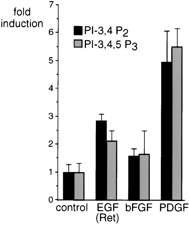
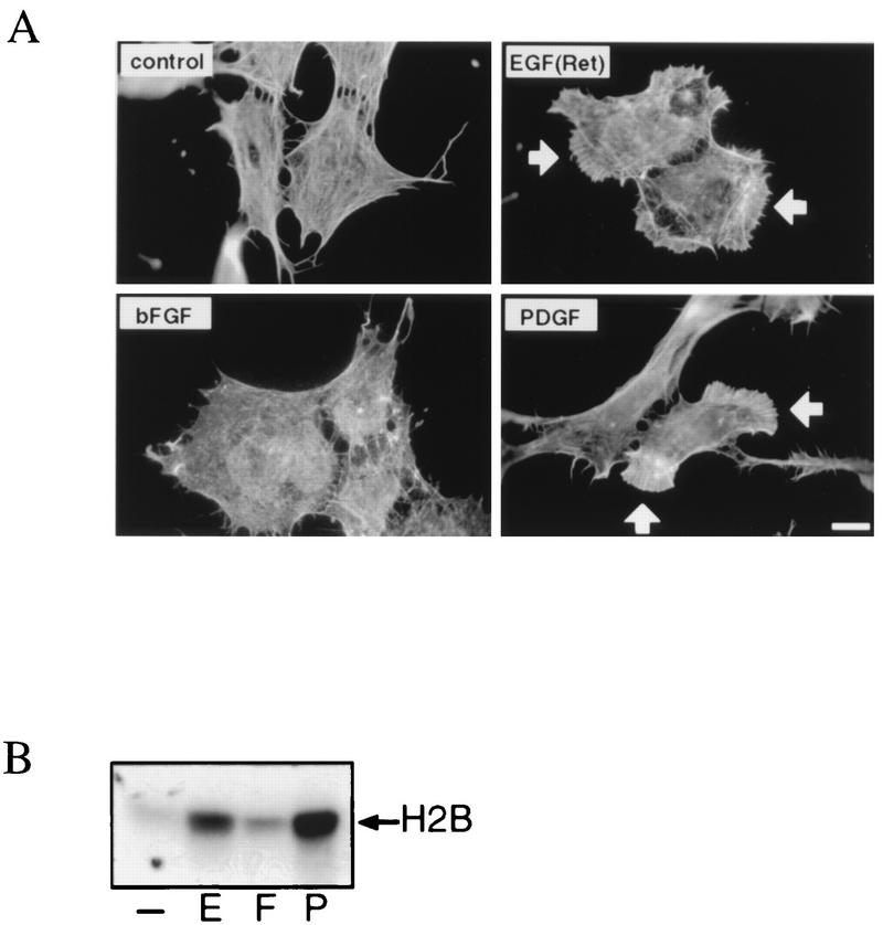
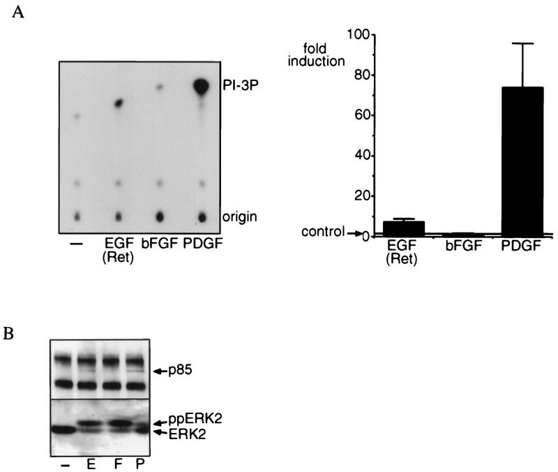
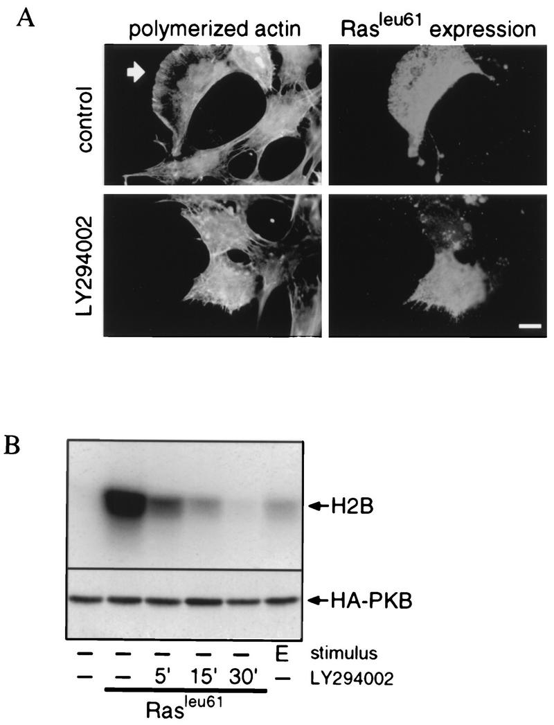
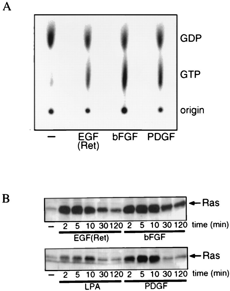
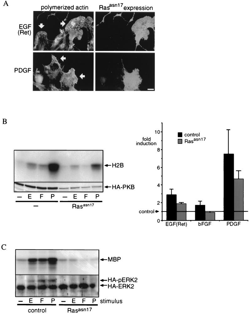
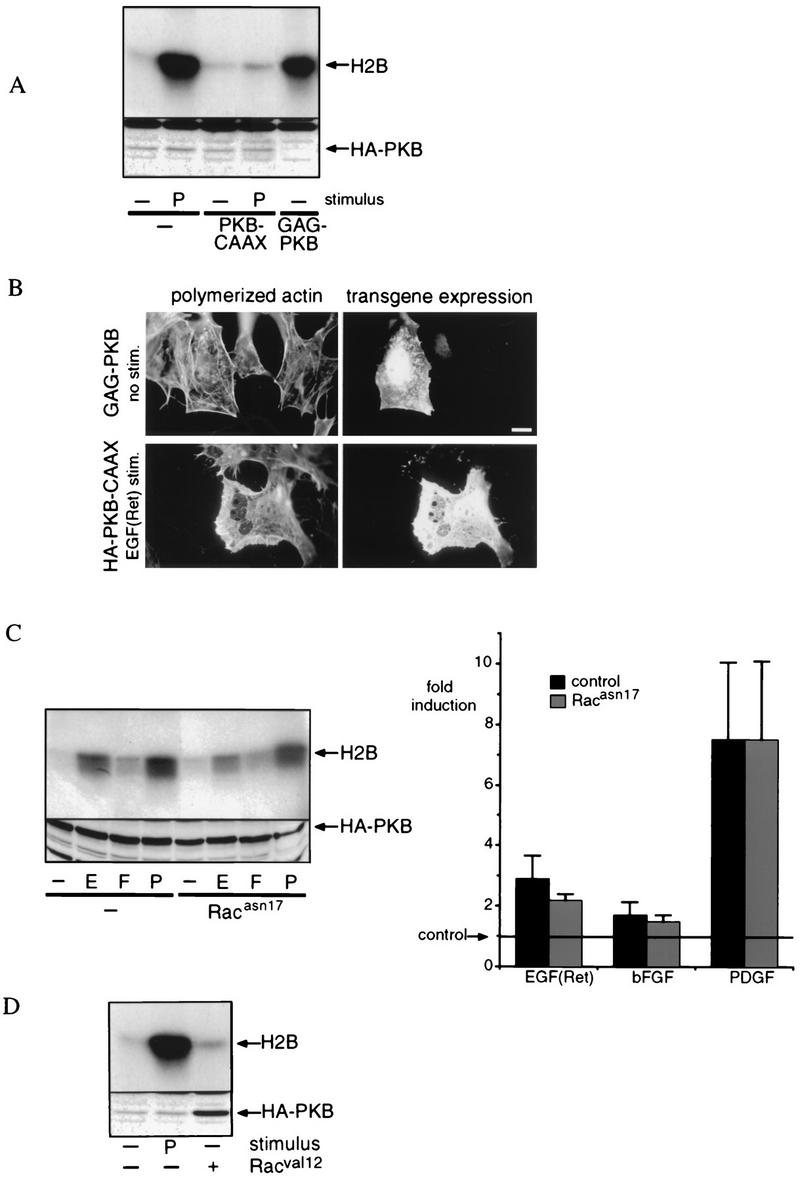
Regulation of phosphoinositide 3-kinase (PI 3-kinase) can occur by binding of the regulatory p85 subunit to tyrosine-phosphorylated proteins and by binding of the p110 catalytic subunit to activated Ras. However, the way in which these regulatory mechanisms act to regulate PI 3-kinase in vivo is unclear. Here we show that several growth factors (basic fibroblast growth factor [bFGF], platelet-derived growth factor [PDGF], and epidermal growth factor [EGF; to activate an EGF receptor-Ret chimeric receptor]) all activate PI 3-kinase in vivo in the neuroectoderm-derived cell line SKF5. However, these growth factors differ in their ability to activate PI 3-kinase-dependent signaling. PDGF and EGF(Ret) treatment induced PI 3-kinase-dependent lamellipodium formation and protein kinase B (PKB) activation. In contrast, bFGF did not induce lamellipodium formation but activated PKB, albeit to a small extent. PDGF and EGF(Ret) stimulation resulted in binding of p85 to tyrosine-phosphorylated proteins and strong Ras activation. bFGF, however, induced only strong activation of Ras. In addition, while RasAsn17 abolished bFGF activation of PKB, PDGF- and EGF(Ret)-induced PKB activation was only partially inhibited and lamellipodium formation was unaffected. Interestingly, in contrast to activation of only endogenous Ras (bFGF), ectopic expression of activated Ras did result in lamellipodium formation. From this we conclude that, in vivo, p85 and Ras synergize to activate PI 3-kinase and that strong activation of only endogenous Ras exerts a small effect on PI 3-kinase activity, sufficient for PKB activation but not lamellipodium formation. This differential sensitivity to PI 3-kinase activation could be explained by our finding that PKB activation and lamellipodium formation are independent PI 3-kinase-induced events.


Figures

FIG. 1
FIG. 1
Growth factor-induced PI 3-kinase activity. EGF(Ret), bFGF, and PDGF induce different amounts of phosphorylated PIs as measured by in vivo labeling and analysis of total lipids. Serum-starved subconfluent cultures of SKF5 cells were labeled in vivo with [32P]orthophosphate and were left untreated (control) or were stimulated for 2 min with 40 ng of EGF per ml or 20 ng of bFGF or PDGF per ml, followed by isolation of total lipids. Radioactive lipids were analyzed by high-pressure liquid chromatography. Shown is the fold induction of the PI-3,4P2 and PI-3,4,5P3 synthesis above control values. Error bars indicate the standard deviations.
FIG. 2
FIG. 2
Growth factor-induced lamellipodium formation and PKB activation in SKF5 cells. (A) EGF(Ret) and PDGF, but not bFGF, induce lamellipodium formation. Serum-starved SKF5 cells grown on coverslips were left untreated (control) or were stimulated for 10 min with 40 ng of EGF per ml or 20 ng of bFGF or PDGF per ml. Polymerized actin was stained with FITC-conjugated phalloidin. Lamellipodia are indicated by white arrows. Bar, 10 μm. (B) EGF(Ret), bFGF, and PDGF differentially induce PKB activity. SKF5 cells were serum starved overnight and were either left untreated (−) or treated for 5 min with 40 ng of EGF per ml (E) or 20 ng of bFGF (F) or PDGF (P) per ml. PKB activity was determined in an in vitro kinase assay on endogenous PKB immunoprecipitated by a rabbit polyclonal anti-PKB serum with histone 2B (H2B) as a substrate. Shown is an autoradiogram of a representative experiment after SDS-PAGE analysis of the kinase reaction.
FIG. 3
FIG. 3
Growth factor-induced, phosphotyrosine-associated PI 3-kinase activity. (A) EGF(Ret) and PDGF, but not bFGF, induce PI 3-kinase activity as measured in an in vitro kinase assay. Serum-starved subconfluent cultures of SKF5 cells were left untreated (−) or were stimulated for 5 min with 40 ng of EGF per ml or 20 ng of bFGF or PDGF per ml, followed by immunoprecipitation of tyrosine-phosphorylated proteins. Immunoprecipitates were subjected to a kinase assay in the presence of [γ-32P]ATP and phosphatidylinositol. Shown is an autoradiogram of the kinase reaction after thin-layer chromatography. The position of the 3′-phosphorylated phosphatidylinositol (PI-3P) is indicated. All experiments were quantified and averaged (±standard error of the mean); the result is represented by bars (fold induction). The number of three independent experiments were used for each point. (B) EGF(Ret) and PDGF, but not bFGF, induce the association of the p85 subunit of PI 3-kinase with tyrosine-phosphorylated proteins. Serum-starved subconfluent cultures of SKF5 cells were left untreated or were stimulated for 5 min with 40 ng of EGF per ml (E) or 20 ng of bFGF (F) or PDGF (P) per ml. In the upper panel, tyrosine-phosphorylated proteins were immunoprecipitated and analyzed by Western blotting with a monoclonal antibody to the p85 subunit of PI 3-kinase. In the lower panel, in the same cell lysates the activity of ERK2 was analyzed by Western blotting with a polyclonal antibody to ERK2. The positions of the active (ppERK2) and inactive (ERK2) forms of the kinase are indicated.
FIG. 4
FIG. 4
Constitutively active Ras induces PI 3-kinase-mediated lamellipodium formation and PKB activation. (A) Constitutively active Ras-induced lamellipodium formation is dependent on PI 3-kinase activity. SKF5 cells were transiently transfected with a RasLeu61 expression plasmid. After overnight serum starvation, the cells were either left untreated (control) or treated for 10 min with LY294002. Polymerized actin was stained with FITC-conjugated phalloidin, and RasLeu61 expression was revealed with a Ras-specific monoclonal antibody. The arrow indicates lamellipodia. Bar, 10 μm. (B) RasLeu61-induced PKB activation is dependent on PI 3-kinase activity. SKF5 cells were transiently transfected with HA-PKB and RasLeu61 expression plasmids. After overnight serum starvation, the cells were either left untreated or treated for the indicated times with LY294002. PKB activity was determined in an in vitro kinase assay on immunoprecipitated HA-PKB with histone 2B (H2B) as a kinase substrate. Shown is an autoradiogram of a representative experiment after SDS-PAGE separation of the proteins (upper panel). Expression of HA-PKB was controlled by 12CA5 immunoblotting of samples taken before immunoprecipitation (lower panel).
FIG. 5
FIG. 5
Growth factor-induced Ras activation. (A) EGF(Ret), bFGF, and PDGF induce Ras activation. Serum-starved subconfluent cultures of SKF5 cells were labeled in vivo with [32P]orthophosphate and then treated for 5 min with 40 ng of EGF per ml or 20 ng of bFGF or PDGF per ml, and Ras-bound guanine nucleotides were isolated. Shown is an autoradiogram of the extracted guanine nucleotides after thin-layer chromatography. The positions of labeled GDP and GTP are indicated. The percentages of Ras in the GTP-bound state are as follows: control, 16%; EGF(Ret), 35%; bFGF, 48%; PDGF, 39%. (B) Kinetics of EGF(Ret)-, bFGF-, PDGF- and LPA-induced Ras activation. Serum-starved subconfluent cultures of SKF5 cells were stimulated for the indicated times with 40 ng of EGF per ml, 20 ng of bFGF or PDGF per ml, or 1 μM LPA. GTP-bound Ras was specifically precipitated with GST-Raf-RBD and analyzed by Western blotting.
FIG. 6
FIG. 6
Role of endogenous Ras in lamellipodium formation and PKB activation. (A) Dominant negative Ras does not inhibit lamellipodium formation. SKF5 cells were transiently transfected with dominant negative RasAsn17. The cells were serum starved and were treated with 40 ng of EGF per ml or 20 ng of PDGF per ml. RasAsn17 expression was revealed with an anti-Ras monoclonal antibody, and polymerized actin was stained with phalloidin-FITC. Arrows indicate lamellipodia. Bar, 10 μm. (B) Dominant negative Ras inhibits bFGF-induced activation but partially affects EGF(Ret)- or PDGF-induced PKB activation. SKF5 cells were transiently transfected with HA-PKB and RasAsn17 expression plasmids. After overnight serum starvation, the cells were either left untreated (−) or treated for 5 min with 40 ng of EGF (E) per ml or 20 ng of bFGF (F) or PDGF (P) per ml. PKB activity was determined in an in vitro kinase assay on immunoprecipitated HA-PKB with histone 2B (H2B) as a substrate. The upper panel shows an autoradiogram of a representative kinase assay after SDS-PAGE analysis of the kinase reaction. Expression of HA-PKB was controlled by 12CA5 immunoblotting of samples taken before immunoprecipitation (lower panel). All experiments were quantified and averaged (±standard error of the mean); the results are represented by the bar diagram (fold induction). Five independent experiments were used for each point. (C) RasAsn17 completely blocks Ras-dependent signaling. SKF5 cells were transiently transfected with HA-MAP kinase and RasAsn17 expression plasmids. After overnight serum starvation, the cells were either left untreated or treated for 5 min with 40 ng of EGF per ml or 20 ng of bFGF or PDGF per ml. HA-MAP kinase activity was determined in an in vitro kinase assay on immunoprecipitated HA-MAP kinase with myelin basic protein (MBP) as a substrate (upper panel). Before immunoprecipitation, a sample was taken to control for the expression of HA-MAP kinase. Proteins were separated with a gel system that reveals an electrophoretic mobility shift of phosphorylated (activated) HA-MAP kinase followed by 12CA5 immunoblotting. The positions of active (HA-pERK2) and inactive (HA-ERK2) MAP kinase are indicated (lower panel).
FIG. 7
FIG. 7
Rac and PKB represent separate PI 3-kinase-dependent signaling pathways. (A) Characterization of constitutively active and dominant negative PKB. SKF5 cells were transiently transfected with GAG-PKB (2 μg) or with HA-PKB (0.5 μg) and either left unstimulated (−) or stimulated with 20 ng of PDGF (P) per ml. To test for dominant negative function, 0.5 μg of HA-PKB was cotransfected with 5 μg of CAAX-PKB (no HA tag). Shown is an autoradiogram representative of three independent experiments. The expression of HA-PKB was controlled by 12CA5 immunoblotting of samples taken before immunoprecipitation (lower panel). (B) Constitutively active PKB does not induce lamellipodium formation, and dominant negative PKB does not interfere with EGF(Ret)-induced lamellipodium formation. SKF5 cells were transiently transfected with constitutively active GAG-PKB or with dominant negative HA-PKB-CAAX, as indicated. The cells were serum starved overnight and were left untreated (GAG-PKB) or stimulated for 10 min with 40 ng of EGF per ml (HA-PKB-CAAX). GAG-PKB expression was revealed with an anti-PKB polyclonal antibody, HA-PKB-CAAX expression was revealed with a monoclonal anti-HA antibody, and polymerized actin was stained with phalloidin-FITC. Bar, 10 μm. (C) Dominant negative Rac does not inhibit growth factor-induced PKB activation. SKF5 cells were transiently transfected with HA-PKB and RacAsn17 expression plasmids. After overnight serum starvation, the cells were either left untreated or treated for 5 min with 40 ng of EGF (E) per ml or 20 ng of bFGF (F) or PDGF per ml. PKB activity was determined in an in vitro kinase assay on immunoprecipitated HA-PKB with histone 2B (H2B) as a substrate. The upper panel shows an autoradiogram of a representative kinase assay after SDS-PAGE analysis of the kinase reaction. Expression of HA-PKB was controlled by 12CA5 immunoblotting of samples taken before immunoprecipitation (lower panel). All experiments were quantified and averaged (±standard error of the mean); the result of this is represented by the bar diagram (fold induction). Four independent experiments were used for each point. (D) Constitutively active Rac does not induce PKB activity. SKF5 cells were transiently transfected with HA-PKB and, as indicated, with a RacVal12 expression plasmid. After overnight serum starvation, the cells were either left untreated (−) or were treated for 5 min with 20 ng of PDGF (P) per ml. PKB activity was determined in an in vitro kinase assay on immunoprecipitated HA-PKB with histone 2B (H2B) as a kinase substrate. The upper panel shows an autoradiogram of a representative kinase assay after SDS-PAGE analysis of the kinase reaction. Expression of HA-PKB was controlled by 12CA5 immunoblotting of samples taken before immunoprecipitation (lower panel).

Similar articles

Cited by

References

    1. Alessi D R, Andjelkovich M, Caudwell B, Cron P, Morrice N, Cohen P, Hemmings B A. Mechanism of activation of protein kinase B by insulin and IGF-1. EMBO J. 1996;15:6541–6551. - PMC - PubMed
    1. Alessi D R, James S R, Downes C P, Holmes A B, Gaffney P R, Reese C B, Cohen P. Characterization of a 3-phosphoinositide-dependent protein kinase which phosphorylates and activates protein kinase B-alpha. Curr Biol. 1997;7:261–269. - PubMed
    1. Burgering, B. M. T., et al. Unpublished data.
    1. Burgering B M T, Coffer P J. Protein kinase B (c-Akt) in phosphatidylinositol-3-OH kinase signal transduction. Nature. 1995;376:599–602. - PubMed
    1. Carpenter C L, Auger K R, Duckworth B C, Hou W M, Schaffhausen B, Cantley L C. A tightly associated serine/threonine protein kinase regulates phosphoinositide 3-kinase activity. Mol Cell Biol. 1993;13:1657–1665. - PMC - PubMed

Publication types

MeSH terms

Substances