. 1998 Apr;18(4):1866-78.
doi: 10.1128/MCB.18.4.1866.

Rex-1, a gene encoding a transcription factor expressed in the early embryo, is regulated via Oct-3/4 and Oct-6 binding to an octamer site and a novel protein, Rox-1, binding to an adjacent site

Affiliations

Rex-1, a gene encoding a transcription factor expressed in the early embryo, is regulated via Oct-3/4 and Oct-6 binding to an octamer site and a novel protein, Rox-1, binding to an adjacent site

E Ben-Shushan et al. Mol Cell Biol. 1998 Apr.

Abstract

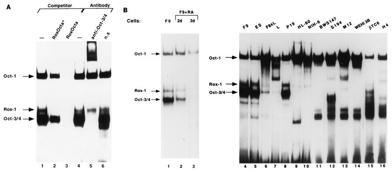
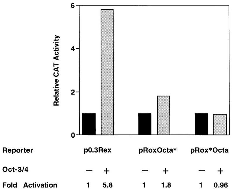
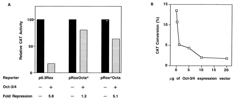
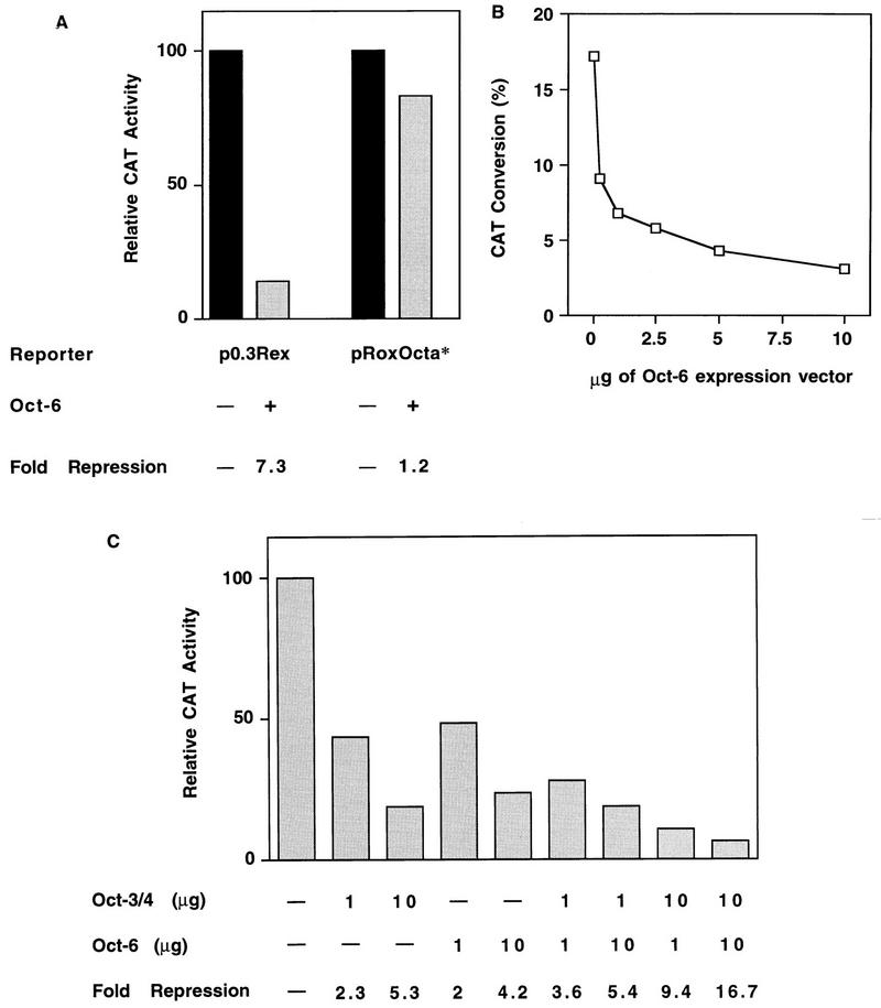
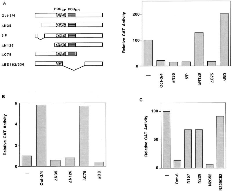
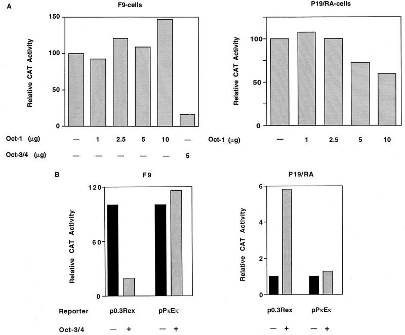
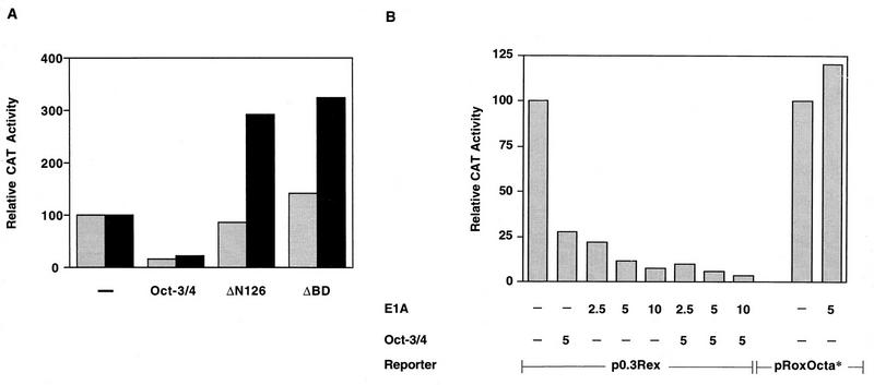
The Rex-1 (Zfp-42) gene, which encodes an acidic zinc finger protein, is expressed at high levels in embryonic stem (ES) and F9 teratocarcinoma cells. Prior analysis identified an octamer motif in the Rex-1 promoter which is required for promoter activity in undifferentiated F9 cells and is involved in retinoic acid (RA)-associated reduction in expression. We show here that the Oct-3/4 transcription factor, but not Oct-1, can either activate or repress the Rex-1 promoter, depending on the cellular environment. Rex-1 repression is enhanced by E1A. The protein domain required for Oct-3/4 activation was mapped to amino acids 1 to 35, whereas the domain required for Oct-3/4 repression was mapped to amino acids 61 to 126, suggesting that the molecular mechanisms underlying transcriptional activation and repression differ. Like Oct-3/4, Oct-6 can also lower the expression of the Rex-1 promoter via the octamer site, and the amino-terminal portion of Oct-6 mediates this repression. In addition to the octamer motif, a novel positive regulatory element, located immediately 5' of the octamer motif, was identified in the Rex-1 promoter. Mutations in this element greatly reduce Rex-1 promoter activity in F9 cells. High levels of a binding protein(s), designated Rox-1, recognize this novel DNA element in F9 cells, and this binding activity is reduced following RA treatment. Taken together, these results indicate that the Rex-1 promoter is regulated by specific octamer family members in early embryonic cells and that a novel element also contributes to Rex-1 expression.


Figures

FIG. 1
FIG. 1
Binding to the Rex-1 promoter region. (A) DNase I footprinting of the murine Rex-1 promoter. A 32P-end-labeled Rex-1 promoter fragment (SpeI-HindIII) was incubated with 75 μg of WCE prepared from F9 cells (lanes 3, 4, 8, and 9) and in the absence of WCE (lanes 1, 2, 6, and 7). Lanes 5 and 10, Maxam-Gilbert A+C and C+T, respectively, sequencing ladders of the sense (lanes 1 to 5) and antisense (lanes 6 to 10) probes. The protected regions are boxed and the corresponding sequences are indicated. (B) The indicated 32P-end-labeled oligonucleotide probes were incubated with WCE prepared from F9 cells. Binding reactions were performed in the absence of competitors (lanes 1, 5, and 6) or in the presence of the indicated competitors (lanes 2 to 4). Binding reactions shown in lanes 2 to 4 were performed in the presence of a 100-fold molar excess of the indicated unlabeled oligonucleotides. The arrows indicate the known Oct-1, Oct-3/4, and Oct-6 complexes and the novel Rox-1 complex.
FIG. 2
FIG. 2
The Rox-1 complex. (A) 32P-end-labeled RoxOcta oligonucleotide was incubated with WCE prepared from F9 cells. Binding reactions were performed in the absence of a competitor (lanes 1 and 4), in the presence of a 100-fold molar excess of the indicated competitors (lanes 2 and 3), or in the presence of 1 μl of anti-Oct-3/4 (lane 5) or nonspecific (n.s.; lane 6) antibody. The oligonucleotides are depicted in Table 1. The arrows indicate the Oct-1, Oct-3/4, and Rox-1 complexes. Oct-6 is detected only in freshly prepared WCEs, and the extracts used in this experiment were frozen and subsequently thawed. (B) 32P-end-labeled RoxOcta probe was incubated with WCE prepared from F9 cells (lanes 1 and 4), F9 cells treated with RA for either 2 days (lane 2) or 3 days (lane 3), ES cells (lane 5), F9 × L somatic cell hybrids (1) (lane 6), L cells (lane 7), P19 cells (lane 8), HL60 cells (lane 9), NIH-9 cells (lane 10), BW5417 T cells (lane 11), S194 myeloma cells (lane 12), M12 lymphoma cells (lane 13), WEHI 3B myeloid cells (lane 14), βTC6 insulinoma cells (lane 15), and H4 hepatoma cells (lane 16).
FIG. 3
FIG. 3
Identification of the Rox-1 binding site. (A) 32P-end-labeled RoxOcta oligonucleotide was incubated with F9 WCE. Binding reactions were done in the absence (−) or presence of Octa*-3′ (an oligonucleotide carrying mutations in the Octa site, same mutations as in the Octa oligomer shown in Fig. 4, and no mutation in the sequence located 3′ of the Octa) (lane 2), and Octa-3′ oligonucleotide containing wild-type Octa and wild-type sequences located 3′ to it (lane 3). (B) 32P-end-labeled RoxOcta oligonucleotide was incubated with F9 WCE. Binding reactions were done in the absence (−) or presence of the indicated oligonucleotides.
FIG. 4
FIG. 4
Functional importance of the Rox-1 binding site for Rex-1 promoter activity. (A) The p0.3Rex-CAT, pRoxOcta*-CAT, pRox*Octa-CAT, pmRox-1 to pmRox6, and pmRox1,5,6 reporter plasmids (10 μg) were cotransfected with β-Gal-containing reference plasmid (2 μg) into F9 stem cells. After 48 h, cells were harvested and lysed, and CAT activities were determined. The percent conversion to the acetylated forms of each separate transfection was normalized to the β-Gal activity. The values for percent conversion, presented as means ± standard deviation, corresponding to p0.3Rex-CAT, pRoxOcta*-CAT, pRox*Oct-CAT, pmRox1, pmRox2, pmRox3, pmRox4, pmRox5, pmRox6, and pmRox1,5,6 are 15 ± 2.06, 1.23 ± 0.4, 5.00 ± 0.14, 5.22 ± 0.2, 15.26 ± 1.5, 21.39 ± 3.2, 5.13 ± 0.4, 28.98 ± 0.3, 3.84 ± 0.07, and 2.63 ± 0.3, respectively. CAT activity of p0.3Rex-CAT was arbitrarily set at 100%. Relative CAT activity represents CAT activity relative to that obtained with p0.3Rex-CAT. (B) Sequences of wild-type RoxOcta and the mutant oligonucleotides. The RoxOcta probe is the sequence of the wild-type Rex-1 promoter from −234 to −204 (21). The octamer motif is boxed, and the mutant bases are underlined.
FIG. 5
FIG. 5
Oct-3/4 activates Rex-1 promoter activity in P19/RA cells. The p0.3Rex-CAT, pRoxOcta*-CAT, and pRox*Octa-CAT reporter plasmids (10 μg) were transfected with β-Gal-containing reference plasmid (2 μg) into P19 cells which had been cultured in the presence of RA for 36 h. Transfections were performed in the absence (−) or presence (+) of 10 μg of wild-type Oct-3/4 expression vector. CAT activities were determined and normalized to β-Gal activity. The values for percent conversion, presented as means ± standard deviations, corresponding to p0.3Rex-CAT, pRoxOcta*-CAT, and pRox*Octa-CAT each in the absence and presence of Oct-3/4 are 8.1 ± 1.5, 47.7 ± 5, 5.4 ± 1.1, 9.72 ± 1.6, 16.9 ± 2.2, and 16.2 ± 1.5, respectively. CAT activity of each construct alone, in the absence of Oct-3/4, was arbitrarily set at 1. Relative CAT activity represents CAT activity relative to that obtained from p0.3Rex-CAT, pRoxOcta*-CAT, and pRox*Octa-CAT, respectively. Fold activation was calculated as the ratio between CAT activities in the absence and presence of Oct-3/4.
FIG. 6
FIG. 6
Oct-3/4 inhibits Rex-1 promoter activity in F9 cells. (A) The p0.3Rex-CAT (containing the wild-type Rex-1 promoter), pRoxOcta*-CAT (containing the Octa-mutated Rex-1 promoter), and pRox*Octa-CAT (harboring mutations in the Rox-1 binding site) reporter plasmids (10 μg) were transfected with β-Gal-containing reference plasmid (2 μg) into F9 stem cells, with (+) or without (−) the Oct-3/4 expression vector. The values for percent CAT conversion, presented as means ± standard deviation, corresponding to p0.3Rex-CAT, pRoxOcta*-CAT, and pRox*Octa-CAT each in the absence or presence of Oct-3/4 are 58.7 ± 9.8, 10.11 ± 1.0, 1.69 ± 0.2, 1.36 ± 0.2, 14.8 ± 1.5, and 12.9 ± 0.3, respectively. CAT activity of each construct alone, in the absence of Oct-3/4, was arbitrarily set at 100. Relative CAT activity represents CAT activity relative to that obtained from p0.3Rex-CAT, pRoxOcta*-CAT, and pRox*Octa-CAT, respectively. Fold repression was calculated as the ratio between CAT activities in the absence and presence of Oct-3/4, and the data are averages of at least four independent experiments. (B) Effect of increasing amounts of Oct-3/4 on Rex-1 promoter activity. F9 stem cells were cotransfected with p0.3Rex-CAT and increasing amounts of Oct-3/4, and CAT activity was measured and assayed as described for panel A. The values for percent CAT conversion corresponding to p0.3Rex-CAT in the absence and presence of 0.25, 1, 5, 10, and 20 μg of Oct-3/4 are 13.46, 10.67, 5.17, 4.33, 2.0, and 1.71, respectively.
FIG. 7
FIG. 7
Oct-6 inhibits Rex-1 promoter activity. (A) The p0.3Rex-CAT and pRoxOcta*-CAT reporter plasmids (10 μg) were transfected with the β-Gal-containing reference plasmid (2 μg) into F9 stem cells with (+) or without (vector backbone only; −) the Oct-6 expression vector. After 48 h, CAT activities were measured and assayed as described for Fig. 6. The values for percent CAT conversion, presented as means ± standard deviation, corresponding to p0.3Rex-CAT and pRoxOcta*-CAT each in the absence or presence of Oct-6 are 18.2 ± 2.5, 2.5 ± 0.2, 0.46 ± 0.09, and 0.38 ± 0.08, respectively. (B) F9 stem cells were cotransfected with p0.3Rex-CAT and increasing amounts of the Oct-6 expression vector. Values for percent CAT conversion corresponding to p0.3Rex-CAT in the absence or presence of 0.25, 1, 2.5, 5, and 10 μg of Oct-6 are 17.2, 9.1, 6.8, 5.8, 4.3, and 3.1, respectively. (C) F9 cells were cotransfected with p0.3Rex-CAT in the absence or the presence of Oct-3/4 (1 or 10 μg), Oct-6 (1 or 10 μg), or both Oct-3/4 and Oct-6 expression vectors at the indicated amounts. The values for percent CAT conversion, presented as means ± standard deviation, corresponding to p0.3Rex-CAT are as follows: in the absence of Oct-3/4 and Oct-6, 23.4 ± 0.2; in the presence of 1.0 and 10 μg of Oct-3/4, 10.2 ± 1 and 4.4 ± 0.5; in the presence of 1 and 10 μg of Oct-6, 11.3 ± 0.15 and 5.5 ± 0.6; in the presence of 1 μg of Oct-3/4 and 1 μg of Oct-6, 6.5 ± 1.2; in the presence of 1 μg of Oct-3/4 and 10 μg of Oct-6, 4.3 ± 0.5; in the presence of 10 μg of Oct-3/4 and 1 μg of Oct-6 2.5 ± 0.3; and in the presence of 10 μg of Oct-3/4 and 10 μg of Oct-6, 1.4 ± 0.2.
FIG. 8
FIG. 8
Deletion mapping of the repressive region of Oct-3/4 and Oct-6. (A) F9 stem cells were cotransfected with p0.3Rex-CAT reporter construct (10 μg) and a β-Gal-containing reference plasmid (1 μg) in the absence (−) or presence of 10 μg of wild-type Oct-3/4, N-terminal-deleted Oct-3/4 (ΔN35, 5′P, and ΔN126), C-terminus-deleted Oct-3/4 (ΔC75), and DNA-binding-domain-deleted Oct-3/4 (ΔDB). CAT activity was measured and quantitated as described in the legend to Fig. 6. The values for percent CAT conversion, presented as means ± standard deviation, corresponding to p0.3Rex-CAT in the absence or presence of Oct-3/4, ΔN35, 5′P, ΔN126, ΔC75, and ΔDB are 34.8 ± 5.1, 6.8 ± 0.7, 5.4 ± 0.6, 43.86 ± 4.5, 6.5 ± 0.7, and 69.4 ± 7.2, respectively. (B) P19/RA cells were cotransfected with 0.3Rex-CAT reporter constructs (10 μg) and a β-Gal-containing reference plasmid (1 μg) in the absence (−) or presence of the expression plasmids described above. The values for percent CAT conversion, presented as means ± standard deviation, corresponding to p0.3Rex-CAT in the absence or presence of Oct-3/4, ΔN35, ΔN126, ΔC75, ΔDB are 8.5 ± 1.1, 49.3 ± 5.1, 5.1 ± 0.4, 6.8 ± 0.7, 48.45 ± 6.3, and 3.4 ± 0.6, respectively. (C) F9 stem cells were cotransfected with p0.3Rex-CAT reporter construct (10 μg) and a β-Gal ΔDB-containing reference plasmid (1 μg) in the absence (−) or presence of 10 μg of wild-type Oct-6, N-terminus-deleted Oct-6 (N157 or N229), C-terminus-deleted Oct-6 (N2C52), and DNA-binding-domain-deleted Oct-6 (N229C52). These vectors were previously described (35). The values for percent CAT conversion, presented as means ± standard deviation, corresponding to p0.3Rex-CAT in the absence or presence of Oct-6, N157, N229, N2C52, and N229C52 are 18.2 ± 0.3, 2.5 ± 0.3, 12.3 ± 0.1, 12.3 ± 0.2, 1.2 ± 0.2, and 16.6 ± 0.3, respectively.
FIG. 9
FIG. 9
Oct-3/4 specifically regulates the Rex-1 promoter. (A) F9 and P19/RA cells were transiently transfected with the reporter plasmid p0.3Rex-CAT (10 μg), a β-Gal-containing reference plasmid (1 μg), in the absence (−) or presence of the indicated increasing amounts of Oct-1 expression vector or Oct-3/4 (5 μg) expression vector. Transfection efficiency and CAT activity were monitored and assayed as described in the legend to Fig. 6. The values for percent CAT conversion in F9 cells, presented as means ± standard deviation, corresponding to p0.3Rex-CAT in the absence or presence of 1, 2.5, 5, and 10 μg of Oct-1 are 37.2 ± 4.1, 34.3 ± 3.5, 45.0 ± 5.0, 40.2 ± 6.1, and 55.4 ± 6.5, respectively; the value in the presence of 5 μg of Oct-3/4 is 6.2 ± 0.5. The values for percent CAT conversion in P19/RA cells (a representative experiment) corresponding to p0.3Rex-CAT in the absence or presence of 1, 2.5, 5, and 10 μg of Oct-1 are 9.0, 9.7, 9, 6.5, and 5.3, respectively. (B) p0.3Rex-CAT and pPκEκ-CAT (containing the Ig κ-chain promoter and intronic enhancer) reporter plasmids were transfected with β-Gal-containing reference plasmid into F9 and P19/RA cells in the absence (−) or presence (+) of the Oct-3/4 expression vector. The values for percent CAT conversion in F9 cells, presented as means ± standard deviation, corresponding to p0.3Rex-CAT and pPκEκ-CAT each in the absence or presence of Oct-3/4 are 52.8 ± 0.6, 10.2 ± 1.2, 5.2 ± 0.6, and 6.0 ± 0.7, respectively. The values for percent CAT conversion in P19/RA cells (a representative experiment) corresponding to p0.3Rex-CAT and pPκEκ-CAT each in the absence or presence of Oct-3/4 are 8.1, 47.7, 2.95, and 3.82, respectively.
FIG. 10
FIG. 10
Modulation of Oct-3/4 activity. (A) ΔN126 and ΔDB expression vectors activate Rex-1 promoter in F9 cells. F9 cells were transiently transfected with the p0.3Rex-CAT reporter plasmid and a β-Gal-containing reference plasmid (1 μg) in the absence (−) or presence of 10 μg (grey bars) and 20 μg (black bars) of Oct-3/4, ΔN126, and ΔDB expression vectors. The values for percent CAT conversion, presented as means ± standard deviation, corresponding to p0.3Rex-CAT in the absence or presence of 10 and 20 μg of Oct-3/4, ΔN126, and ΔDB are 37.2 ± 4.1, 18.2 ± 0.3, 5.46 ± 0.7, 3.87 ± 0.4, 31.43 ± 3.0, 53.14 ± 4.9, 52.37 ± 5.1, and 58.74 ± 6.1, respectively. (B) Effect of E1A on Rex-1 expression. F9 cells were transiently transfected with either the p0.3Rex-CAT or pRoxOcta*-CAT reporter plasmid and a β-Gal-containing reference plasmid (1 μg) in the absence (−) or presence of the indicated E1A and Oct-3/4 expression vectors. The values for percent CAT conversion, presented as means ± standard deviation, corresponding to p0.3Rex-CAT are as follows: in the absence of Oct-3/4 and E1A, 27.1 ± 0.3; in the presence of Oct-3/4, 7.6 ± 0.8; in the presence of 2.5, 5, and 10 μg of E1A, 5.9 ± 0.7, 3.2 ± 0.4, and 2.1 ± 0.2, respectively; in the presence of Oct-3/4 and 2.5, 5, and 10 μg of E1A, 2.6 ± 0.3, 1.6 ± 0.2, and 1.0 ± 0.2, respectively. The values for percent CAT conversion, presented as means ± standard deviation, corresponding to pRoxOcta*-CAT in the absence or presence of E1A are 1.3 ± 0.2 and 1.56 ± 0.2, respectively.

References

    1. Ambrosetti D-C, Basilico C, Dailey L. Synergistic activation of the fibroblast growth factor 4 enhancer by Sox2 and Oct-3 depends on protein-protein interactions facilitated by a specific spatial arrangement of factor binding sites. Mol Cell Biol. 1997;17:6321–6329. - PMC - PubMed
    1. Ben-Shushan E, Pikarsky E, Klar A, Bergman Y. Extinction of Oct-3/4 gene expression in embryonal carcinoma × fibroblast somatic cell hybrids is accompanied by changes in the methylation status, chromatin structure, and transcriptional activity of the Oct-3/4 upstream region. Mol Cell Biol. 1993;13:891–901. - PMC - PubMed
    1. Ben-Shushan E, Sharir H, Pikarsky E, Bergman Y. A dynamic balance between ARP-1/COUP-TFII, EAR-3/COUP-TFI, and retinoic acid receptor:retinoid X receptor heterodimers regulates Oct-3/4 expression in embryonal carcinoma cells. Mol Cell Biol. 1995;15:1034–1048. - PMC - PubMed
    1. Borrelli E, Hen R, Wasylyk C, Wasylyk B, Chambon P. The immunoglobulin heavy chain enhancer is stimulated by the adenovirus type 2 E1A products in mouse fibroblasts. Proc Natl Acad Sci USA. 1986;83:2846–2849. - PMC - PubMed
    1. Bushmeyer S, Park K, Atchison M L. Characterization of functional domains within the multifunctional transcription factor, YY1. J Biol Chem. 1995;270:30213–30220. - PubMed

Publication types

Substances

LinkOut - more resources