. 1998 Apr;18(4):1956-66.
doi: 10.1128/MCB.18.4.1956.

Interactions within the yeast Sm core complex: from proteins to amino acids

Affiliations

Interactions within the yeast Sm core complex: from proteins to amino acids

A Camasses et al. Mol Cell Biol. 1998 Apr.

Abstract

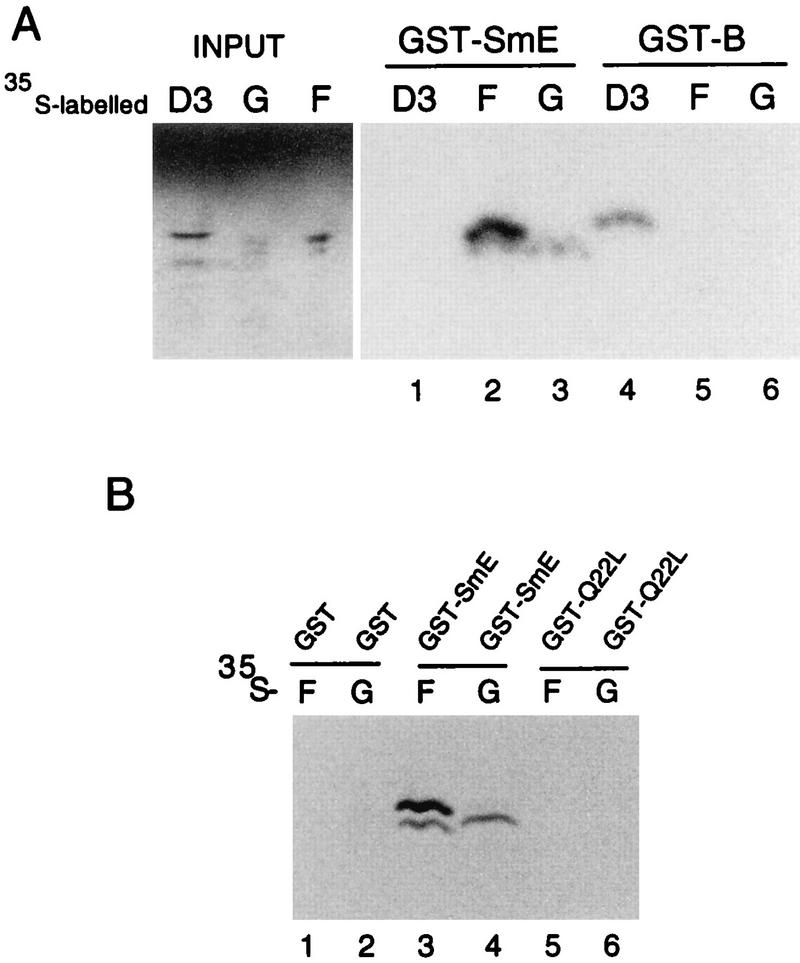
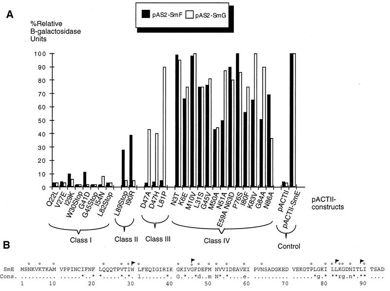
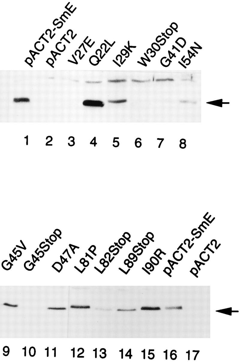
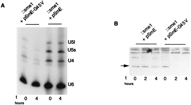
Sm core proteins play an essential role in the formation of small nuclear ribonucleoprotein particles (snRNPs) by binding to small nuclear RNAs and participating in a network of protein interactions. The two-hybrid system was used to identify SmE interacting proteins and to test for interactions between all pairwise combinations of yeast Sm proteins. We observed interactions between SmB and SmD3, SmE and SmF, and SmE and SmG. For these interactions, a direct biochemical assay confirmed the validity of the results obtained in vivo. To map the protein-protein interaction surface of Sm proteins, we generated a library of SmE mutants and investigated their ability to interact with SmF and/or SmG proteins in the two-hybrid system. Several classes of mutants were observed: some mutants are unable to interact with either SmF or SmG proteins, some interact with SmG but not with SmF, while others interact moderately with SmF but not with SmG. Our mutational analysis of yeast SmE protein shows that conserved hydrophobic residues are essential for interactions with SmF and SmG as well as for viability. Surprisingly, we observed that other evolutionarily conserved positions are tolerant to mutations, with substitutions affecting binding to SmF and SmG only mildly and conferring a wild-type growth phenotype.


Figures

FIG. 1
FIG. 1
In vitro binding of yeast Sm proteins. (A) [35S]methionine-labeled SmD3, SmF, and SmG prepared by in vitro transcription and translation were mixed with GST-SmE or GST-SmB produced in E. coli. Proteins bound to glutathione-Sepharose beads were washed, denatured, and separated on gels in Tricine buffer. Input represents aliquots of radioactive proteins corresponding to 25% of that used in each of the binding reactions. The predicted molecular sizes of the proteins are as follows: SmD3, 11.2 kDa; SmG, 8.4 kDa; SmF, 9.6 kDa. Additional bands observed in input lanes correspond to readthrough and premature translation termination products. Exposure times were identical for both panels. (B) [35S]methionine-labeled SmF and SmG were mixed with GST, GST-SmE, and SmE mutant Q22L fused to GST (GST-Q22L) produced in E. coli. Proteins bound to glutathione-Sepharose beads were treated as described for panel A.
FIG. 2
FIG. 2
Protein-protein interactions of mutant SmE fusion proteins. (A) β-Galactosidase activities shown correspond to averages of two independent transformants assayed in duplicate and are presented relative to those of the wild-type pACTII-SmE interactions with pAS2-SmF and pAS2-SmG. (B) The amino acid sequence of SmE protein is given. Cons., consensus inferred from Sm and Sm-like protein sequences (47). The positions of the point mutants are indicated above the sequence by small circles. Stop mutants are represented by flags.
FIG. 3
FIG. 3
Western analysis of mutant GAD-SmE fusion proteins. Similar amounts of cell extracts from Y190 transformants carrying the indicated mutated SmE fusion proteins were fractionated by SDS-PAGE and immunoblotted with anti-SmE antibodies. Control extracts were made from cells carrying the wild-type SmE fusion protein pACTII-SmE (lanes 1 and 16) or pACTII vector alone (lanes 2 and 17).
FIG. 4
FIG. 4
Defects of SmE mutant G45V. (A) Amounts of spliceosomal snRNAs are decreased after shift of SmE mutant G45V to the restrictive temperature. Northern analysis was performed on RNA isolated from strains carrying wild-type (pSmE) or mutant G45V (pSmE-G45V) SmE genes before and 4 h after shift to 37°C. Probes were oligonucleotides complementary to U4, U5, and U6 snRNAs. The US snRNA is represented by two bands corresponding to a long (U5l) and a short (U5S) form. (B) Stability of SmE mutant G45V protein is affected at the restrictive temperature. Strains carrying wild-type (pSmE) or mutant G45V (pSmE-G45V) genes under the natural promoter as the sole source of SmE protein were grown and shifted for the indicated times at 37°C. Similar amounts of cell extracts were separated by SDS-PAGE in Tricine buffer and immunoblotted with anti-SmE antibodies.
FIG. 5
FIG. 5
snRNA stability in SmE mutants. RNA was extracted from endogenous sme1-disrupted strains bearing theGAL1::SME1 allele and either the indicated mutant SmE genes under the natural promoter (plasmid pUN-PrE) (lanes 1 to 8), the wild-type SmE gene (lanes 9 and 10), or vector pUN alone (lanes 11 and 12) before and 12 h after shift to glucose (GAL1-repressing conditions). RNA was separated on a 6% polyacrylamide–8 M urea gel, subjected to Northern analysis, and hybridized with probes specific for the yeast U4, U5, and U6 snRNAs. The poor transfer of the U1 and U2 snRNAs precluded their analyses in this experiment.
FIG. 6
FIG. 6
Decreased 5′ cap hypermethylation of spliceosomal U snRNAs in SmE mutant L81P. RNA blot analysis after immunoprecipitation with anti-TMG antibody. Endogenoussme1-disrupted strains containing theGAL1::SME1 allele and plasmids carrying the wild-type SmE gene, the mutant L81P and I54N genes, or the vector pUN alone were shifted to glucose-containing medium for 0 and 12 h. RNA was extracted and immunoprecipitated with anti-TMG antibodies. Supernatants (S) and pellets (P) were subjected to Northern analysis using U4 and U5 as probes. For mutants I54N and L81P, the amounts of snRNA were increased twofold to compensate for the observed slight decrease in U snRNAs 12 h after shift to glucose (see Fig. 5). Quantitation of the results is presented in Table 4.

References

    1. Altschul S F, Gish W, Miller W, Meyer E W, Lipman D J. Basic local alignment search tool. J Mol Biol. 1990;215:403–410. - PubMed
    1. Bordonné R, Banroques J, Abelson J, Guthrie C. Domains of yeast U4 spliceosomal RNA required for PRP4 protein binding, snRNP-snRNP interactions, and pre-mRNA splicing in vivo. Genes Dev. 1990;4:1185–1196. - PubMed
    1. Bordonné R, Tarassov I. The yeast SME1 gene encodes the homologue of human E core protein. Gene. 1996;176:111–117. - PubMed
    1. Breeden L, Nasmyth K. Regulation of HO gene. Cold Spring Harbor Symp Quant Biol. 1985;50:643–650. - PubMed
    1. Cooper M, Johnston L H, Beggs J D. Identification and characterization of Uss1p (Sdb23p): a novel U6 snRNA-associated protein with significant similarity to core proteins of small nuclear ribonucleoproteins. EMBO J. 1995;14:2066–2075. - PMC - PubMed

Publication types

MeSH terms

Substances