. 1998 Apr;18(4):2077-88.
doi: 10.1128/MCB.18.4.2077.

NF-kappaB2 is a putative target gene of activated Notch-1 via RBP-Jkappa

Affiliations

NF-kappaB2 is a putative target gene of activated Notch-1 via RBP-Jkappa

F Oswald et al. Mol Cell Biol. 1998 Apr.

Abstract

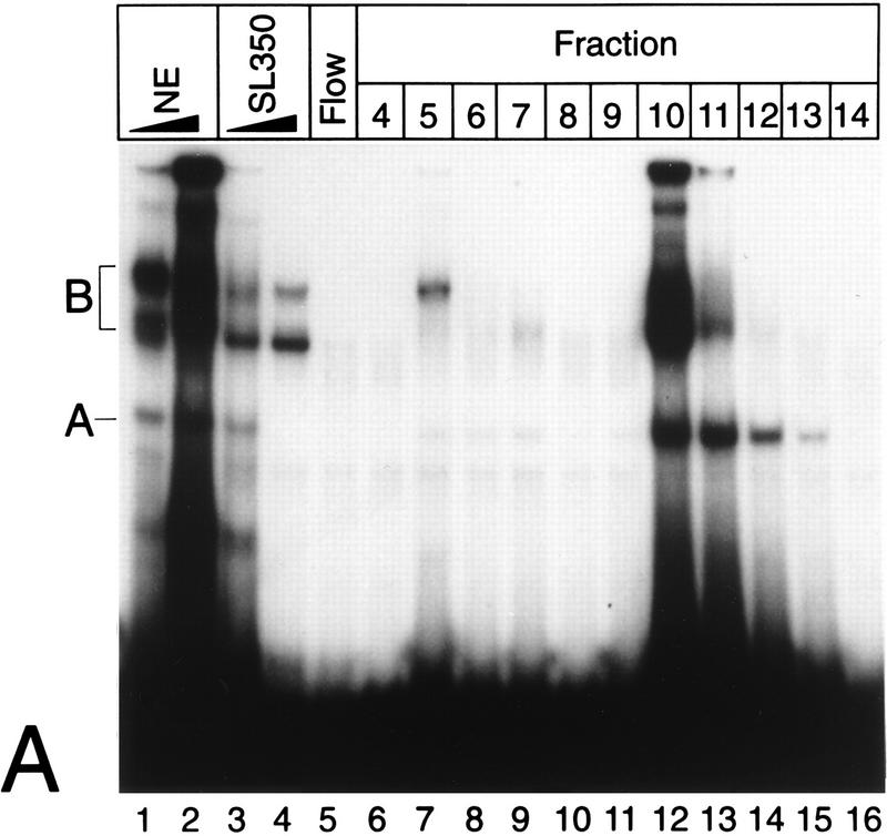
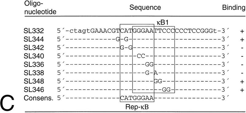
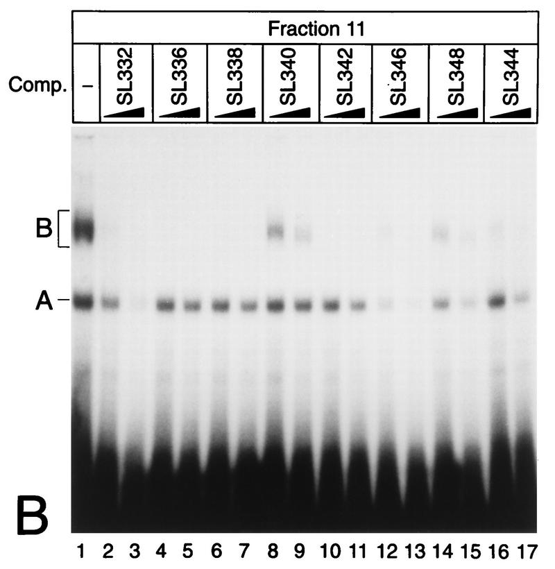
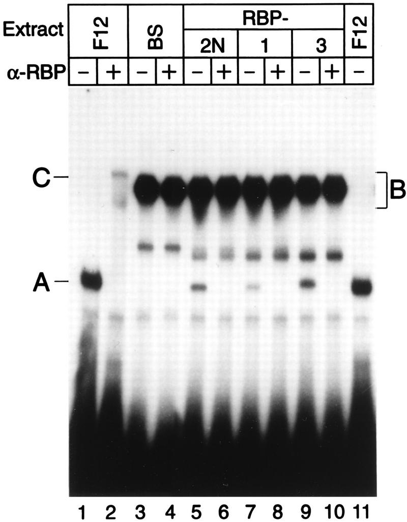
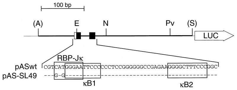
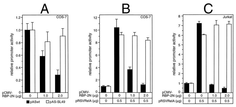
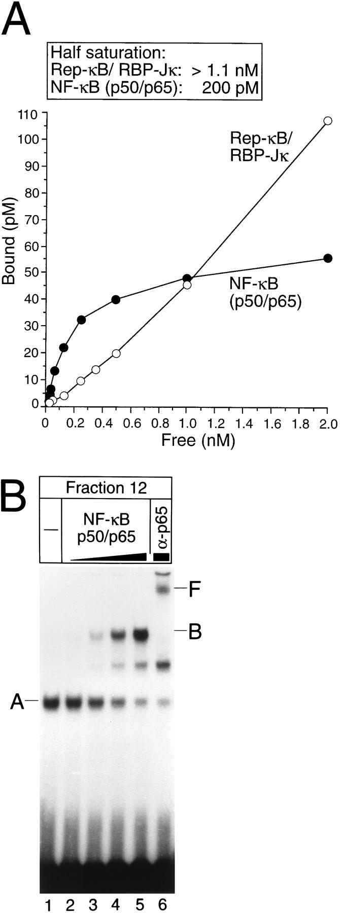
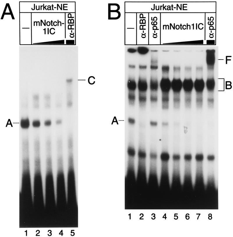
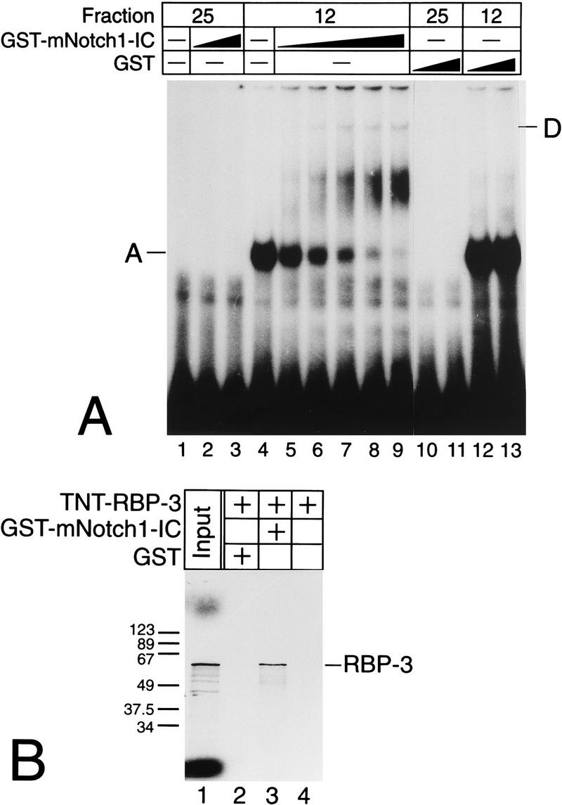
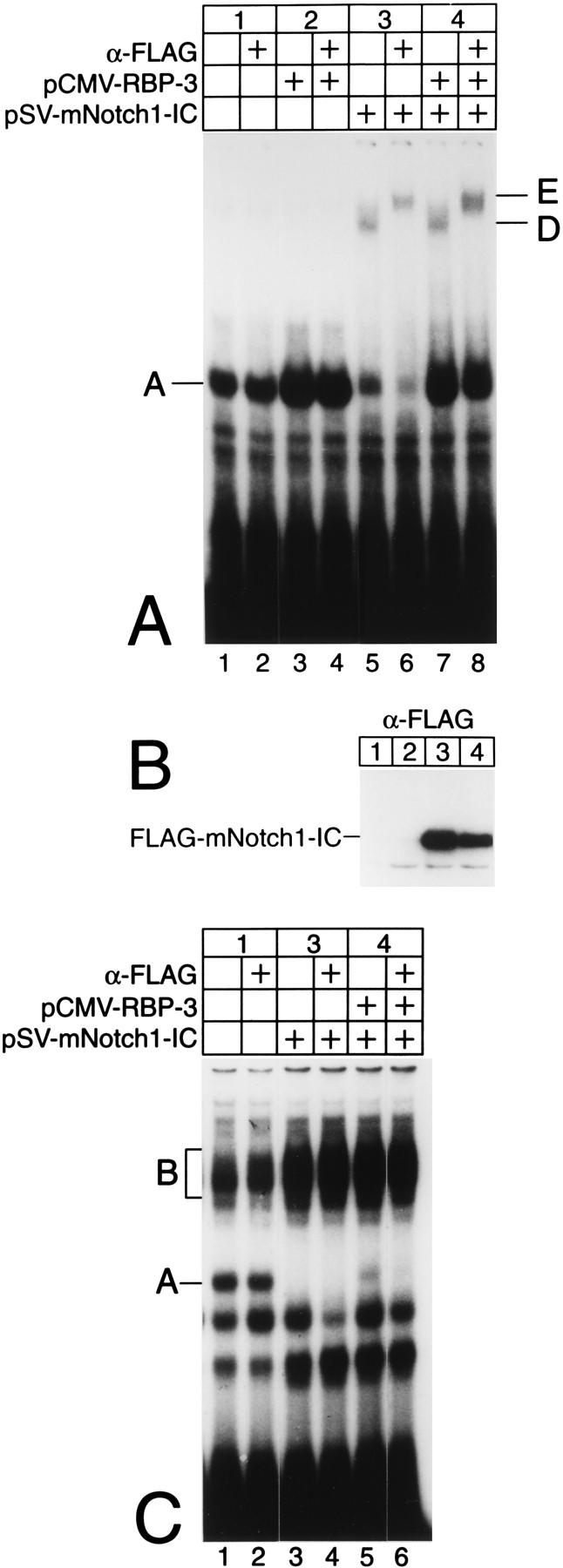
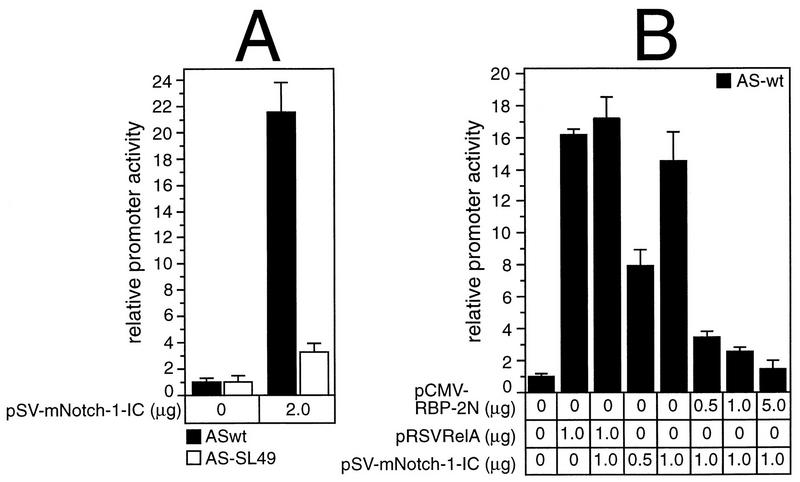
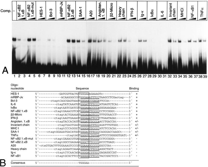
NF-kappaB2 (p100/p52), a member of the NF-kappaB/Rel family of transcription factors, is involved in the regulation of a variety of genes important for immune function. Previously, we have shown that the NF-kappaB2 gene is regulated in a positive and a negative manner. Two kappaB elements within the NF-kappaB2 promoter mediate tumor necrosis factor alpha-inducible transactivation. In addition, we have shown that there exists a transcriptional repression in the absence of NF-kappaB. To identify a DNA binding activity responsible for this transcriptional repression, we have partially purified a nuclear complex, named Rep-kappaB. Here we further analyze this putative repressive binding activity. Detailed examination of Rep-kappaB-DNA interaction revealed the sequence requirements for binding to be almost identical to those of recombination signal binding protein Jkappa (RBP-Jkappa), the mammalian homolog of the protein encoded by Drosophila suppressor of hairless [Su(H)]. In addition, in electromobility shift assays, Rep-kappaB binding activity is recognized by an antibody directed against RBP-Jkappa. By performing transient-transfection assays, we show that human RBP-Jkappa represses basal as well as RelA (p65)-stimulated NF-kappaB2 promoter activity. Studies in Drosophila melanogaster have shown that Su(H) is implicated in the Notch signaling pathway regulating cell fate decisions. In transient-transfection assays we show that truncated Notch-1 strongly induces NF-kappaB2 promoter activity. In summary, our data clearly demonstrate that Rep-kappaB is closely related or identical to RBP-Jkappa. RBP-Jkappa is a strong transcriptional repressor of NF-kappaB2. Moreover, this repression can be overcome by activated Notch-1, suggesting that NF-kappaB2 is a novel putative Notch target gene.


Figures

FIG. 1
FIG. 1
(A) Purification of Rep-κB activity from Jurkat-T cells. Nuclear extracts (NE), flowthrough (Flow), and eluted fractions were tested for binding to the oligonucleotide SL350, which contains both κB elements in the human NF-κB2 promoter and flanking regions (positions −119 to −63). The specificity of binding was analyzed by adding 20- and 60-fold molar excesses of a homologous competitor (lanes 3 and 4). A and B indicate the positions of specific DNA binding complexes. Complex A was identified as Rep-κB activity. (B) Sequence requirements for Rep-κB binding. Competition experiments were performed with eluted fraction 11 (5 μl) and 20- to 60-fold molar excesses of the unlabeled competitor (Comp.) oligonucleotides shown in panel C. As a probe, the 32P-labeled oligonucleotide SL350 was used. −, no competitor added. (C) Sequences and Rep-κB binding abilities of different oligonucleotides derived from the first κB element in the NF-κB2 promoter. Only the positive strand of each oligonucleotide is shown. Lowercase letters represent added linker sequences; dashes indicate identity with the consensus (Consens.) sequence. (D) Rep-κB activity is recognized by an antibody directed against RBP-Jκ. Treatment of eluted fractions 11 and 12 (5 μl) with an RBP-Jκ-specific antibody (α-RBP) specifically abolished band A. Instead, a more slowly migrating complex (band C, lanes 2 and 4) appeared. Another DNA binding activity (complex B) was not affected by the antibody. The probe was a 32P-labeled oligonucleotide, SL332, containing the first κB element (positions −119 to −90) within the NF-κB2 promoter. −, control (antibody not added). (E) Rep-κB binds specifically to the RBP-Jκ site in the murine HES-1 promoter. Nuclear extracts and eluted fractions from Jurkat-T cells were analyzed for specific DNA binding activity, using the 32P labeled oligonucleotide HES-1 as a probe. This oligonucleotide contains the known RBP-Jκ consensus sequence 5′-CTGTGGGAAAGA-3′. As a competitor, a 50-fold molar excess of unlabeled oligonucleotide SL332 (lane 2) or HES-1 (lane 3) was used. Band A represents Rep-κB binding activity. −, control (competitor not added).
FIG. 1
FIG. 1
(A) Purification of Rep-κB activity from Jurkat-T cells. Nuclear extracts (NE), flowthrough (Flow), and eluted fractions were tested for binding to the oligonucleotide SL350, which contains both κB elements in the human NF-κB2 promoter and flanking regions (positions −119 to −63). The specificity of binding was analyzed by adding 20- and 60-fold molar excesses of a homologous competitor (lanes 3 and 4). A and B indicate the positions of specific DNA binding complexes. Complex A was identified as Rep-κB activity. (B) Sequence requirements for Rep-κB binding. Competition experiments were performed with eluted fraction 11 (5 μl) and 20- to 60-fold molar excesses of the unlabeled competitor (Comp.) oligonucleotides shown in panel C. As a probe, the 32P-labeled oligonucleotide SL350 was used. −, no competitor added. (C) Sequences and Rep-κB binding abilities of different oligonucleotides derived from the first κB element in the NF-κB2 promoter. Only the positive strand of each oligonucleotide is shown. Lowercase letters represent added linker sequences; dashes indicate identity with the consensus (Consens.) sequence. (D) Rep-κB activity is recognized by an antibody directed against RBP-Jκ. Treatment of eluted fractions 11 and 12 (5 μl) with an RBP-Jκ-specific antibody (α-RBP) specifically abolished band A. Instead, a more slowly migrating complex (band C, lanes 2 and 4) appeared. Another DNA binding activity (complex B) was not affected by the antibody. The probe was a 32P-labeled oligonucleotide, SL332, containing the first κB element (positions −119 to −90) within the NF-κB2 promoter. −, control (antibody not added). (E) Rep-κB binds specifically to the RBP-Jκ site in the murine HES-1 promoter. Nuclear extracts and eluted fractions from Jurkat-T cells were analyzed for specific DNA binding activity, using the 32P labeled oligonucleotide HES-1 as a probe. This oligonucleotide contains the known RBP-Jκ consensus sequence 5′-CTGTGGGAAAGA-3′. As a competitor, a 50-fold molar excess of unlabeled oligonucleotide SL332 (lane 2) or HES-1 (lane 3) was used. Band A represents Rep-κB binding activity. −, control (competitor not added).
FIG. 2
FIG. 2
The RBP-Jκ splice variants RBP-2N, RBP-1, and RBP-3 bind specifically to the first κB element in the NF-κB2 promoter. Eluted fraction 12 (5 μl) and RBP-Jκ splice variants synthesized in a cell-free system were incubated with (+) or without (−) an antibody directed against RBP-Jκ (α-RBP) prior to gel electrophoresis. Treatment of lysates with the antibody specifically abolished complex A (lanes 2, 6, 8, and 10). This complex was absent from the lysate in which a control plasmid [Bluescript SK(+) (BS)] was used in the translation assay (lane 4). The added reticulocyte lysate (5 μl) contained NF-κB binding activity (complex B) which covered the supershifted antibody-RBP complex (band C) detected in lane 2. The 32P-labeled oligonucleotide SL332, containing the first κB element of the NF-κB2 promoter, was used as a probe.
FIG. 3
FIG. 3
Schematic representation of the luciferase (LUC) reporter constructs used for transfection assays. The constructs pASwt and pAS-SL49 contain a DNA fragment (positions −198 to +165) derived from the human NF-κB2 promoter. The sequences of the κB elements (black boxes) and the overlapping putative RBP-Jκ elements (open boxes) are shown. In the construct pAS-SL49, the putative RBP-Jκ consensus element is mutated as indicated. Abbreviations: A, AvaI; E, EcoRI; N, NruI; Pv, PvuI; S, SalI.
FIG. 4
FIG. 4
Repression of basal as well as RelA (p65)-stimulated NF-κB2 promoter activity by RBP-2N depends on a functional RBP-Jκ consensus sequence. (A) Portions (2 μg) of reporter constructs pASwt (black bars) and pAS-SL49 (open bars) were cotransfected into COS-7 cells with increasing amounts of an RBP-2N expression plasmid. (B and C) Portions (2 μg) of reporter constructs pASwt (black bars) and pAS-SL49 (open bars) were cotransfected with 500 ng of pRSVRelA and increasing amounts of CMV-RBP-2N into COS-7 cells (B) or Jurkat-T cells (C). Luciferase activity was determined from 100 μg of total cell extract, and the basal promoter activity of each construct was set to one. Mean values and standard deviations for at least four independent experiments are shown.
FIG. 5
FIG. 5
(A) Affinity of Rep-κB/RBP-Jκ and NF-κB/Rel (p50/p65) for the first κB element and the overlapping RBP-Jκ site within the NF-κB2 promoter, evaluated by Scatchard analysis. Protein-DNA binding studies were performed with a range of concentrations of labeled oligonucleotide SL332 in a 20-μl reaction volume. Bound and free probe species were quantitated with a PhosphorImager (Molecular Dynamics). Reactions were performed in triplicate. (B) Competitive binding of NF-κB/Rel (complex B) and Rep-κB/RBP-Jκ (complex A). The band shift experiment was performed with fraction 12 of partially purified nuclear extracts from Jurkat-T cells and increasing amounts of cell-free cotranslated NF-κB/Rel (p50/p65) heterodimers. Band F represents a RelA (p65)-containing complex which was supershifted after treatment with antiserum (α-p65). Radiolabeled oligonucleotide SL332 was used as a probe. −, control (no NF-κB/Rel added).
FIG. 6
FIG. 6
Cell-free system-synthesized Notch-1-IC interacts with Rep-κB/RBP-Jκ from Jurkat T cell nuclear extracts (Jurkat-NE) at the HES-1 and NF-κB2 promoters. (A) Extracts (2 μg) were incubated without (lane 1) or with increasing amounts of (lanes 2 to 5) reticulocyte lysate programmed with Notch-1-IC (mNotch-1IC). Volumes were equalized by addition of pure reticulocyte lysate. A 32P-labeled HES-1-specific double-stranded oligonucleotide (SL366) was used as a probe. Rep-κB/RBP-Jκ binding activity (complex A) is evidenced by an RBP-Jκ-specific antibody (α-RBP) supershifting a novel band (complex C). Note that this more slowly migrating band (lane 5) does not represent a Notch-1-IC-containing complex. (B) Extracts (2 μg) were incubated without (lane 1) and with increasing amounts of (lanes 4 to 8) reticulocyte lysate programmed with Notch-1-IC. 32P-labeled NF-κB2-specific oligonucleotide SL332 was used as a probe. In addition to the Rep-κB/RBP-Jκ-specific DNA binding activity (complex A) that was recognized by a specific antibody (α-RBP), a second complex (complex B) was detected in nuclear extracts and was more abundant after addition of reticulocyte lysate. Treatment with an antiserum directed against RelA (p65) (α-p65) interfered with this binding activity and supershifted band F (lanes 3 and 8).
FIG. 7
FIG. 7
(A) GST–Notch-1-IC forms a higher-order complex with Rep-κB/RBP-Jκ bound to the RBP-Jκ site within the NF-κB2 promoter. Fractions 12 and 25 were incubated with the GST proteins as indicated. Only the addition of GST–Notch-1-IC to a Rep-κB/RBP-Jκ-containing fraction resulted in a decrease in RBP-Jκ-specific DNA binding activity (complex A), and a novel, more slowly migrating complex (band D) appeared. (B) GST–Notch-1-IC interacts with recombinant RBP-Jκ in vitro. GST proteins were immobilized with Sepharose beads and incubated with cell-free system-synthesized RBP-3 (TNT-RBP-3). After extensive washing steps, the reaction mixtures were boiled and proteins were separated by SDS-polyacrylamide gel electrophoresis. The positions of molecular size markers (in kilodaltons) are shown on the left.
FIG. 8
FIG. 8
Truncated mammalian Notch-1 interacts with Rep-κB/RBP-Jκ DNA binding activity in transfected HEK-293 cells. (A) In cell extracts from HEK-293 cells transfected with a Notch-1-IC expression plasmid, Rep-κB/RBP-Jκ-specific DNA binding activity bound to the HES-1-specific oligonucleotide (complex A) was decreased and a more slowly migrating complex appeared (complex D). Treatment with an anti-FLAG antibody (α-FLAG) recognized complex D and supershifted a novel complex, labeled E. (B) Cell extracts were prepared 48 h after transfection of 5 μg of pCMV-RBP-3 and/or 5 μg pSV-mNotch-1-IC and assayed for Notch-1-IC expression by Western blotting. (C) In cell lysates transfected with a Notch-1-IC expression plasmid, Rep-κB/RBP-Jκ-specific DNA binding activity bound to the NF-κB2-specific oligonucleotide (complex A) was abolished, but no higher-order complex, could be identified; it was probably masked by additional NF-κB-specific DNA binding activity (complex B).
FIG. 9
FIG. 9
(A) Stimulation of the NF-κB2 promoter by activated Notch-1. Portions (2 μg) of reporter construct pASwt (black bars) and pAS-SL49 (open bars) were cotransfected into COS-7 cells with the indicated amounts of plasmid DNA expressing truncated Notch-1. (B) The RelA (p65)-mediated promoter activity is not further increased by the addition of Notch-1-IC, but Notch-1-IC-stimulated NF-κB2 promoter activity is abrogated by RBP-2N. Portions (2 μg) of reporter construct pASwt were cotransfected into COS-7 cells with the indicated amounts of plasmid DNA expressing RelA (p65), Notch-1-IC, and RBP-2N. Luciferase activity in 100 μg of total cellular extract was determined. The basal promoter activity of each construct was set to one. Mean values and standard deviations for at least four independent experiments are shown.
FIG. 10
FIG. 10
Rep-κB/RBP-Jκ binds to a subset of κB elements. (A) Competition experiments were performed with fraction 12 of partially purified nuclear extract from Jurkat-T cells; a radiolabeled oligonucleotide (SL332) encompassing the first κB element of the human NF-κB2 promoter (positions −119 to −90) was used as a probe. As competitors, the unlabeled double-stranded oligonucleotides (shown in panel B) were added at 20- and 60-fold molar excesses. (B) Oligonucleotides used for competition analysis. The sequences were derived from the promoters-enhancers of the genes indicated. In each case, the putative κB element (underlined) and the overlapping RBP-Jκ site are marked. Their GenBank accession numbers are as follows: A20, M96756; angiotensinogen (Angioten.), M31673; Bcl-3, M31731; β2-microglobulin (β2-Micro), M12485; immunoglobulin heavy chain, U17387; HES-1, D16464; IFN-β, V00534; Igκ, X00268; IκBα, U08468; IL-6, M22111; invariant chain, M35872; MHC-I, M11847; NF-κB1, X63942; RBP-Jκ, X58337; SAA-1, M64088; and TNF-α, X59352. Symbols: +, strong binding; +/−, intermediate-level binding; −, weak or no binding.

References

    1. Amakawa R, Jing W, Ozawa K, Matsunami N, Hamaguchi Y, Matsuda F, Kawaichi M, Honjo T. Human Jκ recombination signal binding protein gene (IGKJRB): comparison with its mouse homologue. Genomics. 1993;17:306–315. - PubMed
    1. Artavanis-Tsakonas S, Matsuno K, Fortini M E. Notch signaling. Science. 1995;268:225–232. - PubMed
    1. Baeuerle P A, Baltimore D. NF-κB: ten years after. Cell. 1996;87:13–20. - PubMed
    1. Baeuerle P A, Henkel T. Function and activation of NF-κB in the immune system. Annu Rev Immunol. 1994;12:141–179. - PubMed
    1. Baldwin A S., Jr The NF-κB and IκB proteins: new discoveries and insights. Annu Rev Immunol. 1996;14:649–683. - PubMed

Publication types

LinkOut - more resources