. 1998 Apr;18(4):2153-63.
doi: 10.1128/MCB.18.4.2153.

A conserved negative regulatory region in alphaPAK: inhibition of PAK kinases reveals their morphological roles downstream of Cdc42 and Rac1

Affiliations

A conserved negative regulatory region in alphaPAK: inhibition of PAK kinases reveals their morphological roles downstream of Cdc42 and Rac1

Z S Zhao et al. Mol Cell Biol. 1998 Apr.

Abstract

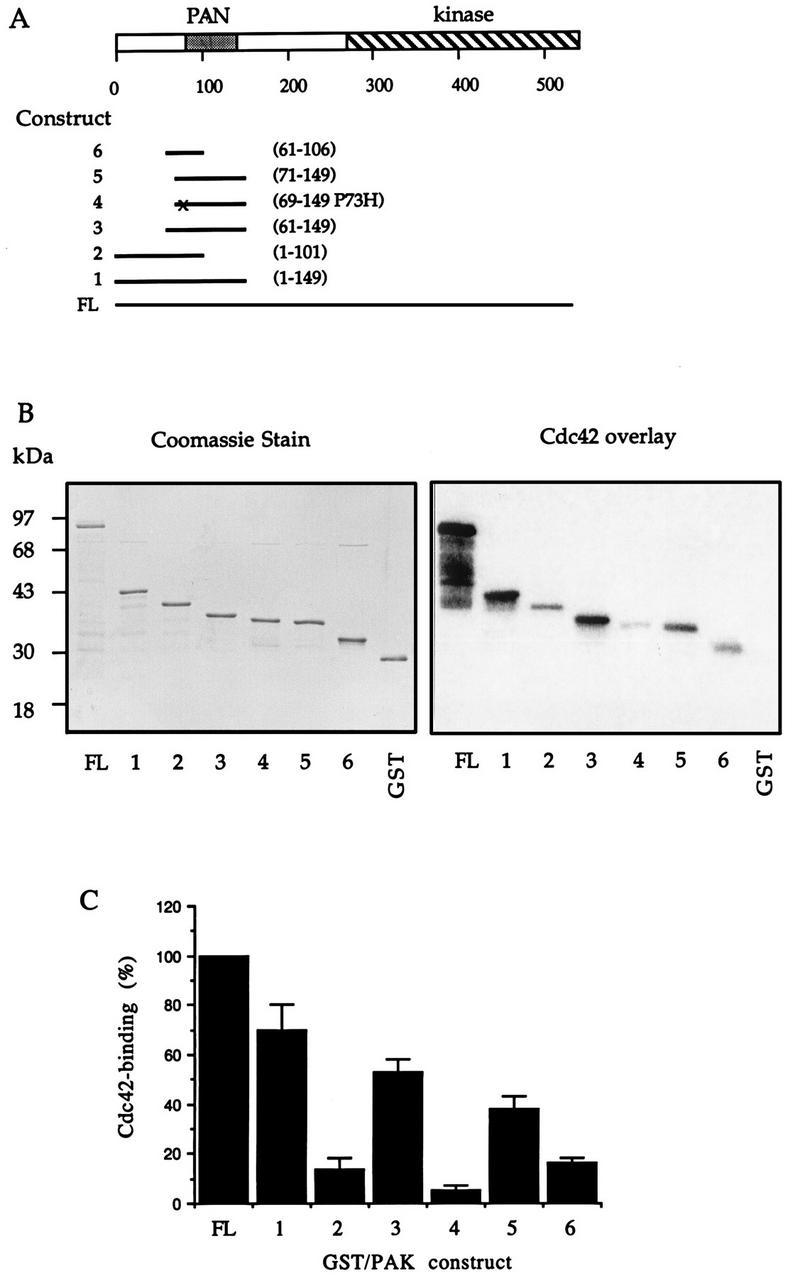
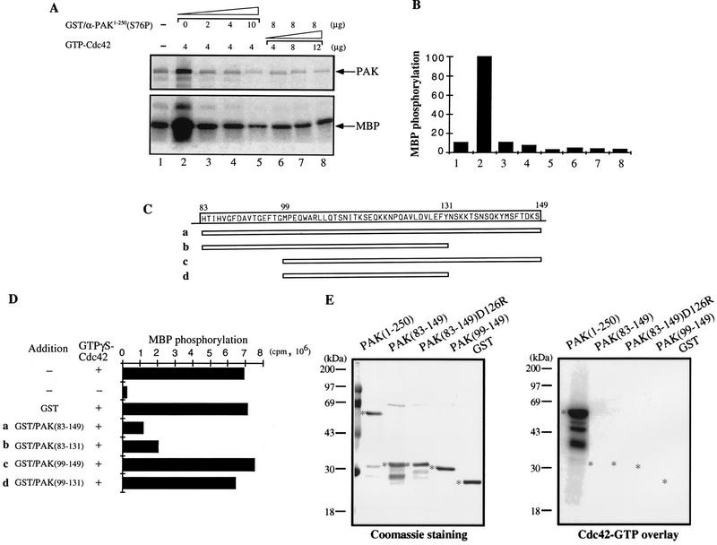
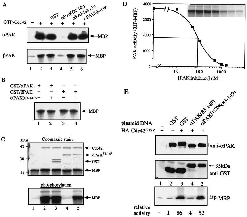
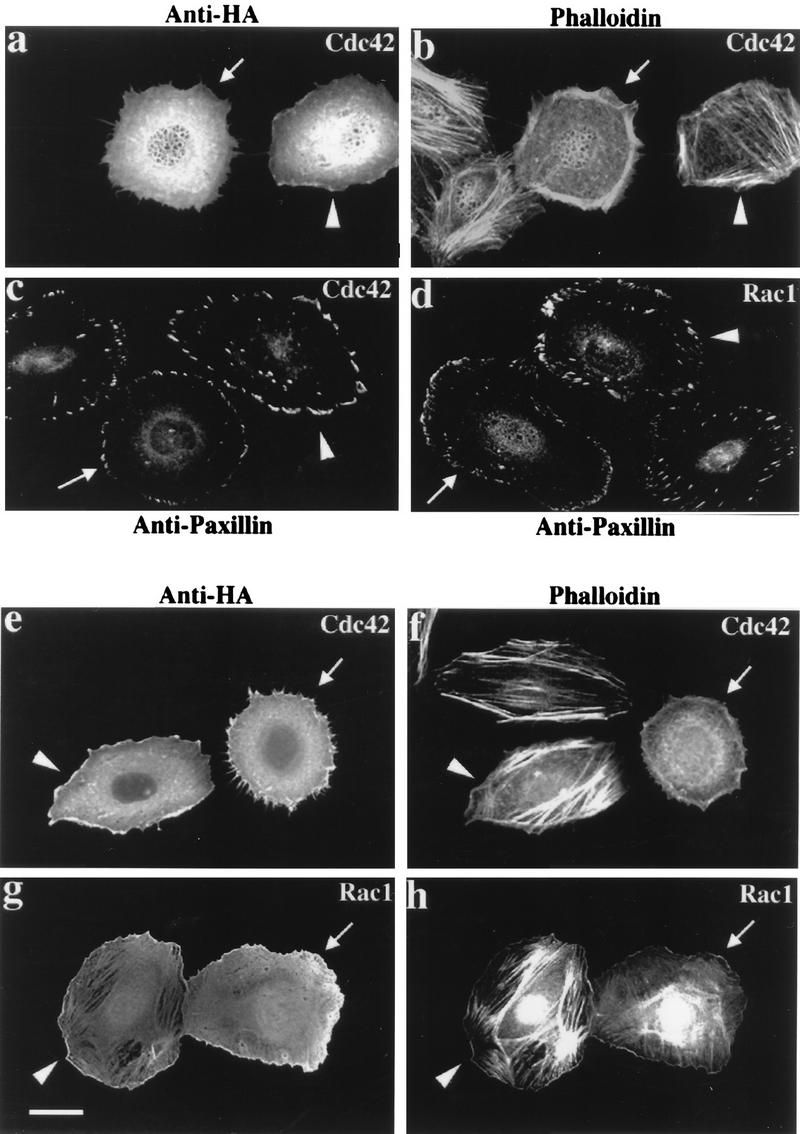
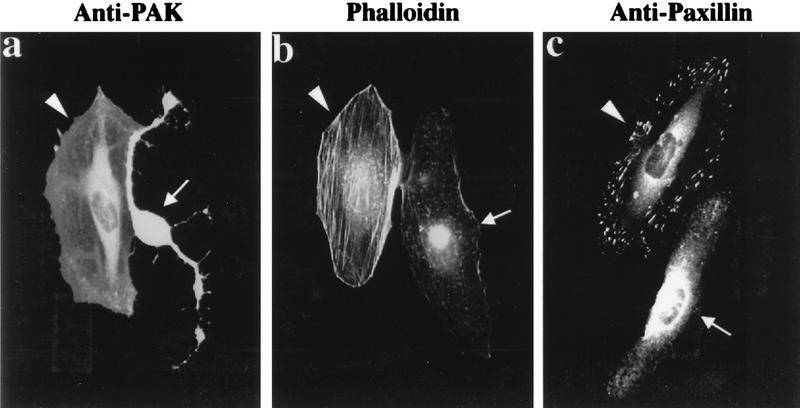
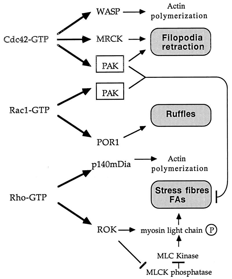
AlphaPAK in a constitutively active form can exert morphological effects (E. Manser, H.-Y. Huang, T.-H. Loo, X.-Q. Chen, J.-M. Dong, T. Leung, and L. Lim, Mol. Cell. Biol. 17:1129-1143, 1997) resembling those of Cdc42G12V. PAK family kinases, conserved from yeasts to humans, are directly activated by Cdc42 or Rac1 through interaction with a conserved N-terminal motif (corresponding to residues 71 to 137 in alphaPAK). alphaPAK mutants with substitutions in this motif that resulted in severely reduced Cdc42 binding can be recruited normally to Cdc42G12V-driven focal complexes. Mutation of residues in the C-terminal portion of the motif (residues 101 to 137), though not affecting Cdc42 binding, produced a constitutively active kinase, suggesting this to be a negative regulatory region. Indeed, a 67-residue polypeptide encoding alphaPAK83-149 potently inhibited GTPgammaS-bound Cdc42-mediated kinase activation of both alphaPAK and betaPAK. Coexpression of this PAK inhibitor with Cdc42G12V prevented the formation of peripheral actin microspikes and associated loss of stress fibers normally induced by the p21. Coexpression of PAK inhibitor with Rac1G12V also prevented loss of stress fibers but not ruffling induced by the p21. Coexpression of alphaPAK83-149 completely blocked the phenotypic effects of hyperactive alphaPAKL107F in promoting dissolution of focal adhesions and actin stress fibers. These results, coupled with previous observations with constitutively active PAK, demonstrate that these kinases play an important role downstream of Cdc42 and Rac1 in cytoskeletal reorganization.


Figures

FIG. 1
FIG. 1
Sequences affecting efficiency of p21 binding to αPAK. (A) Schematic diagram representing regions of αPAK expressed and purified from E. coli. Amino acid numbers at the beginning and the end of each fragment are indicated on the right. The point mutation present in construct 4 is marked by “x.” FL, full length. (B) GST/αPAK constructs were expressed and purified as GST fusion proteins, and 1 μg of each protein was resolved by SDS-PAGE (12% gel) and stained with Coomassie brilliant blue (left); 10 pmol of each of these proteins was analyzed by an overlay binding assay using [γ-32P]GTP-Cdc42 (right). (C) The Cdc42-binding signals shown in panel B (right) were quantified on a PhosphorImager (Molecular Dynamics). The means of data from two independent experiments are shown.
FIG. 2
FIG. 2
Mutations in the N terminus of the PAN motif abolish p21 binding. (A) Sequence alignment of PAN motifs of PAK-related proteins from rat (α-, β-, and γ-PAK), Drosophila (DPAK), Caenorhabditis elegans (Ce-PAK), S. cerevisiae (Ste20p, Cla4p, and Sc-PAK), and Schizosaccharomyces pombe (Shk1p). Accession numbers are given elsewhere (27). The conserved residues are boxed in black, and a consensus of these is shown below. Amino acid substitutions corresponding to each of the mutant constructs are shown. (B) The first 250 amino acids of each αPAK mutant and wild-type (Wt) construct were purified as GST fusion proteins, and 1 μg of each protein was resolved by SDS-PAGE (11% gel) and stained with Coomassie brilliant blue (top); p21 binding to bands containing 0.4 μg of each protein was determined by overlays with [γ-32P]GTP-Cdc42 (bottom). (C) The Cdc42 binding signals in panel B (bottom) were quantified on a PhosphorImager; the means of two independent experiments are shown. (D) Expression constructs encoding HA-αPAK mutants as indicated were transfected into HeLa cells alone or with FLAG-Cdc42G12V. Typical cells stained for αPAK are shown; in all cases, the cells were also stained with antipaxillin to confirm that peripherally located PAK was in FCs. Bar, 10 μm.
FIG. 3
FIG. 3
Mutations in the C-terminal portion of the PAN motif activate αPAK. (A) Activity of bacterially expressed GST/αPAK and GST/αPAKL404S in the presence or absence of GTP-Cdc42. Purified GST/αPAK or GST/αPAKL404S was assayed for kinase activity with MBP at 20 μM [γ-33P]ATP in the presence or absence of GTP-Cdc42. The kinase reaction was carried out at 30°C for 30 min. WT, wild type. (B) The GST/αPAKL404S mutants containing additional mutations in the PAN motif were expressed and purified from E. coli. The GST/PAK bands (arrows) correspond to 1 μg of the purified proteins stained with Coomassie brilliant blue. (C) Proteins shown in panel B were assayed for kinase activity to MBP in the presence or absence of GTP-Cdc42. Arrows indicate positions of autophosphorylated PAK and phosphorylated MBP bands. (D) Quantification of the MBP phosphorylation shown in panel C.
FIG. 4
FIG. 4
The C terminus of the PAN motif inhibits PAK activation by GTP-Cdc42. (A) The PAK N-terminal fusion protein GST/αPAK1-250(S76P) inhibits PAK activation by GTP-Cdc42. The kinase activity of bacterially expressed GST/αPAKL404S was assayed in the absence (lane 1) or presence (lane 2 to 8) of the indicated amounts of GTP-Cdc42. The autoradiograph shows inhibition due to the indicated amounts of GST/αPAK1-250(S76P) added during the kinase reactions. Signals of αPAK autophosphorylation and MBP phosphorylation are indicated by arrows. (B) Quantification of the MBP phosphorylation shown in panel A. (C) Schematic diagram of four peptides in the PAN motif region. The corresponding amino acid sequence is shown at the top. These peptides were expressed as GST fusion proteins. SDS-PAGE analysis of 1 μg of each purified protein showed single appropriately sized bands (not shown). (D) One microgram of bacterially expressed GST/αPAKL404S was assayed for kinase activity to MBP in the presence of excess GTP-Cdc42 (4 μg) and 4 μg of each inhibitory peptide. The MBP phosphorylation signals were quantified on a PhosphorImager. (E) One microgram each of GST/αPAK and GST proteins was resolved by SDS-PAGE (11% gel) and blotted onto a polyvinylidene difluoride membrane. The proteins were stained with Coomassie blue (left), and their p21 binding was analyzed by overlay with [γ-32P]GTP-Cdc42 (right). Asterisks indicate positions of protein bands.
FIG. 5
FIG. 5
Specific inhibition of αPAK and βPAK activation by the GST/αPAK83-149 polypeptide. (A) In each assay, 0.2 μg of αPAK or βPAK purified from COS-7 cells was assayed for kinase activity toward MBP in the absence (lane 1) or presence of 2 μg of GTPγS-Cdc42 (lanes 2 to 6) and 2 μg of indicated PAK-derived polypeptides (lanes 4 to 6). GST was used as a control (lane 3). Kinase reactions were carried out with 10 μM [γ-33P]ATP and 10 μg of MBP in a final volume of 30 μl; half of the reaction was analyzed on 12% polyacrylamide gels. The MBP region of the autoradiograph is shown. (B) Bacterially expressed (active) GST/αPAK (1 μg) and GST/βPAK (1 μg) were assayed for MBP kinase activity (as described above) in the absence (lanes 1 and 3) or presence (lanes 2 and 4) of 5 μg of GST/αPAK83-149. (C) A mutation (D126R) in GST/αPAK83-148 interferes with its inhibitory activity. Top, Coomassie blue-stained gel (12%); bottom, corresponding autoradiograph of the MBP region. Each reaction was carried out with 0.2 μg of αPAK which was activated in the presence of 2 μg of Cdc42-GTPγS. Lanes 4 and 5 contained 1.5 μg of GST/αPAK83-148 or GST/αPAK83-148(D126R), a level ∼15-fold higher than the calculated Ki of the wild-type inhibitor (D). GST alone has no effect on activation (the larger GST form contains attached polylinker-derived sequence). Kinase reactions were as for panel A. (D) Concentration dependence of αPAK inhibition was determined by using 50 ng of kinase (=18 nM) under activation conditions as for panel A while varying the GST/αPAK83-148 concentration. MBP phosphorylation was quantified on a PhosphorImager. For reference, the data of MBP phosphorylation is shown in the inset. (E) Inhibition of αPAK activation by coexpression of GST/αPAK83-149 in COS-7 cells. Cells (in 100-mm-diameter dishes) were transfected with 3 μg of FLAG-αPAK (lanes 2 to 5) with or without 3 μg of HA-Cdc42G12V expression plasmid as indicated. Expression of GST, GST/αPAK83-149, or the mutant (D126R) version (6 μg of plasmid per dish) was detected in the total extract by using anti-GST antibodies. The activity of the anti-FLAG immunoprecipitates, recovering equivalent amounts of the kinase (top), was determined by phosphorylation of MBP (counts quantified at the bottom).
FIG. 6
FIG. 6
Morphological effects of Cdc42 and Rac are blocked by inhibitory PAK83-149. HeLa cells were examined 2 h (a to d) or 4 h (e to h) after plasmid microinjection. In each case, a constitutively active mutant (G12V) form of Cdc42 or Rac1 was used. The top panels show that coinjection of the PAK inhibitor (arrowheads) blocks the reorganization of actin stress fibers normally seen with Cdc42 (arrows). Analysis of FCs by paxillin staining (c and d) shows production of peripherally located FCs to occur in the presence of the PAK inhibitor (arrowheads), but the resultant structures are thicker and there is no apparent loss of the existing internal focal adhesions. Even after 4 h, Cdc42G12V-expressing cells (e) do not form filopodia or retraction fibers when injected with the PAK inhibitor: with both Cdc42G12V and RacG12V, PAK inhibition leads to an overall increase in the intensity of stress fiber staining (f and h).
FIG. 7
FIG. 7
The PAK inhibitor can block effects of constitutively active PAK in vivo. The effects of microinjected αPAKL107F on HeLa cells (arrows) include cell retraction (a) seen in cells stained for expressed PAK, loss of actin stress fibers (b), and dissolution of paxillin-stained focal adhesions (c). All three effects could be blocked by coinjection of an expression construct encoding αPAK83-149 (arrowheads).
FIG. 8
FIG. 8
A model of PAK function in relation to known effectors of Rho-p21s. The targets of Cdc42, Rac, and Rho which have known morphological roles are shown. MRCK is the myotonin kinase-related Cdc42-binding kinase (26); p140mDia is the mammalian homolog of Drosophila diaphanous (58); the other Rho-binding kinase, PKN (59), has no known morphological function. PAK acts downstream of both Cdc42 and Rac to break down actin cytoskeletal structures and focal adhesions (FAs). This function could be particularly important in remodeling the cytoskeleton to allow dynamic events such as filopodial extension and subsequent formation of lamellipodia, ultimately leading to cell migration or growth cone extension (20). MLC, myosin light chain; MLCK, myosin light-chain kinase.

References

    1. Aspenstrom P, Lindberg U, Hall A. Two GTPases, Cdc42 and Rac, bind directly to a protein implicated in the immunodeficiency disorder Wiskott-Aldrich syndrome. Curr Biol. 1995;6:70–75. - PubMed
    1. Bagrodia S, Derijard B, Davis R J, Cerione R A. Cdc42 and PAK-mediated signalling leads to JNK and p38 mitogen-activated protein kinase activation. J Biol Chem. 1995;270:27995–27998. - PubMed
    1. Bagrodia S, Taylor S J, Creasy C L, Chernoff J, Cerione R A. Identification of a mouse p21cdc42/rac activated kinase. J Biol Chem. 1995;270:22731–22737. - PubMed
    1. Benner G E, Dennis P B, Masaracchia R A. Activation of an S6/H4 kinase (PAK 65) from human placenta by intramolecular and intermolecular autophosphorylation. J Biol Chem. 1995;270:21121–21128. - PubMed
    1. Bokoch G M, Wang Y, Bohl B P, Sells M A, Quilliam L A, Knaus U G. Interaction of the Nck adapter protein with p21-activated kinase (PAK1) J Biol Chem. 1996;271:25746–25749. - PubMed

Publication types

MeSH terms