The microsatellite sequence (CT)n x (GA)n promotes stable chromosomal integration of large tandem arrays of functional human U2 small nuclear RNA genes
- PMID: 9528797
- PMCID: PMC121475
- DOI: 10.1128/MCB.18.4.2262
The microsatellite sequence (CT)n x (GA)n promotes stable chromosomal integration of large tandem arrays of functional human U2 small nuclear RNA genes
Abstract
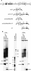
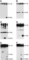
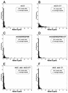
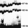
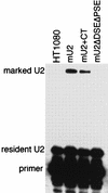
The multigene family encoding human U2 small nuclear RNA (snRNA) is organized as a single large tandem array containing 5 to 25 copies of a 6.1-kb repeat unit (the RNU2 locus). Remarkably, each of the repeat units within an individual U2 tandem array appears to be identical except for an irregular dinucleotide tract, known as the CT microsatellite, which exhibits minor length and sequence polymorphism. Using a somatic cell genetic assay, we previously noticed that the CT microsatellite appeared to stabilize artificial tandem arrays of U2 snRNA genes. We now demonstrate that the CT microsatellite is required to establish large tandem arrays of transcriptionally active U2 genes, increasing both the average and maximum size of the resulting arrays. In contrast, the CT microsatellite has no effect on the average or maximal size of artificial arrays containing transcriptionally inactive U2 genes that lack key promoter elements. Our data reinforce the connection between recombination and transcription. Active U2 transcription interferes with establishment or maintenance of the U2 tandem array, and the CT microsatellite opposes these effects, perhaps by binding GAGA or GAGA-related factors which alter local chromatin structure. We speculate that the mechanisms responsible for maintenance of tandem arrays containing active promoters may differ from those that maintain tandem arrays of transcriptionally inactive sequences.
Figures

Similar articles
-
Concerted evolution of the tandemly repeated genes encoding primate U2 small nuclear RNA (the RNU2 locus) does not prevent rapid diversification of the (CT)n.(GA)n microsatellite embedded within the U2 repeat unit.Genomics. 1995 Dec 10;30(3):583-93. doi: 10.1006/geno.1995.1280. Genomics. 1995. PMID: 8825646
-
Striking bimodal methylation of the repeat unit of the tandem array encoding human U2 snRNA (the RNU2 locus).Genomics. 1999 Dec 15;62(3):508-18. doi: 10.1006/geno.1999.6052. Genomics. 1999. PMID: 10644450
-
Adenovirus type 12-induced fragility of the human RNU2 locus requires U2 small nuclear RNA transcriptional regulatory elements.Mol Cell Biol. 1995 Nov;15(11):6246-55. doi: 10.1128/MCB.15.11.6246. Mol Cell Biol. 1995. PMID: 7565777 Free PMC article.
-
Concerted evolution of the tandemly repeated genes encoding human U2 snRNA (the RNU2 locus) involves rapid intrachromosomal homogenization and rare interchromosomal gene conversion.EMBO J. 1997 Feb 3;16(3):588-98. doi: 10.1093/emboj/16.3.588. EMBO J. 1997. PMID: 9034341 Free PMC article.
-
Concerted evolution of the tandem array encoding primate U2 snRNA occurs in situ, without changing the cytological context of the RNU2 locus.EMBO J. 1995 Jan 3;14(1):169-77. doi: 10.1002/j.1460-2075.1995.tb06987.x. EMBO J. 1995. PMID: 7828589 Free PMC article.
Cited by
-
d(GA x TC)(n) microsatellite DNA sequences enhance homologous DNA recombination in SV40 minichromosomes.Nucleic Acids Res. 2000 Dec 1;28(23):4617-22. doi: 10.1093/nar/28.23.4617. Nucleic Acids Res. 2000. PMID: 11095670 Free PMC article.
-
Misconception of Schizophyllum commune strain 20R-7-F01 origin from subseafloor sediments over 20 million years old.Sci Rep. 2025 Feb 24;15(1):6586. doi: 10.1038/s41598-024-84457-2. Sci Rep. 2025. PMID: 39994251 Free PMC article.
-
A tandem array of minimal U1 small nuclear RNA genes is sufficient to generate a new adenovirus type 12-inducible chromosome fragile site.J Virol. 1998 May;72(5):4205-11. doi: 10.1128/JVI.72.5.4205-4211.1998. J Virol. 1998. PMID: 9557709 Free PMC article.
-
Characterization of Microsatellites in the Akebia trifoliata Genome and Their Transferability and Development of a Whole Set of Effective, Polymorphic, and Physically Mapped Simple Sequence Repeat Markers.Front Plant Sci. 2022 Mar 18;13:860101. doi: 10.3389/fpls.2022.860101. eCollection 2022. Front Plant Sci. 2022. PMID: 35371184 Free PMC article.
-
Emerging roles of macrosatellite repeats in genome organization and disease development.Epigenetics. 2017 Jul 3;12(7):515-526. doi: 10.1080/15592294.2017.1318235. Epub 2017 Apr 20. Epigenetics. 2017. PMID: 28426282 Free PMC article. Review.
References
-
- Ares M, Jr, Chung J-S, Giglio L, Weiner A M. Distinct factors with Sp1 and NF-A specificities bind to adjacent functional elements of the human U2 snRNA gene enhancer. Genes Dev. 1987;1:808–817. - PubMed
-
- Armour J A, Crosier M, Malcolm S, Chan J C, Jeffreys A J. Human minisatellite loci composed of interspersed GGA-GGT triplet repeats. Proc R Soc Lond Ser B. 1995;261:345–349. - PubMed
Publication types
MeSH terms
Substances
Grants and funding
LinkOut - more resources
Full Text Sources
