. 1998 Apr;18(4):2298-308.
doi: 10.1128/MCB.18.4.2298.

Shc and Enigma are both required for mitogenic signaling by Ret/ptc2

Affiliations

Shc and Enigma are both required for mitogenic signaling by Ret/ptc2

K Durick et al. Mol Cell Biol. 1998 Apr.

Abstract

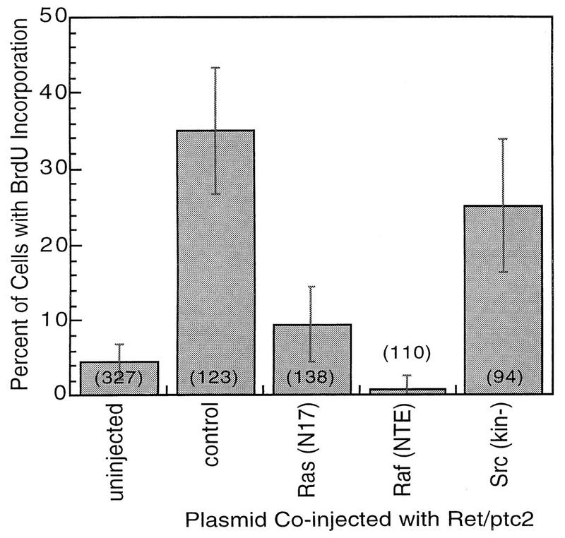
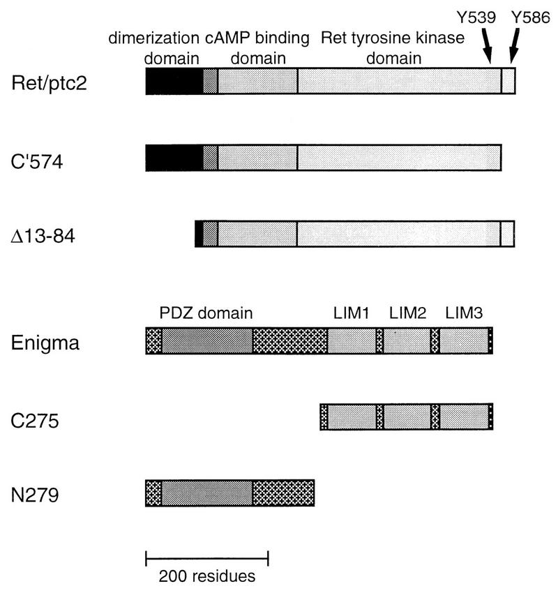
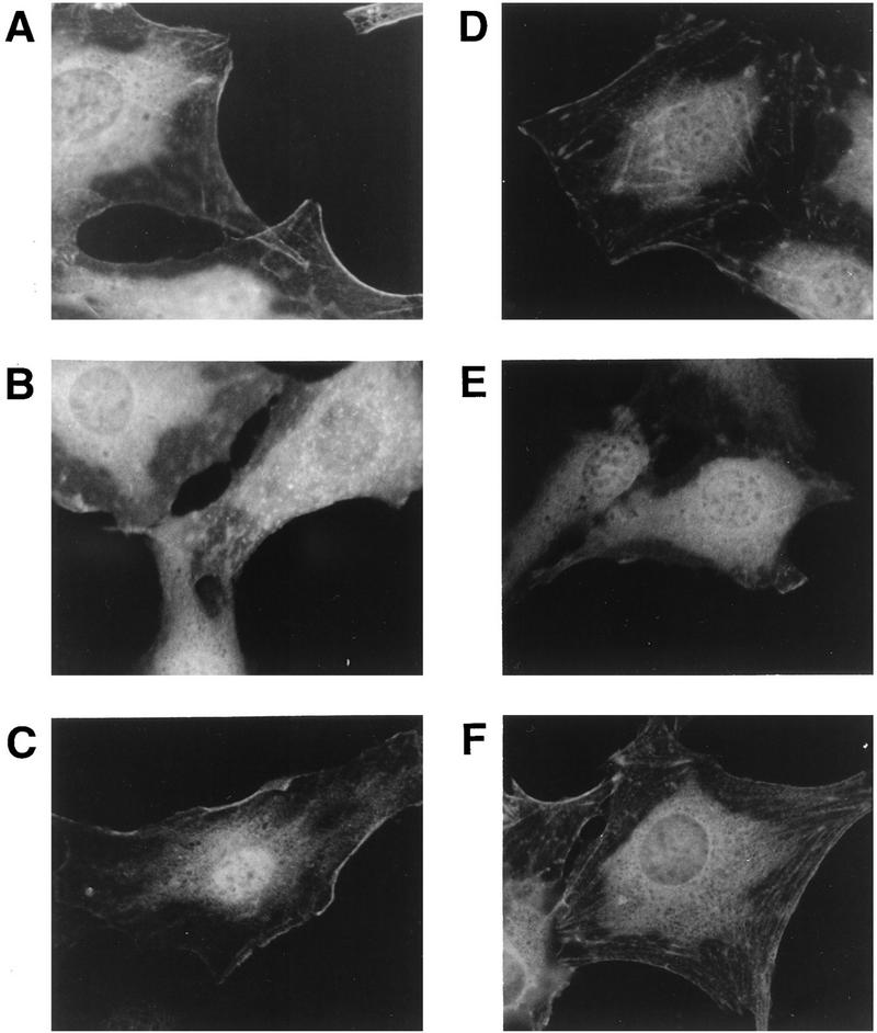
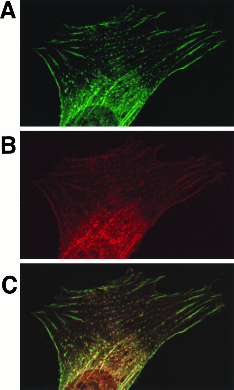
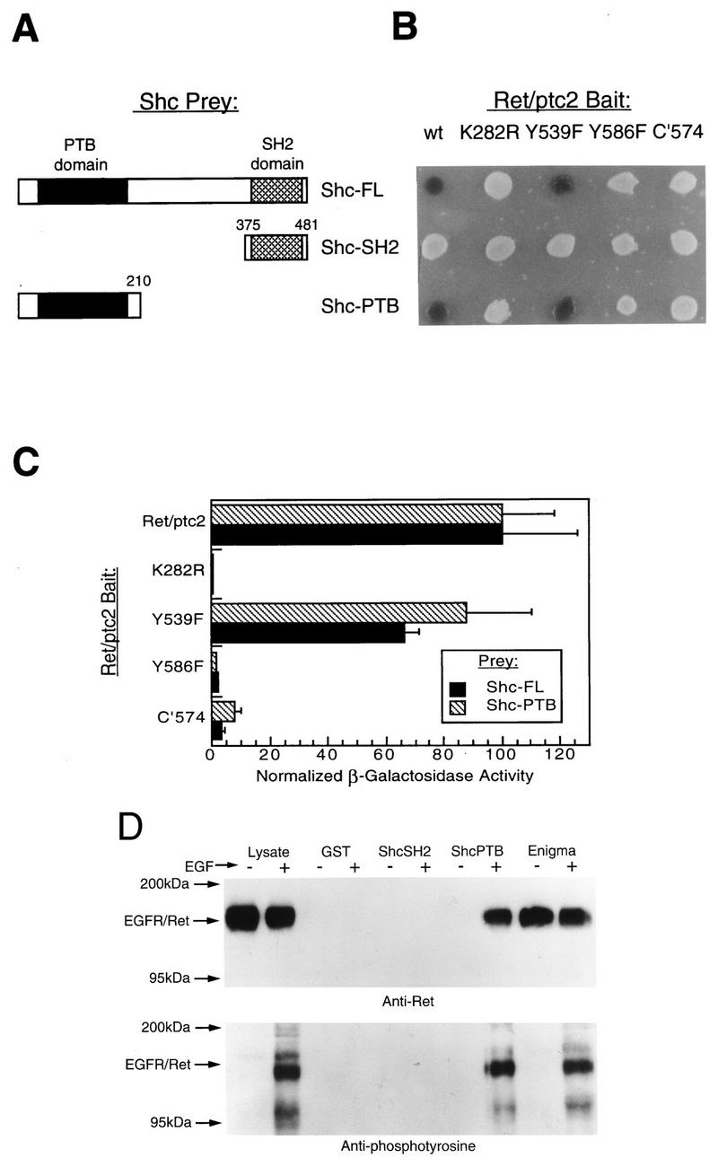
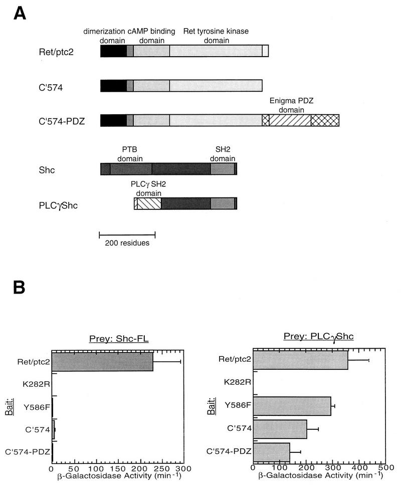
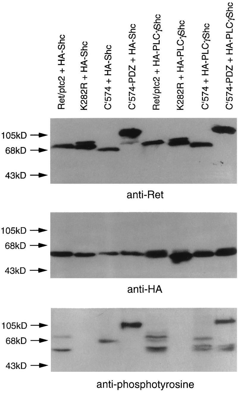
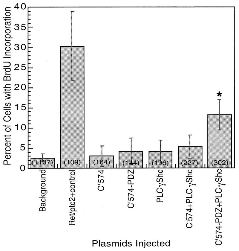
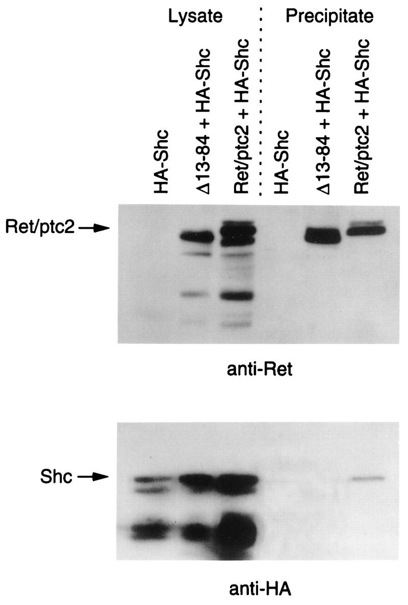
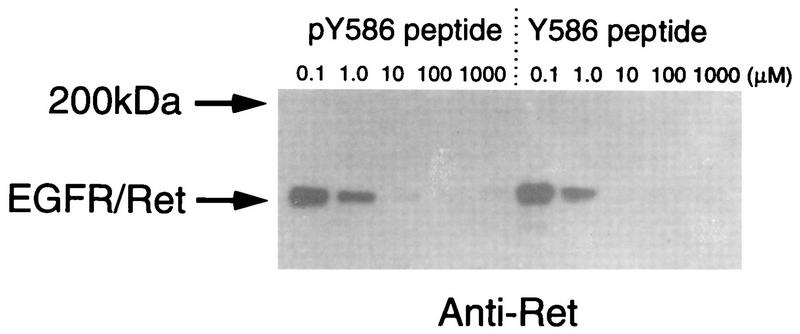
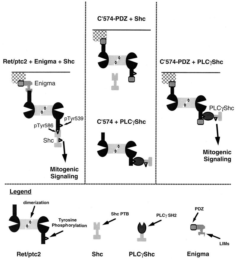
Ret/ptc2 is a constitutively active, oncogenic form of the c-Ret receptor tyrosine kinase. Like the other papillary thyroid carcinoma forms of Ret, Ret/ptc2 is activated through fusion of the Ret tyrosine kinase domain to the dimerization domain of another protein. Investigation of requirements for Ret/ptc2 mitogenic activity, using coexpression with dominant negative forms of Ras and Raf, indicated that these proteins are required for mitogenic signaling by Ret/ptc2. Because activation of Ras requires recruitment of Grb2 and SOS to the plasma membrane, the subcellular distribution of Ret/ptc2 was investigated, and it was found to localize to the cell periphery. This localization was mediated by association with Enigma via the Ret/ptc2 sequence containing tyrosine 586. Because Shc interacts with MEN2 forms of Ret, and because phosphorylation of Shc results in Grb2 recruitment and subsequent signaling through Ras and Raf, the potential interaction between Ret/ptc2 and Shc was investigated. The PTB domain of Shc also interacted with Ret/ptc2 at tyrosine 586, and this association resulted in tyrosine phosphorylation of Shc. Coexpression of chimeric proteins demonstrated that mitogenic signaling from Ret/ptc2 required both recruitment of Shc and subcellular localization by Enigma. Because Shc and Enigma interact with the same site on a Ret/ptc2 monomer, dimerization of Ret/ptc2 allows assembly of molecular complexes that are properly localized via Enigma and transmit mitogenic signals via Shc.


Figures

FIG. 1
FIG. 1
Effect of coexpression with dominant negative Ras or Raf on mitogenic activity of Ret/ptc2. Serum-starved mouse fibroblasts (10T1/2) were microinjected with a mixture of Ret/ptc2 expression plasmid along with one of the following: a control empty plasmid (control); plasmid expressing mutant Ras with serine 17 replaced by asparagine [Ras(N17)]; plasmid expressing the NTE, residues 1 through 290, of Raf-1 [Raf(NTE)]; or plasmid expressing a catalytically inactive form of Src [Src(kin−)]. In each case constructs were injected at 100 μg/ml. Cells were then assessed for entry into S phase by immunofluorescent detection of BrdU incorporation. The fraction of injected cells positive for BrdU incorporation is shown, with error bars displaying the 95% confidence intervals, which were calculated by using the standard error of proportion. The numbers in parentheses are the total numbers of injected cells.
FIG. 2
FIG. 2
Schematic representation of Ret/ptc2 and Enigma constructs. Ret/ptc2 is 596 residues in length, with the N-terminal 236 amino acids from the type Iα regulatory subunit of cAMP-dependent protein kinase (RIα). The dimerization domain of RIα is located within the first 84 residues. Enigma is 455 residues in length, with an N-terminal PDZ domain and three C-terminal LIM domains. An HA tag was added to the amino termini of the Enigma constructs to facilitate detection by anti-HA monoclonal antibodies.
FIG. 3
FIG. 3
Determinants of subcellular localization of Ret/ptc2 and Enigma. (A, B, and C) Sequence requirements for the subcellular targeting of Ret/ptc2. Fibroblasts microinjected with various Ret/ptc2 expression plasmids were fixed 4 h after injection and immunofluorescently stained with anti-Ret antibodies. Constructs injected coded for the following: (A) wild-type Ret/ptc2; (B) C′574, a mutant where the last 22 amino acids are missing, including the Enigma binding site; and (C) Ret/ptc2Δ(13-84), a dimerization domain deletion mutant. (D, E, and F) Sequence requirements for the subcellular targeting of Enigma. Cells injected with constructs expressing HA-tagged forms of Enigma were stained for immunofluorescence with anti-HA tag monoclonal antibodies. Plasmids expressed the following HA-tagged proteins: (D) full-length Enigma; (E) the C-terminal 275 residues of Enigma, which contain the three LIM domains; and (F) the N-terminal 279 residues of Enigma, which contain the PDZ domain.
FIG. 4
FIG. 4
Codistribution of Ret/ptc2 and Enigma. Mouse fibroblasts coinjected with expression plasmids for Ret/ptc2 and HA-tagged Enigma were fixed 4 h after injection, subjected to immunofluorescent staining, and then imaged by confocal microscopy. (A) Enigma distribution shown by fluorescein linked to anti-HA tag monoclonal antibody; (B) Ret/ptc2 distribution shown by fluorescence of Cy-5 linked to anti-Ret antibody; (C) Digital overlay of the two fluorescent signals.
FIG. 5
FIG. 5
Characterization of the interaction of Shc with Ret/ptc2. The yeast two-hybrid system was used to map the interaction determinants of Ret/ptc2 and Shc by using various Shc-VP16 (Prey) and Ret/ptc2-LexA (Bait) fusion constructs, and results were verified by GST affinity precipitation. (A) Schematic representation of the Shc deletion mutants used. Numbers delineate amino acid sequences included in the constructs. (B) Qualitative β-galactosidase activity of yeast cotransformants. Single colonies were plated on solid media containing X-Gal, and results shown are for 10 h after plating. Bait constructs coded for wild-type Ret/ptc2 (wt), various point mutants, or a C-terminal-truncation mutant (C′574). (C) Solution assay of yeast cotransformant β-galactosidase activity. Cultures of yeast cotransformed with plasmids expressing various Ret/ptc2-LexA and Shc-VP16 fusion proteins were assayed for β-galactosidase activity. Results shown are averages obtained for four assays, with error bars representing the standard deviations. For comparison of results between the full-length (FL) and PTB Shc constructs, normalized values were plotted. The actual activity for full-length Shc with wild-type Ret/ptc2 was 250 U, while the activity for the Shc PTB prey with wild-type Ret/ptc2 was 160 U. Units are per minute, as described for the β-galactosidase solution assay (3). (D) Clonal NIH 3T3 cells expressing an EGFR-Ret chimeric receptor were treated (+) or not treated (−) with 100 nM EGF for 10 min at 37°C before lysis. Aliquots of lysates were incubated with either GST or GST fusion proteins bound to glutathione agarose. The protein fragments expressed as GST fusions were the Shc SH2 and PTB domains and Enigma LIM domains 2 and 3. Western blots of EGFR-Ret that bound to the indicated GST fusion proteins are shown. Gels were run in parallel, blotted to PVDF membranes, and probed with anti-Ret or antiphosphotyrosine antibodies.
FIG. 6
FIG. 6
Characterization of interactions between Ret/ptc2 and Shc or PLCγ-Shc. The yeast two-hybrid system was used to monitor interactions between various Ret/ptc2-LexA constructs and either Shc-VP16 or PLCγ-Shc-VP16 fusions. (A) Schematic representation of the fusion proteins constructed. C′574 was fused to the N-terminal 279 residues of Enigma to make C′574-PDZ. A fragment of the PLCγ sequence encoding the N-terminal SH2 domain was fused to the C-terminal two-thirds of Shc, replacing the Shc PTB domain (PLCγ-Shc). (B) β-Galactosidase activity of yeast cotransformed with the Ret/ptc2 and Shc constructs. Cultures of yeast cotransformed with plasmids expressing various Ret/ptc2-LexA and either Shc-VP16 or PLCγ-Shc-VP16 fusion proteins were assayed for β-galactosidase activity. Results shown are averages of units of activity from four solution assays (3), with error bars representing the standard deviations.
FIG. 7
FIG. 7
Phosphorylation of Shc and PLCγ-Shc by Ret/ptc2 constructs. Kidney 293 cells were cotransfected with plasmids expressing a form of Ret/ptc2 and either HA-tagged Shc (HA-Shc) or PLCγ-Shc (HA-PLCγShc). The Ret/ptc2 constructs expressed wild-type Ret/ptc2, a kinase-inactive point mutant (K282R), a C-terminal-truncation mutant (C′574), or a mutant in which the C terminus was replaced with the PDZ domain of Enigma (C′574-PDZ). Twenty-four hours after transfection, the cells were harvested, and lysates were subjected to SDS-PAGE. Proteins were then transferred to PVDF membranes and detected with either anti-Ret, anti-HA tag, or antiphosphotyrosine antibodies.
FIG. 8
FIG. 8
Mitogenic activity of Ret/ptc2 constructs coexpressed with PLCγ-Shc. Serum-starved mouse fibroblasts (10T1/2) were microinjected with combinations of expression plasmids. In each case, constructs were injected at 100 μg/ml; “background” represents uninjected cells. Cells were then assessed for entry into S phase by immunofluorescent detection of BrdU incorporation. The fraction of injected cells positive for BrdU incorporation is shown, with error bars displaying the 95% confidence intervals, which were calculated by using the standard error of proportion. The numbers in parentheses are the total numbers of injected cells. The asterisk indicates that cells coinjected with plasmids for C′574-PDZ and PLCγ-Shc were significantly above background in BrdU incorporation (P < 0.001).
FIG. 9
FIG. 9
Characterization of the Ret/ptc2-Shc-Enigma complex. Lysates from 293 cells expressing HA-tagged Shc either alone, with wild-type Ret/ptc2, or Ret/ptc2Δ(13-84) were incubated with GST-LIM2 of Enigma bound to glutathione agarose. After extensive washing, the agarose beads were boiled in SDS sample buffer, and bound proteins were resolved by SDS-PAGE. Gels were run in parallel, blotted to PVDF membranes, and probed with anti-Ret or anti-HA antibodies. Lysate samples, run in the first three lanes, were approximately one-fourth of the total amount of lysate used in each incubation.
FIG. 10
FIG. 10
Effects of competitor phospho- and dephosphopeptides on the interaction between Ret and Enigma. Lysates from NIH 3T3 cells expressing the EGFR-Ret chimeric receptor were incubated with GST-LIM2 of Enigma bound to glutathione agarose beads. The incubation was carried out in the presence of various concentrations of peptide containing the last 20 residues of Ret/ptc2, residues 577 to 596. The peptide was synthesized to contain either phosphotyrosine 586 (pY586) or tyrosine 586 (Y586). After incubation the beads were washed extensively, and bound EGFR-Ret was visualized by Western blotting with anti-Ret antibody.
FIG. 11
FIG. 11
Proposed model for wild-type Ret/ptc2 mitogenic signaling. The left panel illustrates functions of Enigma and Shc in Ret/ptc2 mitogenic signaling. The center panel summarizes results indicating that functions of either Enigma or Shc alone are not sufficient for Ret/ptc2 activity. The right panel shows restoration of Ret/ptc2 mitogenic activity by reconstitution of both Enigma and Shc functions via chimeric molecules.

References

    1. Asai N, Murakami H, Iwashita T, Takahashi M. A mutation at tyrosine 1062 in MEN2A-Ret and MEN2B-Ret impairs their transforming activity and association with Shc adaptor proteins. J Biol Chem. 1996;271:17644–17649. - PubMed
    1. Attie T, Pelet A, Sarda P, Edery P, Eng C, Mulligan L M, Ponder B A J, Munnich A, Lyonnet S. A 7bp deletion of the RET proto-oncogene mutations in familial Hirschsprung’s disease. Hum Mol Genet. 1994;3:1439–1440. - PubMed
    1. Ausubel F, Brent R, Kingston R, Moore D, Seidman J, Smith J, Struhl K, editors. Current protocols in molecular biology. New York, N.Y: John Wiley & Sons, Inc.; 1994.
    1. Bongarzone I, Butti M G, Coronelli S, Borrello M G, Santoro M, Mondellini P, Pilotti S, Fusco A, Della Porta G, Pierotti M A. Frequent activation of ret protooncogene by fusion with a new activating gene in papillary thyroid carcinomas. Cancer Res. 1994;54:2979–2985. - PubMed
    1. Bongarzone I, Monzini N, Borrello M G, Carcano C, Ferraresi G, Arighi E, Mondellini P, Della Porta G, Pierotti M A. Molecular characterization of a thyroid tumor-specific transforming sequence formed by the fusion of ret tyrosine kinase and the regulatory subunit RIα of cyclic AMP-dependent protein kinase A. Mol Cell Biol. 1993;13:358–366. - PMC - PubMed

Publication types

MeSH terms

Substances