. 1998 Apr;18(4):2324-33.
doi: 10.1128/MCB.18.4.2324.

Notch1 and Notch2 inhibit myeloid differentiation in response to different cytokines

Affiliations

Notch1 and Notch2 inhibit myeloid differentiation in response to different cytokines

A Bigas et al. Mol Cell Biol. 1998 Apr.

Abstract

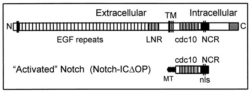
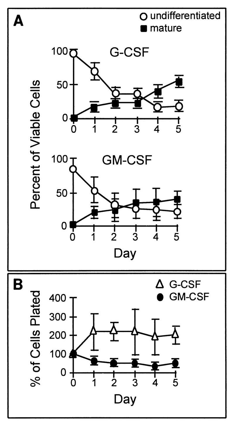
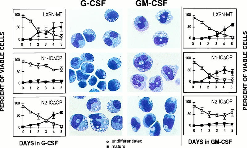
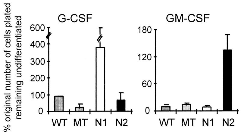
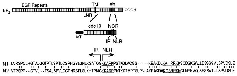
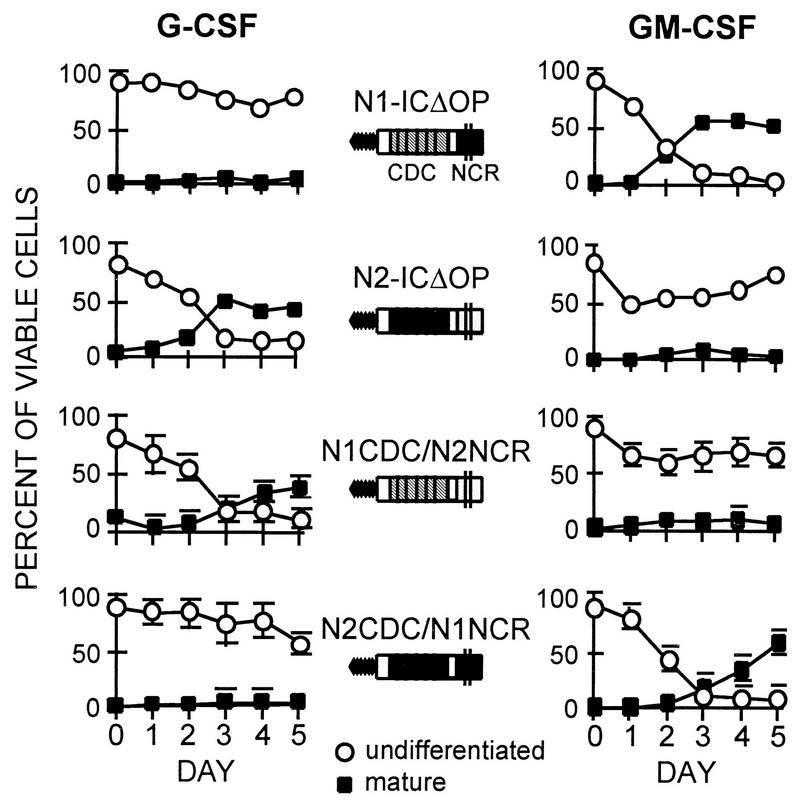
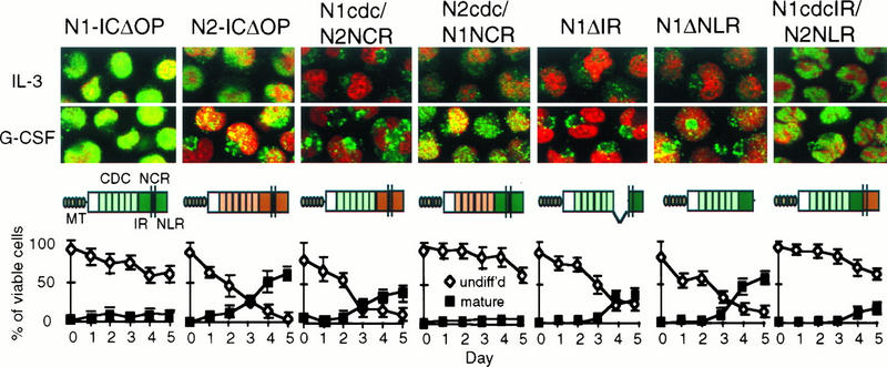
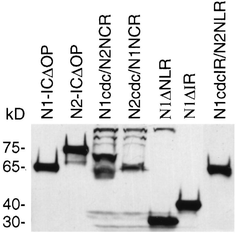
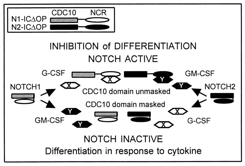
We have compared the ability of two mammalian Notch homologs, mouse Notchl and Notch2, to inhibit the granulocytic differentiation of 32D myeloid progenitor cells. 32D cells undergo granulocytic differentiation when stimulated with either granulocyte colony-stimulating factor (G-CSF) or granulocyte-macrophage colony-stimulating factor (GM-CSF). Expression of the activated intracellular domain of Notch1 inhibits the differentiation induced by G-CSF but not by GM-CSF; conversely, the corresponding domain of Notch2 inhibits differentiation in response to GM-CSF but not to G-CSF. The region immediately C-terminal to the cdc10 domain of Notch confers cytokine specificity on the cdc10 domain. The cytokine response patterns of Notch1 and Notch2 are transferred with this region, which we have termed the Notch cytokine response (NCR) region. The NCR region is also associated with differences in posttranslational modification and subcellular localization of the different Notch molecules. These findings suggest that the multiple forms of Notch found in mammals have structural differences that allow their function to be modulated by specific differentiation signals.


Figures

FIG. 1
FIG. 1
Diagram of the full-length Notch molecule and the activated intracellular Notch construct. Both Notch1 and Notch2 consist of an extracellular domain containing 36 epidermal growth factor (EGF) repeats and 3 LNR; there is a single transmembrane domain (TM). The intracellular domain contains six cdc10/ankyrin repeats (cdc10) and the newly defined NCR region, which contains a putative bipartite NLS. The activated Notch constructs (Notch-ICΔOP) consist of the region of the intracellular domain including the cdc10 repeats and the NCR region. Constructs also encode N-terminal myc epitope tags (MT) to facilitate detection of protein expression.
FIG. 2
FIG. 2
Growth and differentiation characteristics of parental 32D Cl3 cells in the presence of G-CSF or GM-CSF. (A) Granulocytic differentiation in response to G-CSF (upper graph) and to GM-CSF (lower graph) is illustrated by plotting the relative percentage of viable cells maintaining an undifferentiated blast morphology or having attained a terminally differentiated (bands and segmented neutrophils) mature phenotype after successive days in culture. In the presence of either cytokine, there is a continuous fall in the proportion of undifferentiated cells and a concomitant rise in the proportion of differentiated cells. (B) The total number of cells, relative to the original number of cells plated, present after successive days of culture in G-CSF or GM-CSF is shown. There is a slight increase in the cell number in G-CSF and a slight decline in GM-CSF. In all three graphs, the values shown are the averages of three independent experiments; error bars denote standard errors of the mean (SEM).
FIG. 3
FIG. 3
Differentiation of 32D cells expressing activated Notch1 (N1-ICΔOP) or Notch2 (N2-ICΔOP) molecules in response to either G-CSF or GM-CSF stimulation. The percentages of viable cells that are either undifferentiated or differentiated after successive days in culture in G-CSF (left) or GM-CSF (right) are shown in the graphs, and photomicrographs of Wright-stained cells from cultures on the final day are shown beside the corresponding graph. Control clones (LXSN-MT) are shown for comparison. As previously demonstrated (26), Notch1, but not Notch2, inhibits the differentiation induced by G-CSF. The converse effect is noted when the cells are induced with GM-CSF: Notch2 inhibits differentiation, but Notch1 does not. A representative experiment is shown; graphs represent the averages of results obtained with two (LXSN-MT) or three (N1-ICΔOP and N2-ICΔOP) clones, with error bars representing SEM. The same clones were used for the G-CSF and GM-CSF cultures. In this experiment, 1% WCM was included in the medium for G-CSF-induced differentiation to improve the uniformity of cell survival; we have previously reported that the addition of 1% WCM does not interfere with G-CSF-induced differentiation (26).
FIG. 4
FIG. 4
Effects of Notch1 and Notch2 activity on the maintenance of undifferentiated 32D cells in the presence of G-CSF or GM-CSF stimulation. Parental 32D cells (WT) and individual clones transduced with a control myc tag (MT) retroviral construct or activated forms of Notch1 (N1; mNotch1-ICΔOP) or Notch2 (N2; mNotch2-ICΔOP) were evaluated for proliferation and differentiation in the presence of 10 ng of G-CSF or GM-CSF per ml. The total number of cells remaining undifferentiated on day 5 (or the last day when at least 40% of the original number of cells were still viable) is expressed relative to the original number of cells plated. The G-CSF graph represents a single experiment involving three independent clones for each construct; the results were comparable to those reported previously (26). The GM-CSF graph represents combined data from four experiments involving the same three independent clones for each construct. Error bars represent the SEM. Note that different scales are used in the two graphs, because of the lower overall proliferation of 32D cells in GM-CSF (Fig. 2).
FIG. 5
FIG. 5
NCR region. The NCR region is shown as part of the full-length (top) and activated (middle) Notch molecules. At the bottom, the amino acid sequences of the Notch1 and Notch2 (N1 and N2) molecules are compared and the demarcation of the IR and NLR of the NCR is denoted. The putative NLS are underlined.
FIG. 6
FIG. 6
Differentiation induced by G-CSF (left) and GM-CSF (right) in 32D cell lines expressing N1CDC/N2NCR and N2CDC/N1NCR hybrid molecules, compared to 32D cell lines expressing the activated Notch1 (N1-ICΔOP) or Notch2 (N2-ICΔOP) proteins. Graphs show the relative percentages of viable differentiated and undifferentiated cells present in the cultures on successive days. Inhibition of differentiation in G-CSF occurs when the expressed Notch molecule contains the NCR region from Notch1. In GM-CSF, differentiation is inhibited when the NCR region is derived from Notch2. The same clones were used in the G-CSF and GM-CSF experiments. Values in the G-CSF graphs each represent the average for three different clones expressing N1-CDC/N2-NCR or N2-CDC/N1-NCR, with error bars denoting SEM. Values in the GM-CSF graphs represent the average for two of the three clones for each construct; the third clones did not survive in GM-CSF. Representative clones expressing N1-ICΔOP and N2-ICΔOP were used in this experiment (see Fig. 3 for more extensive data on N1-ICΔOP and N2-ICΔOP).
FIG. 7
FIG. 7
Correlation of subcellular localization and activity of Notch molecules in 32D cells stimulated with G-CSF. 32D cells transduced with the Notch1/Notch2 constructs indicated were evaluated by immunofluorescent staining and confocal microscopy for subcellular distribution of the Notch construct when the cells were grown in IL-3 or after 48 h in G-CSF. The cells were doubly stained with the nuclear stain propidium iodide (PI, red) and fluorescein isothiocyanate (green) to detect the myc epitope tags. Immunostained cells were visualized by confocal microscopy, and digital images (magnification, ×60) were reproduced and combined with Adobe Photoshop software. Below each set of micrographs is the corresponding graph of Notch activity, showing the percentages of cells remaining undifferentiated or differentiating into mature granulocytes over successive days in culture with G-CSF. Abbreviations for the Notch constructs are described in the text.
FIG. 8
FIG. 8
Western blot analysis of 32D cells expressing Notch1, Notch2, hybrid, and mutant Notch molecules. Whole-cell lysates from 32D cells expressing the indicated Notch constructs were subjected to SDS-polyacrylamide gel electrophoresis, and construct expression was detected by immunoblotting with an anti-myc tag antibody. Molecular masses (in kilodaltons) are shown on the left.
FIG. 9
FIG. 9
Model of cytokine specificity mediated by the NCR domain. In this model, the cdc10 domain is required for inhibitory activity, but this activity is masked by the NCR region. Cytokine stimulation activates signal transduction pathways, represented by X for G-CSF and Y for GM-CSF. The product of the X pathway is able to dissociate the Notch1 NCR from the cdc10 domain, thereby unmasking its activity, but is unable to act on the Notch2 NCR. Conversely, the product of the Y pathway can dissociate the Notch2 NCR but has no effect on the Notch1 NCR. The result is an inhibition of differentiation which is conditional on both Notch activation and cytokine stimulation.

Similar articles

Cited by

References

    1. Artavanis-Tsakonas S, Matsuno K, Fortini M E. Notch signaling. Science. 1995;268:225–232. - PubMed
    1. Artavanis-Tsakonas S, Simpson P. Choosing a cell fate: a view from the Notch locus. Trends Genet. 1991;7:403–408. - PubMed
    1. Austin J, Kimble J. glp-1 is required in the germ line for regulation of the decision between mitosis and meiosis in C. elegans. Cell. 1987;51:589–599. - PubMed
    1. Cagan R L, Ready D F. Notch is required for successive cell decisions in the developing Drosophila retina. Genes Dev. 1989;3:1099–1112. - PubMed
    1. Chitnis A, Henrique D, Lewis J, Ish-Horowicz D, Kintner C. Primary neurogenesis in Xenopus embryos regulated by a homologue of the Drosophila neurogenic gene Delta. Nature. 1995;375:761–766. - PubMed

Publication types

LinkOut - more resources