. 1998 Apr;18(4):2406-15.
doi: 10.1128/MCB.18.4.2406.

A nuclear matrix protein interacts with the phosphorylated C-terminal domain of RNA polymerase II

Affiliations

A nuclear matrix protein interacts with the phosphorylated C-terminal domain of RNA polymerase II

M Patturajan et al. Mol Cell Biol. 1998 Apr.

Abstract

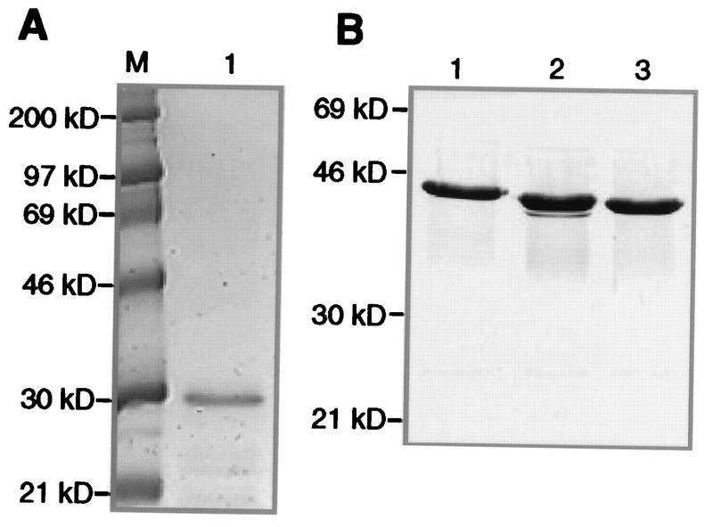
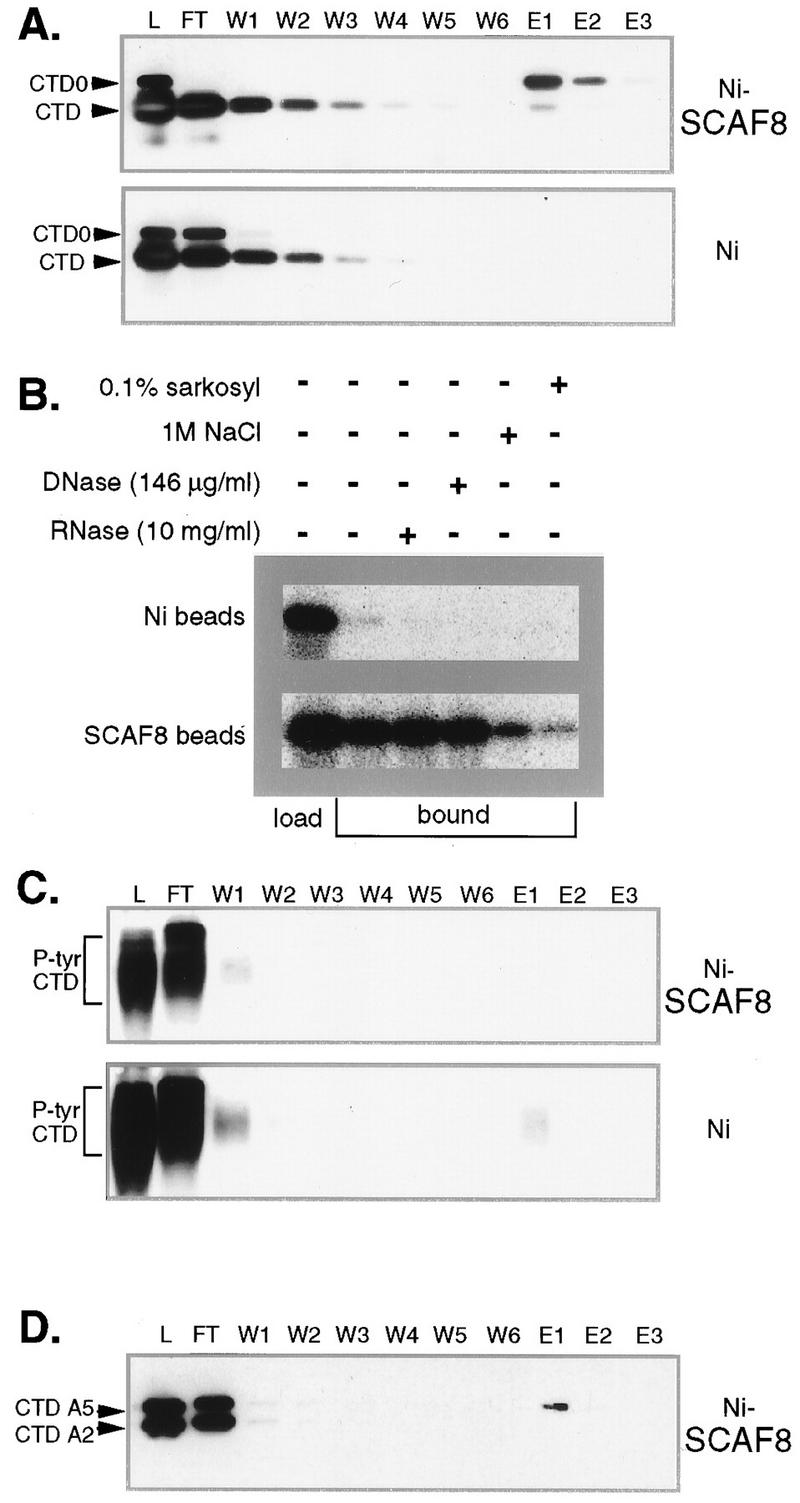
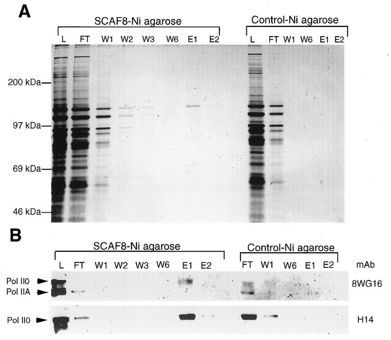
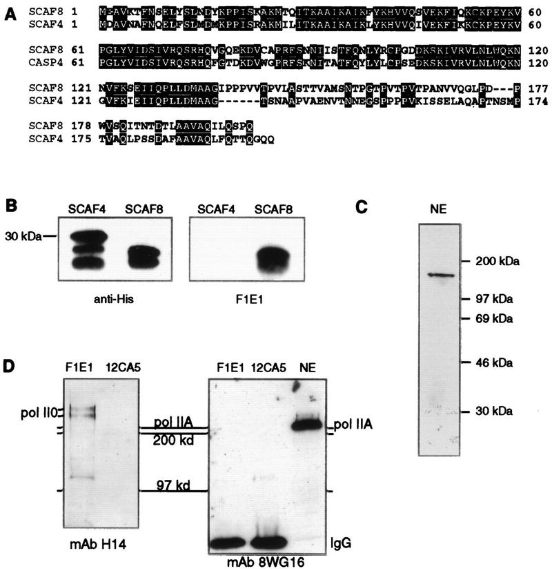
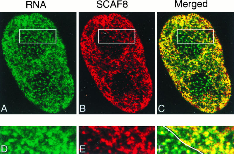
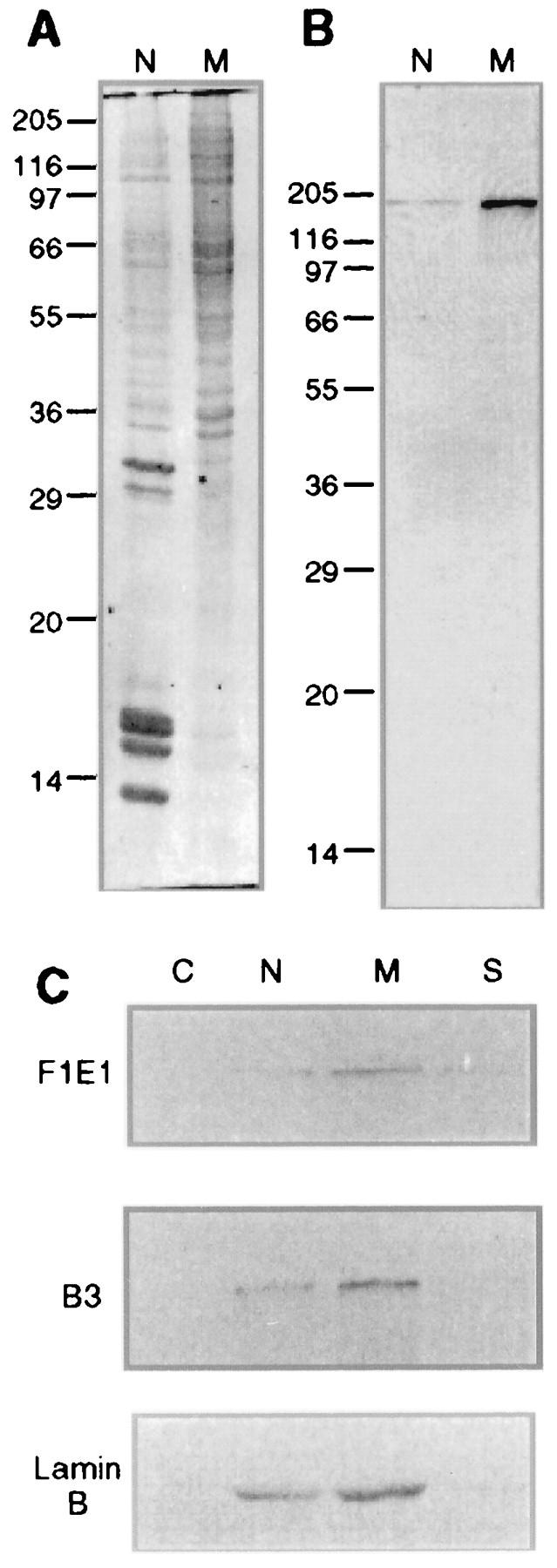
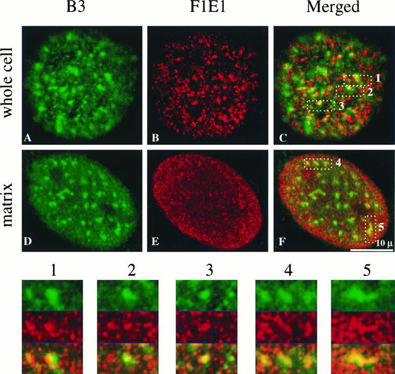
Yeast two-hybrid screening has led to the identification of a family of proteins that interact with the repetitive C-terminal repeat domain (CTD) of RNA polymerase II (A. Yuryev et al., Proc. Natl. Acad. Sci. USA 93:6975-6980, 1996). In addition to serine/arginine-rich SR motifs, the SCAFs (SR-like CTD-associated factors) contain discrete CTD-interacting domains. In this paper, we show that the CTD-interacting domain of SCAF8 specifically binds CTD molecules phosphorylated on serines 2 and 5 of the consensus sequence Tyr1Ser2Pro3Thr4Ser5Pro6Ser7. In addition, we demonstrate that SCAF8 associates with hyperphosphorylated but not with hypophosphorylated RNA polymerase II in vitro and in vivo. This result suggests that SCAF8 is not present in preinitiation complexes but rather associates with elongating RNA polymerase II. Immunolocalization studies show that SCAF8 is present in granular nuclear foci which correspond to sites of active transcription. We also provide evidence that SCAF8 foci are associated with the nuclear matrix. A fraction of these sites overlap with a subset of larger nuclear speckles containing phosphorylated polymerase II. Taken together, our results indicate a possible role for SCAF8 in linking transcription and pre-mRNA processing.


Figures

FIG. 1
FIG. 1
Fusion proteins used in binding studies. (A) Coomassie blue staining of recombinant SCAF8 CID. Five micrograms of purified SCAF8 CID was run on SDS-PAGE (10% polyacrylamide) and stained with Coomassie blue. Lane 1, marker proteins; lane 2, SCAF8 CID. (B) Coomassie blue staining of GST-CTD fusion proteins. Five micrograms of the affinity-purified proteins was run on SDS-PAGE (10% polyacrylamide) and stained with Coomassie blue. Lane 1, wild-type CTD (16 repeats); lane 2, A5 mutant CTD (15 repeats); lane 3, A2 mutant CTD (14 repeats).
FIG. 2
FIG. 2
SCAF8 CTD binding assays. (A) SCAF8 interacts with phosphorylated CTD fusion proteins. The binding assay for SCAF8 CID and the different CTD fusion proteins is described in Materials and Methods. All column fractions were separated by SDS-PAGE (10% polyacrylamide) and then subjected to Western blot analysis. Proteins were detected with monoclonal antibody 12CA5, which detects both phosphorylated and unphosphorylated CTD fusion proteins (indicated at left). The phosphorylated fusion protein can be distinguished by its retarded mobility. The column matrix is indicated to the right. Lane L, protein load; lane FT, flowthrough; lanes W1 to W6, column washes; lanes E1 to E3, SDS sample buffer eluates. (B) Binding controls. A binding assay similar to that described for panel A was performed in the presence of additional components indicated above the figure. In this experiment, the wild-type CTD fusion protein was phosphorylated with Cdc2 kinase in the presence of [γ-32P]ATP, and the bound fractions eluted with SDS sample buffer were pooled. (C) SCAF8 does not bind tyrosine-phosphorylated CTD (P-tyr CTD). A binding assay similar to that described for panel A was performed with tyrosine-phosphorylated CTD (Materials and Methods). Lanes are as indicated for panel A. (D) SCAF8 does not bind mutant CTDs. A binding reaction similar to that for panel A was performed with a mixture of phosphorylated A5 and A2 mutant CTD fusion proteins. The identity of the fusion protein (indicated at left) was determined by running them individually. Lanes are as described for panel A.
FIG. 3
FIG. 3
SCAF8 interacts with phosphorylated pol II. The binding experiment using partially purified pol II is described in Materials and Methods. Proteins were separated by SDS-PAGE (5% polyacrylamide) and then subjected to either silver staining (A) or Western blotting (B). Lane L, column load; lane FT, column flowthrough; lanes W1 to W6, column washes; lanes E1 and E2, SDS sample buffer eluates.
FIG. 4
FIG. 4
SCAF8 interacts with phosphorylated pol II in vivo. (A) Alignment of SCAF8 and SCAF4 CTD interacting domains. These sequences were expressed as 6His-tagged fusion proteins as described in Materials and Methods. (B) MAb F1E1 detects the SCAF8 but not the SCAF4 CID. Partially purified 6His-tagged SCAF CID fusion proteins were separated by SDS gel electrophoresis and immunoblotted with anti-His tag antibody (BABCO) or with MAb F1E1. (C) Western blot analysis of HeLa nuclear extract (NE). SCAF8 was detected with MAb F1E1. (D) Immunoprecipitation of SCAF8 complexes. Immunoprecipitation of the SCAF8 complexes was carried out as described in Materials and Methods. The same blot was probed with MAb H14 (left panel) or with 8WG16 (right panel). The immunoprecipitating antibody is indicated above the lane, while the blotting antibody is indicated below the panel. Markers are indicated to both sides and between the blots. The right lane in the right panel contains nuclear extract to indicate the position of the unphosphorylated RNA pol II largest subunit (IIA).
FIG. 5
FIG. 5
SCAF8 is associated with transcription sites. Mouse Swiss 3T3 cells were permeabilized and incubated with BrUTP to label transcription sites in situ (Materials and Methods). Transcription sites were detected with rat anti-BrUTP antibody (Sera-Lab) and visualized with green (A). SCAF8 was detected with MAb F1E1 and then visualized with red (B), and the merged images are displayed in panel C. The boxed area, including both extranucleolar and nucleolar regions, is further enlarged and displayed in panels D (transcription sites), E (SCAF8), and F (merged images). The line through F indicates the boundary between the nucleolus (left) and the nucleus (right).
FIG. 6
FIG. 6
SCAF8 fractionates with nuclear matrix proteins. (A) Coomassie blue staining of rat liver nuclear (N) and nuclear matrix (M) proteins. Ten micrograms of protein from each sample was run on SDS-PAGE (12.5% polyacrylamide). The molecular mass markers (in kilodaltons) are displayed on the left side. (B) Western blot of rat liver nuclear and nuclear matrix proteins with FIEI antibody. (C) The distribution of the SCAF8 protein, RNA pol II0, and lamin B in the HeLa cytoplasmic fraction (C), purified nuclear fraction (N), nuclear matrix fraction (M), and salt-extracted nuclear proteins (S) was determined by Western blot analysis.
FIG. 7
FIG. 7
Double labeling of 3T3 cells and in situ-prepared nuclear matrix with B3 and FIEI antibodies. The hyperphosphorylated RNA pol II large subunit was recognized with the B3 antibody (green). SCAF8 antigen was recognized with the FIEI antibody (red). The merged image is displayed on the right. Five speckle regions indicated by the B3 staining were magnified and displayed underneath the whole images. The single red and green channel images for the magnified areas of the nucleus in the whole cell are also displayed above the merged magnified regional images. The 0.5-μm optical sections are displayed.

Similar articles

Cited by

References

    1. Akhtar A, Faye G, Bentley D L. Distinct activated and non-activated RNA polymerase II complexes in yeast. EMBO J. 1996;15:4654–4664. - PMC - PubMed
    1. Allison L A, Moyle M, Shales M, Ingles C J. Extensive homology among the largest subunits of eukaryotic and prokaryotic RNA polymerases. Cell. 1985;42:599–610. - PubMed
    1. Allison L A, Wong J K-C, Fitzpatrick V D, Moyle M, Ingles C J. The C-terminal domain of the largest subunit of RNA polymerase II of Saccharomyces cerevisiae, Drosophila melanogaster, and mammals: a conserved structure with an essential function. Mol Cell Biol. 1988;8:321–329. - PMC - PubMed
    1. Bartolomei M S, Halden N F, Cullen C R, Corden J L. Genetic analysis of the repetitive carboxyl-terminal domain of the largest subunit of mouse RNA polymerase II. Mol Cell Biol. 1988;8:330–339. - PMC - PubMed
    1. Baskaran R, Dahmus M E, Wang J Y. Tyrosine phosphorylation of mammalian RNA polymerase II carboxyl-terminal domain. Proc Natl Acad Sci USA. 1993;90:11167–11171. - PMC - PubMed

Publication types