. 1998 Apr;66(4):1349-55.
doi: 10.1128/IAI.66.4.1349-1355.1998.

Immune responses of specific-pathogen-free mice to chronic Helicobacter pylori (strain SS1) infection

Affiliations

Immune responses of specific-pathogen-free mice to chronic Helicobacter pylori (strain SS1) infection

R L Ferrero et al. Infect Immun. 1998 Apr.

Abstract

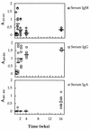
A model permitting the establishment of persistent Helicobacter pylori infection in mice was recently described. To evaluate murine immune responses to H. pylori infection, specific-pathogen-free Swiss mice (n = 50) were intragastrically inoculated with 1.2 x 10(7) CFU of a mouse-adapted H. pylori isolate (strain SS1). Control animals (n = 10) received sterile broth medium alone. Animals were sacrificed at various times, from 3 days to 16 weeks postinoculation (p.i.). Quantitative culture of gastric tissue samples from inoculated mice demonstrated bacterial loads of 4.0 x 10(4) to 8 x 10(6) CFU per g of tissue in the animals. Infected mice had H. pylori-specific immunoglobulin M (IgM) and IgG antibodies in serum (at day 3 p.i.) and IgG and IgA antibodies in their gastric contents (weeks 4 and 16 p.i.) and saliva (week 16 p.i.). Mucosal IgM antibodies were not detected. Histological examination of the gastric mucosae from control and infected mice revealed mild chronic gastritis, characterized by the presence of polymorphoneutrophil cell infiltrates and submucosal lymphoid aggregates, in infected animals at 16 weeks p.i. Differences in the quantities of IgG1 and IgG2a subclass antibodies detected in the sera of mouse strains (Swiss, BALB/c, and C57BL/6) infected by H. pylori suggested that host factors influence the immune responses induced against this bacterium in the host. In conclusion, immune responses to H. pylori infection in mice, like those in chronically infected humans, appear to be ineffective in resolving the infection.


Figures

FIG. 1
FIG. 1
Enumeration of H. pylori colonies from mouse gastric biopsies by a combined culture and urease activity technique. Homogenates of mouse gastric samples were serially diluted in saline and then used to inoculate serum plates (see Materials and Methods). After incubation, a sloppy urea overlay was applied to the plates, permitting the detection of urease-positive H. pylori colonies, which appear as fuchsia-colored zones on a yellow background.
FIG. 2
FIG. 2
The numbers of H. pylori bacteria in gastric biopsies from Swiss mice over time. Mice (n = 50) that had been inoculated with a suspension of H. pylori SS1 were divided into five groups (each containing 10 mice) that were sacrificed at different times. Noninfected control mice (n = 2) were also sacrificed at each time point. The numbers of CFU in homogenates of gastric samples were determined as described in the text. Each point represents the mean of two estimations for a single mouse. Horizontal bars represent the geometric means for mice (n = 10) sacrificed at each time point.
FIG. 3
FIG. 3
Anti-H. pylori antibodies in the sera of Swiss mice infected by H. pylori SS1. Specific IgM, IgG, and IgA class antibodies in the sera of H. pylori-infected mice (Fig. 1) were detected by ELISA. The results are presented as A405–492 readings which correspond to the values given by diluted serum samples (1:100). Each point represents the mean of triplicate determinations for a single mouse. Sera from noninfected control mice (n = 10) were also tested and gave OD values of ≤0.05 (data not shown). Horizontal bars represent the geometric means for mice (n = 10) sacrificed at each time point.
FIG. 4
FIG. 4
Anti-H. pylori antibodies in the gastric contents of Swiss mice infected by H. pylori SS1. Specific IgG and IgA class antibodies in the gastric contents of H. pylori-infected mice (Fig. 1) were detected by ELISA. The results are presented as described for Fig. 3, except that gastric content samples were diluted 1:10. Each point represents the mean of triplicate determinations for a single mouse. Samples from noninfected control mice (n = 10) were also tested and gave OD values of ≤0.05 (data not shown). Horizontal bars represent the geometric means for mice (n = 10) sacrificed at each time point.
FIG. 5
FIG. 5
Anti-H. pylori antibodies in the saliva of Swiss mice infected by H. pylori SS1. Specific IgA class antibodies in the saliva of H. pylori-infected mice (Fig. 1) were detected by ELISA. The results are presented as described for Fig. 3, except that saliva samples were diluted 1:50. Each point represents the mean of triplicate determinations for a single mouse. Saliva from noninfected control mice (n = 10) were also tested and gave OD values of ≤0.05 (data not shown). Horizontal bars represent the geometric means for mice (n = 10) sacrificed at each time point.
FIG. 6
FIG. 6
Hematoxylin and eosin-stained sections of mouse gastric mucosae from uninfected and infected mice at 16 weeks p.i. (A) Normal gastric mucosa. (B and C) Gastric mucosa from an H. pylori-infected mouse, showing a mild diffuse gastritis in the mucosal and submucosal regions of the tissue. Lymphocyte (B) and polymorphoneutrophil (C) cell infiltrates are indicated. (D) Gastric mucosa from another H. pylori-infected mouse, showing a submucosal lymphoid aggregate. Bars = 100 μm.
FIG. 7
FIG. 7
Warthin-Starry silver stain of mouse gastric mucosa showing the presence of H. pylori bacteria in gastric body tissue. (A) A region of dense colonization with H. pylori bacteria present in the gastric glands and pits of the tissue. The bacteria often appear as clumps (arrows). (B) High-power magnification of a gastric gland colonized by spiral-shaped bacteria (arrows). Bars = 10 μm.

Similar articles

Cited by

References

    1. Atherton J C, Tham K T, Peek R M, Jr, Cover T L, Blaser M J. Density of Helicobacter pylori infection in vivo as assessed by quantitative culture and histology. J Infect Dis. 1996;174:552–556. - PubMed
    1. Blanchard T G, Nedrud J G, Czinn S J. Qualitative and quantitative differences in the local immune response to Helicobacter in mice and humans following immunization or infection. Gastroenterology. 1996;110:A867.
    1. Callard R E, Gearing A J H. The cytokine facts book. London, United Kingdom: Academic Press; 1994.
    1. Crabtree, J. E. 1993. Mucosal immune responses to Helicobacter pylori. Eur. J. Gastroenterol. Hepatol. 5(Suppl. 2):S30–S32.
    1. Crabtree J E, Wyatt J I, Sobala G M, Miller G, Tompkins D S, Primrose J N, Morgan A G. Systematic and mucosal humoral responses to Helicobacter pylori in gastric cancer. Gut. 1993;34:1339–1343. - PMC - PubMed

Publication types