. 1998 Apr;66(4):1400-7.
doi: 10.1128/IAI.66.4.1400-1407.1998.

Endotoxin-neutralizing protein protects against endotoxin-induced endothelial barrier dysfunction

Affiliations

Endotoxin-neutralizing protein protects against endotoxin-induced endothelial barrier dysfunction

D D Bannerman et al. Infect Immun. 1998 Apr.

Abstract

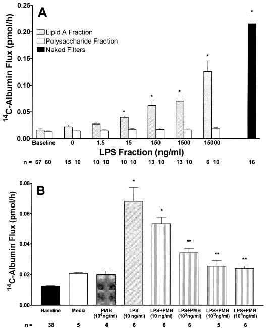
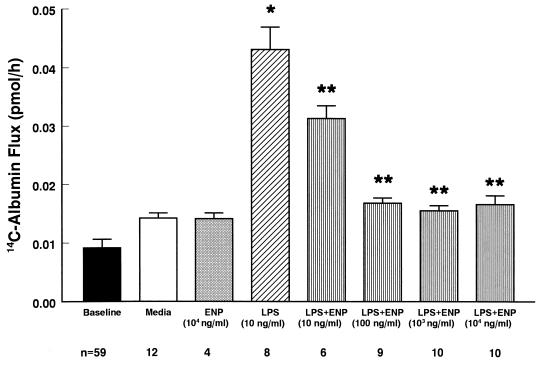
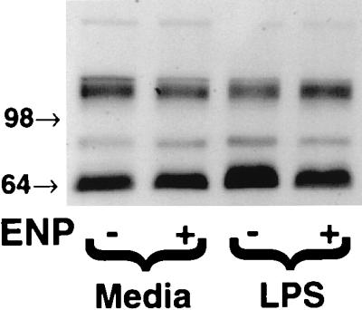
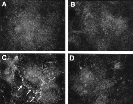
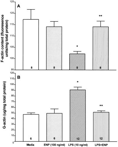
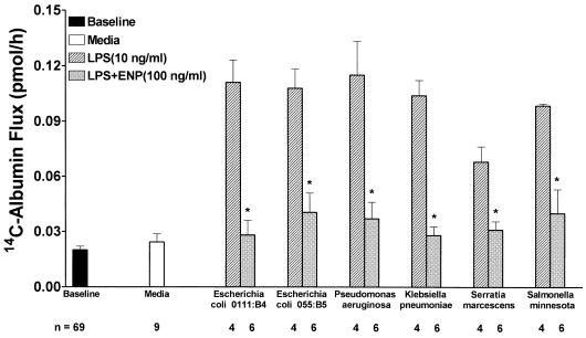
Bacterial lipopolysaccharide induces tyrosine phosphorylation of paxillin, actin reorganization, and opening of the transendothelial paracellular pathway through which macromoles flux. In this study, lipid A was shown to be the bioactive portion of the lipopolysaccharide molecule responsible for changes in endothelial barrier function. We then studied whether endotoxin-neutralizing protein, a recombinant peptide that is derived from Limulus antilipopolysaccharide factor and targets lipid A, could block the effects of lipopolysaccharide on protein tyrosine phosphorylation, actin organization, and movement of 14C-bovine serum albumin across bovine pulmonary artery endothelial cell monolayers. In the presence of serum, a 6-h exposure to lipopolysaccharide (10 ng/ml) increased transendothelial 14C-albumin flux compared to the simultaneous media control. Coadministration of endotoxin-neutralizing protein (> or =10 ng/ml) with lipopolysaccharide (10 ng/ml) protected against lipopolysaccharide-induced barrier dysfunction. This protection was dose dependent, conferring total protection at endotoxin-neutralizing protein/lipopolysaccharide ratios of > or =10:1. Similarly, endotoxin-neutralizing protein was capable of blocking the lipopolysaccharide-induced endothelial cell responses that are prerequisite to barrier dysfunction, including tyrosine phosphorylation of paxillin and actin depolymerization. Finally, endotoxin-neutralizing protein cross-protected against lipopolysaccharide derived from diverse gram-negative bacteria. Thus, endotoxin-neutralizing protein offers a novel therapeutic intervention for the vascular endothelial dysfunction of gram-negative sepsis and its attendant endotoxemia.


Figures

FIG. 1
FIG. 1
Effects of LPS fractions on transendothelial 14C-BSA flux. (A) Transendothelial 14C-BSA flux across monolayers was assayed after exposure for 6 h to increasing concentrations of either the lipid A fraction or the O-specific polysaccharide fraction derived from E. coli O111:B4 LPS. Each bar represents mean (± SE) transendothelial 14C-BSA flux. Pretreatment baseline 14C-BSA flux across monolayers exposed to either lipid A or O-specific polysaccharide fractions as well as 14C-BSA flux across naked filters are also shown. ∗, significantly increased compared with simultaneous medium control. n, number of monolayers studied. (B) EC monolayers were assayed for transendothelial 14C-BSA flux immediately after 6-h exposures to medium alone, PMB, LPS, or LPS (10 ng/ml) coadministered with increasing concentrations of PMB. Mean (± SE) pretreatment baseline 14C-BSA flux is also shown ∗, significantly increased compared to medium control; ∗∗, significantly decreased compared to LPS. n, number of monolayers studied.
FIG. 2
FIG. 2
Dose-dependent effects of ENP on LPS-induced barrier dysfunction. Baseline barrier function was determined for all monolayers prior to treatment. Transendothelial 14C-BSA flux across monolayers was assayed after exposure for 6 h to medium, ENP, LPS, or LPS coadministered with increasing concentrations of ENP. Each bar represents mean (± SE) transendothelial 14C-BSA flux. ∗, significantly increased compared to medium control; ∗∗, significantly decreased compared to LPS alone. n, number of monolayers studied.
FIG. 3
FIG. 3
Effect of ENP on LPS-induced tyrosine phosphorylation of a 66-kDa EC protein. For Western blot analysis of protein tyrosine phosphorylation, EC monolayers were exposed to medium, ENP (1.0 μg/ml), LPS (100 ng/ml), or LPS coadministered with ENP for 1 h. The EC lysates were resolved by SDS-PAGE, transferred to PVDF, and probed for phosphotyrosines. Molecular weights (in thousands) are indicated by arrows on the left. The blot is representative of three separate experiments.
FIG. 4
FIG. 4
Effect of ENP on phosphotyrosine-containing proteins in LPS-exposed EC. EC monolayers grown on filters were exposed for 1 h to medium (A), ENP (1.0 μg/ml; B), LPS (100 ng/ml; C), or LPS coadministered with ENP (D). The monolayers were fixed, probed with FITC-conjugated antiphosphotyrosine antibody, and photographed through an epifluorescence microscope. Arrows indicate phosphotyrosine signal at intercellular boundaries. Magnification, ×600.
FIG. 5
FIG. 5
Effects of ENP on LPS-induced changes in the F- and G-actin pools. For G- and F-actin measurements, monolayers were exposed for 6 h to medium, ENP, LPS, or LPS coadministered with ENP. (A) For the F-actin studies, monolayers were fixed, permeabilized, incubated with NBD-phallicidin, and extracted with methanol. The extracts were spectrofluorimetrically assayed, and F-actin concentrations were expressed as mean (± SE) fluorescent units per milligram of total EC protein. ∗, significantly decreased compared to medium control; ∗∗, significantly increased compared to LPS alone but not significantly decreased compared to medium alone. (B) For quantitation of the G-actin pool, EC were permeabilized and the G-actin-containing supernatants were tested in the DNase I inhibition assay standardized to pure G-actin. Each bar represents mean (± SE) G-actin expressed. ∗, significantly increased compared to medium control; ∗∗, significantly decreased compared to LPS alone but not significantly increased compared to medium alone. n for each experimental group is indicated in each bar.
FIG. 6
FIG. 6
ENP cross-protects against a wide variety of endotoxins. Transendothelial 14C-BSA flux was assayed immediately following 6-h exposures to medium (open bar), equivalent concentrations based on KDO content of LPS derived from E. coli O111:B4 (10 ng/ml), E. coli 055:B5, P. aeruginosa, K. pneumoniae, S. marcescens, or S. minnesota (cross-hatched bars), or these same LPS preparations coadministered with ENP (100 ng/ml) (gray bars). Each bar represents mean (± SE) transendothelial 14C-BSA flux. Baseline barrier function for all monolayers studied is also indicated. ∗, significantly decreased compared to LPS alone at P < 0.05 but not significantly increased compared to medium control.

Similar articles

Cited by

References

    1. Alpert G, Baldwin G, Thompson C, Wainwright N, Novitsky T J, Gillis Z, Parsonnet J, Fleisher G R, Siber G R. Limulus antilipopolysaccharide factor protects rabbits from meningococcal endotoxin shock. J Infect Dis. 1992;165:494–500. - PubMed
    1. Arditi M, Zhou J, Torres M, Durden D L, Stins M, Kim K S. Lipopolysaccharide stimulates the tyrosine phosphorylation of mitogen-activated protein kinase p44, p42, and p41 in vascular endothelial cells in a soluble CD14-dependent manner. Role of protein tyrosine phosphorylation in lipopolysaccharide-induced stimulation of endothelial cells. J Immunol. 1995;155:3994–4003. - PubMed
    1. Bannerman D D, Goldblum S E. Endotoxin induces endothelial barrier dysfunction through protein tyrosine phosphorylation. Am J Physiol. 1997;273:L217–L226. - PubMed
    1. Battafaraono R J, Dahlberg P S, Ratz C A, Johnston J W, Gray B H, Haseman J R, Mayo K H, Dunn D L. Peptide derivatives of three distinct lipopolysaccharide binding proteins inhibit lipopolysaccharide-induced tumor necrosis factor-alpha secretion in vitro. Surgery. 1995;118:318–324. - PubMed
    1. Brigham K L, Meyrick B. Endotoxin and lung injury. Am Rev Respir Dis. 1986;133:913–927. - PubMed

Publication types

MeSH terms