. 1998 Apr;66(4):1427-31.
doi: 10.1128/IAI.66.4.1427-1431.1998.

M protein of the group A Streptococcus binds to the seventh short consensus repeat of human complement factor H

Affiliations

M protein of the group A Streptococcus binds to the seventh short consensus repeat of human complement factor H

T K Blackmore et al. Infect Immun. 1998 Apr.

Abstract

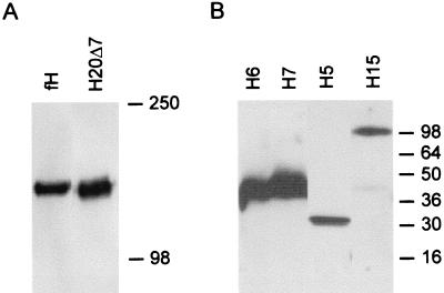
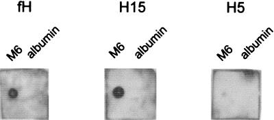
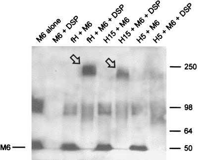
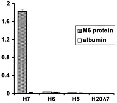
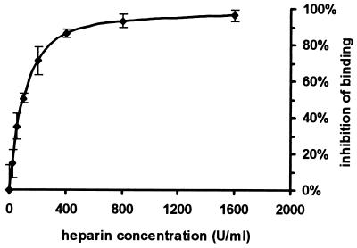
Streptococcus pyogenes evades complement by binding the complement-regulatory protein factor H (fH) via the central conserved C-repeat region of M protein. However, the corresponding binding region within fH has not previously been precisely localized. fH is composed of 20 conserved modules called short consensus repeats (SCRs), each of which contains approximately 60 amino acids. A series of fH truncated and deletion mutants were prepared, and their interaction with M6 protein was examined. The M protein binding site was initially localized to SCRs 6 to 15 as demonstrated by ligand dot blotting, chemical cross-linking, and enzyme-linked immunosorbent assay. SCR 7 was then shown to contain the M protein binding site, as a construct consisting of the first seven SCRs bound M protein but a construct containing the first six SCRs did not bind. In addition, deletion of SCR 7 from full-length fH abolished binding to M protein. SCR 7 is known to contain a heparin binding domain, and binding of fH to M6 protein was almost totally inhibited in the presence of 400 U of heparin per ml. These results localize the M6 protein binding site of fH to SCR 7 and indicate that it is in close proximity to the heparin binding site.


Figures

FIG. 1
FIG. 1
Western blots of fH and fH mutant proteins. CHO cells were transfected with the appropriate cDNA, and supernatants were collected and separated by SDS-PAGE under nonreducing conditions. H20 and H20Δ7 were separated by SDS–6% PAGE (A). H5His, H6His, H7His, and H15 His were separated by SDS–12% PAGE (B). Recombinant proteins were detected by Western blotting, using polyclonal anti-fH antibodies as described in Materials and Methods. Apparent molecular masses (in kilodaltons) are indicated.
FIG. 2
FIG. 2
Ligand dot blotting of binding of fH, H15, and H5 to immobilized M6 protein. M6 protein and albumin were dried onto nitrocellulose, blocked, and incubated with approximately 0.5 μg of the indicated fH-derived protein per ml. Bound protein was detected with anti-fH polyclonal antibodies and HRP-conjugated protein A followed by chemiluminescence.
FIG. 3
FIG. 3
Chemical cross-linking of fH, H15, and H5 to M6 protein. Biotinylated M6 protein and fH, H15, or H5 were incubated in 50 mM bicarbonate buffer (pH 7.4) with or without DSP cross-linker, as described in Materials and Methods. Proteins were separated by SDS–6% PAGE under nonreducing conditions, transferred to nitrocellulose, and then incubated in streptavidin-HRP. M6 protein and cross-linked M6 protein were detected by chemiluminescence. Cross-linking of M6 protein to fH and H15 is indicated by the arrows.
FIG. 4
FIG. 4
Binding of C-terminal truncation mutants of fH to immobilized M6 protein. fH mutant proteins were added to ELISA wells on which M6 protein or albumin had been immobilized. After washing, bound protein was detected with polyclonal goat anti-fH antibodies followed by protein A-HRP and substrate. Amounts of fH mutant proteins equivalent to those shown in Fig. 1 were used. Results shown are the mean optical densities at 490 nm for four experiments, and error bars represent one standard deviation.
FIG. 5
FIG. 5
Effect of heparin on binding of H7 to M6 protein. H7 in 50 mM phosphate buffer (pH 7.4) containing between 0 and 1,600 U of heparin per ml was incubated with immobilized M6 protein. Binding of H7 was measured by the same ELISA method described in the legend to Fig. 4. Results are expressed as [1 − (optical density at 490 nm of test wells/optical density at 490 nm of wells without heparin)] × 100. Error bars represent one standard deviation.

References

    1. Avery V M, Gordon D L. Characterization of factor H binding to human polymorphonuclear leukocytes. J Immunol. 1993;151:5545–5553. - PubMed
    1. Barlow P N, Campbell I D. Strategy for studying modular proteins: application to complement modules. Methods Enzymol. 1994;239:465–485. - PubMed
    1. Bisno A L. Alternate complement pathway activation by group A streptococci: role of M protein. Infect Immun. 1979;26:1172–1176. - PMC - PubMed
    1. Bisno A L, Stevens D L. Streptococcal infections of skin and soft tissues. N Engl J Med. 1996;334:240–245. - PubMed
    1. Blackmore T K, Hellwage J, Zipfel P F, Gordon D L. Identification of an additional heparin binding domain in the C-terminal region of human factor H. Exp Clin Immunogenet. 1997;14:30.

Publication types

LinkOut - more resources