. 1998 Apr;66(4):1492-9.
doi: 10.1128/IAI.66.4.1492-1499.1998.

Sequence analysis of the mip gene of the soilborne pathogen Legionella longbeachae

Affiliations

Sequence analysis of the mip gene of the soilborne pathogen Legionella longbeachae

R M Doyle et al. Infect Immun. 1998 Apr.

Abstract

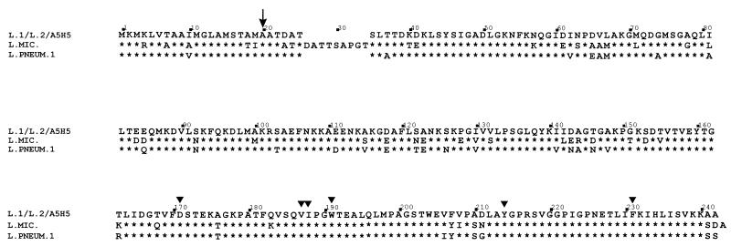
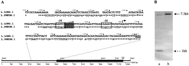
To understand the basis of pathogenesis by Legionella longbeachae serogroup 1, the importance of the Mip protein in this species was examined. Amino-terminal analysis of the purified, cloned L. longbeachae serogroup 1 ATCC 33462 Mip protein confirmed that the cloned gene protein was expressed and processed in an Escherichia coli background. DNA sequence analysis of plasmid pIMVS27, containing the entire L. longbeachae serogroup 1 mip gene, revealed a high degree of homology to the mip gene of Legionella pneumophila serogroup 1, 76% homology at the DNA level and 87% identity at the amino acid level. Primer extension analysis determined that the start site of transcription was the same for both species, with some differences observed for the -10 and -35 promoter regions. Primers designed from the mip gene sequence obtained for L. longbeachae serogroup 1 ATCC 33462 were used to amplify the mip genes from L. longbeachae serogroup 2 ATCC 33484 and an Australian clinical isolate of L. longbeachae serogroup 1 A5H5. The mip gene from A5H5 was 100% identical to the type strain sequence. The serogroup 2 strain of L. longbeachae differed by 2 base pairs in third-codon positions. Allelic exchange mutagenesis was used to generate an isogenic mip mutant in ATCC 33462 and strain A5H5. The ATCC mip mutant was unable to infect a strain of Acanthamoebae sp. both in liquid and in a potting mix coculture system, while the A5H5 mip mutant behaved in a manner siilar to that of L. pneumophila serogroup 1, i.e., it displayed a reduced capacity to infect and multiply within Acanthamoebae. To determine if this mutation resulted in reduced virulence in the guinea pig animal model, the A5H5 mip mutant and its parent strain were assessed for their abilities to establish an infection after aerosol exposure. Unlike the virulent parent strain, the mutant strain did not kill any animals under two different dose regimes. The data indicate that the Mip protein plays an important role in the intracellular life cycle of L. longbeachae serogroup 1 species and is required for full virulence.


Figures

FIG. 1
FIG. 1
Amino acid comparison of the Mip proteins of L. longbeachae serogroup 1 ATCC 33462 (L.1), L. longbeachae serogroup 1 A5H5 (A5H5), L. longbeachae serogroup 2 ATCC 33484 (L.2), L. pneumophila serogroup 1 (L. PNEUM. 1), and L. micdadei (L. MIC.). Asterisks indicate amino acids identical to those of L. longbeachae; triangles indicate amino acids predicted to form part of the active site for PPIase activity of Mip. The arrow indicates the site of signal peptidase cleavage.
FIG. 2
FIG. 2
(A) Line diagram depicts the DNA sequence determined from sequencing pIMVS27. The solid box shows the mip gene from L. longbeachae serogroup 1 ATCC 33462, selected restriction sites in the mip gene, and the stem loop structure at the end of the ORF. The inset sequence is the DNA sequence upstream of the ATG start site for translation in L. longbeachae serogroup 1 and L. pneumophila serogroup 1, showing the −10 and −35 promoter regions determined by primer extension analysis. The shaded box indicates the −35 promoter region proposed for L. longbeachae. The nonshaded box is the −35 region proposed in reference . The start site for transcription is shown with a solid arrow. (B) Southern hybridization demonstrating mutagenesis by allelic exchange of the L. longbeachae serogroup 1 mip gene. DNA was digested with KpnI and probed with DIG-labeled pIMVS27. Lanes: a, L. longbeachae serogroup 1 A5H5; b, B8. The solid arrow indicates the 7.3-kb fragment generated in B8 due to the loss of an internal KpnI site which generates 1- and 7-kb fragments in the parent strain. A similar pattern was observed for L. longbeachae serogroup 1 ATCC 33462 (data not shown).
FIG. 3
FIG. 3
Coculture of Acanthomoebae with strains of Legionella. (A) Amoeba liquid cocultures were set up in saline with approximately 104 amoebae/ml and 103 CFU (each) of L. pneumophila serogroup 1 (Philadelphia) (⧫), L. longbeachae serogroup 1 ATCC 33462 (▪), B10 (▵), A5H5 (•), and B8 (×) per ml. Samples were taken at various time intervals, and the number of Legionella organisms was determined by plating on selective media. Each time point represents the mean number of CFU recovered, and the vertical bars indicate standard deviations. (B) Amoebae were cocultured in an artificial potting mix system with strains of Legionella as indicated above. Numbers of viable Legionella organisms were determined at various time points by treatment of the soil sample with acid and plating on selective media. The experiments shown are representative of two independent experiments.
FIG. 4
FIG. 4
Percentage weight gain or loss in guinea pigs exposed to an aerosol of different strains of Legionella longbeachae serogroup 1. (A) Animals exposed to a dose of 109 L. longbeachae serogroup 1 A5H5 organisms; (B) animals exposed to a dose of 109 B8 organisms; (C) animals exposed to a dose of 1010 B8 organisms; (D) animals exposed to a dose of 109 B8.22 organisms. Guinea pig death is indicated by the termination of the ribbon graph prior to the end of the experiment on day 7.

Similar articles

Cited by

References

    1. Aiba H, Adhya S, de Crombrugghe B. Evidence for two functional gal promoters in intact Escherichia coli cells. J Biol Chem. 1981;256:11905–11910. - PubMed
    1. Bangsborg J M, Cianciotto N P, Hindersson P. Nucleotide sequence analysis of the Legionella micdadei mip gene, encoding a 30-kilodalton analog of the Legionella pneumophila Mip protein. Infect Immun. 1991;59:3836–3840. - PMC - PubMed
    1. Baskerville A, Fitzgeorge R B, Broster M, Hambleton P. Histopathology of experimental Legionnaires’ disease in guinea pigs, rhesus monkeys and marmosets. J Pathol. 1983;139:349–362. - PubMed
    1. Baskerville A, Dowsett A B, Fitzgeorge R B, Hambleton P, Broster M. Ultrastructure of pulmonary alveoli and macrophages in experimental Legionnaires’ disease. J Pathol. 1983;140:77–90. - PubMed
    1. Bellinger-Kawahara C, Horwitz M A. Complement component C3 fixes selectively to the major outer membrane protein (MOMP) of Legionella pneumophila and mediates phagocytosis of liposome-MOMP complexes by human monocytes. J Exp Med. 1990;172:1201–1210. - PMC - PubMed

Publication types

MeSH terms

Associated data

LinkOut - more resources