. 1998 Apr;66(4):1570-8.
doi: 10.1128/IAI.66.4.1570-1578.1998.

Role of intimin and bundle-forming pili in enteropathogenic Escherichia coli adhesion to pediatric intestinal tissue in vitro

Affiliations

Role of intimin and bundle-forming pili in enteropathogenic Escherichia coli adhesion to pediatric intestinal tissue in vitro

S Hicks et al. Infect Immun. 1998 Apr.

Abstract

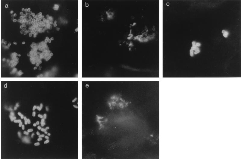
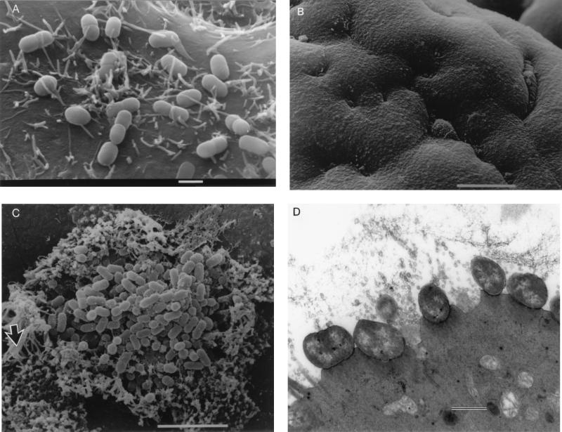
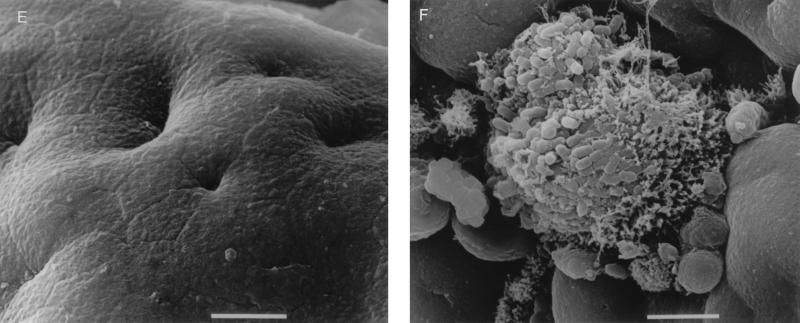
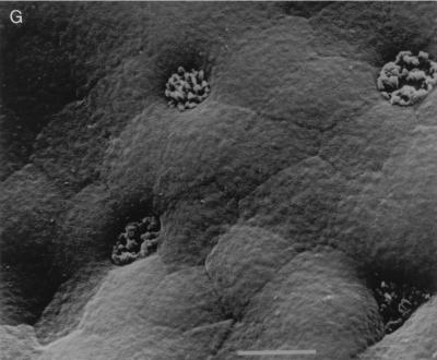
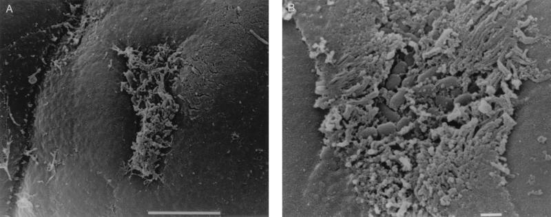
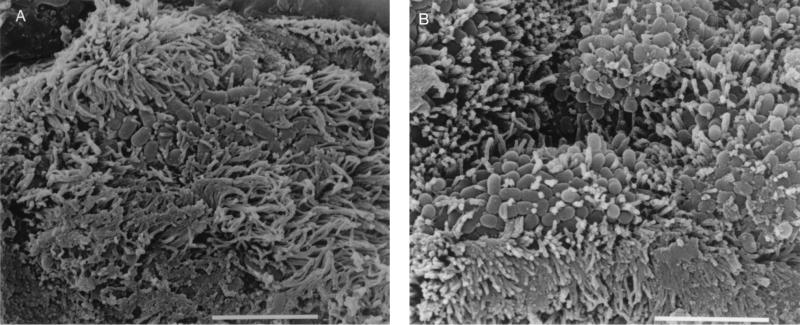
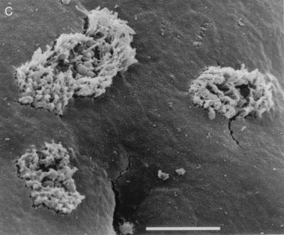
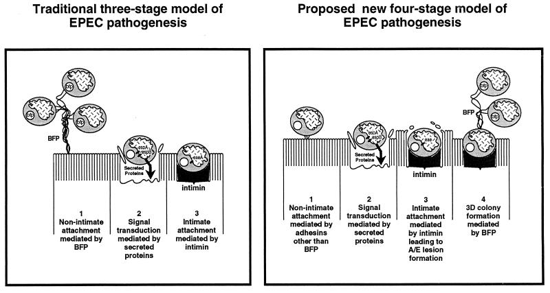
Attaching and effacing (A/E) lesion formation is central to enteropathogenic Escherichia coli (EPEC) pathogenesis. In vitro experiments with human epithelial cell lines have implicated virulence plasmid-encoded bundle-forming pili (BFP) in initial binding and intimin in intimate attachment and A/E lesion formation. This study investigated the role of BFP and intimin in EPEC interactions with pediatric small intestinal biopsy tissue in in vitro organ culture. Organ culture infections (2 to 8 h) were performed with E2348/69 (a wild-type EPEC O127:H6 clinical isolate) and E2348/69 derivatives including CVD206 (eae deficient), CVD206(pCVD438) (eae-complemented CVD206), CVD206(pCVD438/01) (expressing intimin, which is nonfunctional due to a single amino acid substitution), JPN15 (spontaneous EPEC adherence factor virulence plasmid-cured E2348/69), and 31-6-1(1) (E2348/69 with a TnphoA insertion inactivation mutation in the virulence plasmid-encoded bfpA gene). Scanning and transmission electron microscopy revealed that after 8 h E2348/69 and CVD206 (pCVD438) (both Int+ BFP+) adhered to all specimens, causing A/E lesions with surrounding microvillous elongation. JPN15 and 31-6-1(1) (both Int+ BFP-) adhered and caused A/E lesions although bacteria adhered in "flat," two-dimensional groups. CVD206 and CVD206(pCVD438/01) (both Int- BFP+) did not adhere to any sample, and no pathological tissue changes were seen. Thus, in human intestinal organ culture, BFP do not appear to be involved in the initial stages of EPEC nonintimate adhesion but are implicated in the formation of complex, three-dimensional colonies via bacterium-bacterium interactions. Intimin appears to play an essential role in establishing colonization of EPEC on pediatric small intestinal tissue.


Figures

FIG. 1
FIG. 1
Panel of immunofluorescence micrographs of HEp-2 cell-adherent bacteria stained with anti-intimin or anti-BFP. (a) E2348/69 stained with anti-intimin, (b) E2348/69 stained with anti-BFP, (c) CVD206(pCVD438/01) stained with anti-intimin, (d) bfpA-mutated 31-6-1(1) stained with anti-intimin, (e) CVD206 stained with anti-BFP.
FIG. 2
FIG. 2
(A) Scanning electron micrograph of CVD206 adhering to HEp-2 cells after 3 h of incubation. Bar = 1 μm. (B) Scanning electron micrograph of uninoculated small intestinal tissue after 8 h in organ culture. Bar = 10 μm. (C) Pediatric ileal biopsy tissue incubated with E2348/69 for 8 h. Bacteria adhere in localized colonies causing extreme microvillous elongation (arrow) and vesiculation. Bar = 5 μm. (D) Transmission electron micrograph showing A/E lesions with microvillous vesiculation caused by E2348/69. Bar = 0.5 μm. (E) CVD206 causing no tissue damage or adhesion to small intestinal tissue. Bar = 5 μm. (F) CVD206(pCVD438) adhering in localized colonies to small intestinal mucosa with an appearance similar to that of E2348/69 shown in panel C. Bar = 5 μm. (G) CVD206(pCVD438/01) with an appearance similar to that of CVD206 shown in panel E. Bar = 5 μm.
FIG. 2
FIG. 2
(A) Scanning electron micrograph of CVD206 adhering to HEp-2 cells after 3 h of incubation. Bar = 1 μm. (B) Scanning electron micrograph of uninoculated small intestinal tissue after 8 h in organ culture. Bar = 10 μm. (C) Pediatric ileal biopsy tissue incubated with E2348/69 for 8 h. Bacteria adhere in localized colonies causing extreme microvillous elongation (arrow) and vesiculation. Bar = 5 μm. (D) Transmission electron micrograph showing A/E lesions with microvillous vesiculation caused by E2348/69. Bar = 0.5 μm. (E) CVD206 causing no tissue damage or adhesion to small intestinal tissue. Bar = 5 μm. (F) CVD206(pCVD438) adhering in localized colonies to small intestinal mucosa with an appearance similar to that of E2348/69 shown in panel C. Bar = 5 μm. (G) CVD206(pCVD438/01) with an appearance similar to that of CVD206 shown in panel E. Bar = 5 μm.
FIG. 3
FIG. 3
Proximal small intestinal mucosa incubated with E2348/69. (A) Small group of adherent bacteria associated with minor microvillous elongation after 2 h of culture. Bar = 5 μm. (B) Larger bacterial group seen after 4 h of culture. Bar = 1 μm.
FIG. 4
FIG. 4
(A) JPN15 after 8 h in IVOC showing two-dimensional flat group with surrounding microvillous elongation. Bar = 5 μm. (B) 31-6-1(1) showing a similar appearance to JPN15 in panel A. Bar = 5 μm. (C) JPN15 after 4 h in IVOC showing small clusters of adherent bacteria and minor microvillous elongation. Bar = 5 μm.
FIG. 4
FIG. 4
(A) JPN15 after 8 h in IVOC showing two-dimensional flat group with surrounding microvillous elongation. Bar = 5 μm. (B) 31-6-1(1) showing a similar appearance to JPN15 in panel A. Bar = 5 μm. (C) JPN15 after 4 h in IVOC showing small clusters of adherent bacteria and minor microvillous elongation. Bar = 5 μm.
FIG. 5
FIG. 5
Proposed model of EPEC infection of pediatric small intestinal tissue in vitro in comparison to the model based primarily on studies using HEp-2 cells. The latter model is from Donnenberg and Kaper (6).

References

    1. Baldini M M, Kaper J B, Levine M M, Candy D C A, Moon H W. Plasmid-mediated adhesion in enteropathogenic Escherichia coli. J Pediatr Gastroenterol Nutr. 1983;2:534–538. - PubMed
    1. Bokete T N, Whittam T S, Wilson R A, Clausen C R, O’Callahan C M, Moseley S L, Fritsche T R, Tarr P I. Genetic and phenotypic analysis of Escherichia coli with enteropathogenic characteristics isolated from Seattle children. J Infect Dis. 1997;175:1382–1389. - PubMed
    1. Cravioto A, Gross R J, Scotland S M, Rowe B. An adhesive factor found in strains of Escherichia coli belonging to traditional infantile enteropathogenic serotypes. Curr Microbiol. 1979;3:95–99.
    1. Cravioto A, Tello A, Navarro A, Ruiz J, Villafán H, Uribe F, Eslava C. Association of Escherichia coli HEp-2 adherence patterns with type and duration of diarrhoea. Lancet. 1991;337:262–264. - PubMed
    1. Donnenberg M S, Kaper J B. Construction of an eae deletion mutant of enteropathogenic Escherichia coli by using a positive-selection vector. Infect Immun. 1991;59:4310–4317. - PMC - PubMed

Publication types

LinkOut - more resources