. 1998 Apr;66(4):1594-600.
doi: 10.1128/IAI.66.4.1594-1600.1998.

Maturation of the arginine-specific proteases of Porphyromonas gingivalis W50 is dependent on a functional prR2 protease gene

Affiliations

Maturation of the arginine-specific proteases of Porphyromonas gingivalis W50 is dependent on a functional prR2 protease gene

J Aduse-Opoku et al. Infect Immun. 1998 Apr.

Abstract

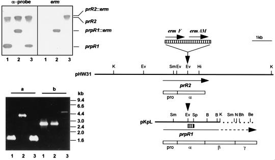
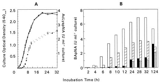
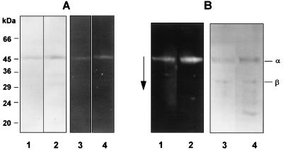
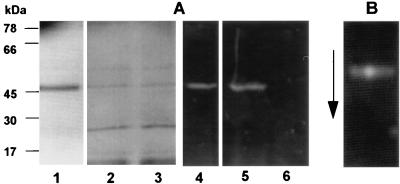
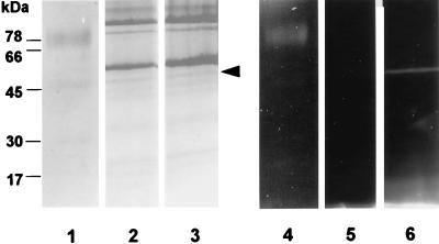
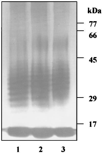
The prpR1 of Porphyromonas gingivalis codes for three distinct enzymes with specificity for arginyl peptide bonds termed RI, RIA, and RIB. These three isoforms comprise the majority of the extracellular, arginine-specific protease activity in P. gingivalis W50. RI is a heterodimer in which the catalytic alpha chain is noncovalently associated with a second chain involved in adherence phenomena. RIA and RIB are both monomeric species. RIA represents the free alpha chain, and RIB is a highly posttranslationally modified form of the alpha chain which is exclusively vesicle or membrane associated and migrates as a diffuse band on sodium dodecyl sulfate-polyacrylamide gel electrophoresis. In previous studies, insertional inactivation of the prpR1 demonstrated that arginine-specific protease activity can also arise from a closely related second gene, prR2. In the present work, the prR2 was insertionally inactivated in P. gingivalis W50 in order to establish the contribution of this locus to the arginine-specific protease activity of this periodontal bacterium. Loss of prR2 function had several effects on prpR1-derived enzymes. First, the total Arg-X activity was reduced by approximately 50% relative to that of the parent strain. The reduction in total activity was a consequence of decreased concentrations of the monomeric enzymes derived from the prpR1, while the heterodimeric enzyme, RI, was unaffected by this mutation. Second, the chromatographic behavior of both the soluble and vesicle- or membrane-associated monomeric enzymes was radically different from the behavior of RIA and RIB from the parent strain. Finally, the vesicle- or membrane-associated enzyme in the prR2 mutant strain lacked the extensive posttranslational additions which are found on RIB in P. gingivalis W50. These data suggest that the product(s) of the prR2 plays a significant role in the maturation pathway of prpR1-derived enzymes, and this may contribute to the coconservation of these two genes in P. gingivalis.


Figures

FIG. 1
FIG. 1
Genomic organization of the major arginine-specific protease genes prR2 and prpR1 and Southern and PCR analyses of P. gingivalis W50 strains. The two bold lines represent an ∼8.5-kb-HindIII (pHW31) or Sau3AI-KpnI (pKpL, nucleotide 1 to 3471 of the sequence with accession no. X82680) fragment of P. gingivalis W50 genomic DNA bearing prR2 and truncated prpR1, respectively. Unique restriction sites are shown as bars above the fragments. Note that both pKpL and prpR1 have been extended for comparative purposes, and this is shown as broken lines. Insertion of the Erm cassette, encoding ermF and ermAM (hatched box above restriction maps), is at the EcoRV site (indicated by down-pointing arrows) for generation of P. gingivalis W501 or W50D7 via electrotransformation. Positions and direction of genes are indicated by arrows under the DNA fragments. Empty boxes are schematic representations of translated sequences (PrRII and PrpRI) with their domain characteristics (pro, α, β, and γ); pro and α domains are common to the two gene products. The α-probe, used in the Southern hybridization experiment, is shown as a small hatched box under pKpL. In the Southern hybridization (upper inserted panel), P. gingivalis chromosomal DNA was restricted with SmaI and membranes were probed with either the 270-bp α-probe or the 2.1-kb Erm cassette. The positions of the 3.2-kb prpR1 and ≥12-kb prR2 together with the corresponding erm inserted loci are shown. Lanes: 1, W50 (wt); 2, W501 (prpR1 mutant); 3, W50D7 (prR2 mutant). The PCR panel (lower insert) shows the agarose electrophoresis of amplicons generated with primers specific for prpR1 (a) or prR2 (b) by using P. gingivalis chromosomal DNA as templates. The lane numbers are the same as for the Southern hybridization experiment. B, BamHI; Be, BstEII; Bh, BssHII; Ev, EcoRV; Hi, HincII; K, KpnI; N, NcoI; Sm, SmaI; Sp, SphI.
FIG. 2
FIG. 2
(A) Growth curves and Lys-X protease activity of P. gingivalis W50 (x - x) and W50D7 (prR2 mutant) (• - •). A 10% inoculum of overnight cultures of both strains was used to inoculate brain heart infusion broth supplemented with hemin (5 mg liter−1). Samples were withdrawn throughout growth for absorbance measurements (solid lines), and total Lys-X protease activity (dotted lines) was measured following sonication of whole culture mixed with an equal volume of 25 mM MOPS (morpholinepropanesulfonic acid) pH 6.0 buffer (10 mM CaCl2, 0.2 M NaCl). Units of enzyme activity are expressed as increase in absorbance at 405 nm/min. (B) Arg-X activity in culture supernatant and in sonicated cultures was measured as described in the text. Open bars indicate cell-bound activity, and solid bars indicate supernatant of P. gingivalis W50. Broad hatched bars indicate cell-bound activity, and fine hatched bars indicate supernatant in W50D7 (prR2 mutant). Units of enzyme activity are expressed as increase in absorbance at 405 nm/min.
FIG. 3
FIG. 3
Gel electrophoresis of DNS-EGR-CK-labelled RI from P. gingivalis W50 and W50D7 (prR2 mutant). Enzymes from strains W50 and W50D7 were labelled with the fluorescent irreversible inhibitor DNS-EGR-CK as described in the text and subjected to SDS-PAGE on 12.5% gels (A) or 8 M urea-PAGE on 7.5% gels (B) followed by blotting onto polyvinylidene difluoride membranes. Gels and blots were viewed under UV light to visualize labelled proteins and then stained for total protein with Coomassie brilliant blue. (A) Lanes: 1 and 2, stained with Coomassie blue; 3 and 4, viewed under UV light; 1 and 3, RI-W50; 2 and 4, RI-W50D7. The molecular masses of marker proteins are indicated alongside the gel. (B) Lanes: 1 and 2, viewed under UV light; 3 and 4, stained with Coomassie blue; 1 and 3, RI-W50; 2 and 4, RI-W50D7 (prR2 mutant). The arrow indicates the direction of migration. α and β indicate the α-catalytic chain and the β-adhesin chain of RI.
FIG. 4
FIG. 4
Gel electrophoresis of DNS-EGR-CK-labelled RIA from P. gingivalis W50 and monomeric enzyme from the S-fraction of W50D7 (RIAs-W50D7). Enzymes from strains W50 and W50D7 were labelled with DNS-EGR-CK as described in the text and subjected to SDS-PAGE (A) or 8 M urea-PAGE (B) and detected as described in the legend to Fig. 3. (A) Lanes: 1 to 3, stained with Coomassie blue; 4 to 6, viewed under UV light; 1 and 4, RIA-W50; 2 and 5, RIAs-W50D7; 3 and 6, RIAs-W50D7 labelled in the presence of 50 μM leupeptin. (B) A mixture of RIA-W50 and RIAs-W50D7 labelled with DNS-EGR-CK and run on an 8 M urea-PAGE gel.
FIG. 5
FIG. 5
Gel electrophoresis of DNS-EGR-CK-labelled RIB from P. gingivalis W50 and monomeric enzyme from the P-fraction of W50D7 (RIAv-W50D7). Enzymes from strains W50 and W50D7 were labelled as described in the legend to Fig. 3 and subjected to SDS-PAGE on 12.5% gels. Lanes: 1 to 3, stained with Coomassie blue; 4 to 6, viewed under UV light; 1 and 4, RIB-W50; 2 and 5, RIAv-W50D7 labelled in the presence of 50 μM leupeptin; 3 and 6, RIAv-W50D7. The arrowhead next to lane 3 indicates the protein which is labelled with DNS-EGR-CK.
FIG. 6
FIG. 6
Silver-stained SDS-PAGE of LPS preparations from P. gingivalis W50 (lane 1), W501 (prpR1 mutant) (lane 2), and W50D7 (prR2 mutant) (lane 3). Proteinase K digests of stationary phase cells were subjected to SDS-PAGE on 12.5% gels followed by silver staining. No Coomassie blue staining material was present.

References

    1. Aduse-Opoku J, Muir J, Slaney J M, Rangarajan M, Curtis M A. Characterization, genetic analysis, and expression of a protease antigen (PrpRI) of Porphyromonas gingivalis W50. Infect Immun. 1995;63:4744–4754. - PMC - PubMed
    1. Aduse-Opoku J, Slaney J M, Rangarajan M, Muir J, Young K A, Curtis M A. The Tla protein of Porphyromonas gingivalis W50: a homolog of the RI protease precursor (PrpRI) is an outer membrane receptor required for growth on low levels of hemin. J Bacteriol. 1997;179:4778–4788. - PMC - PubMed
    1. Allaker R P, Aduse-Opoku J, Batten J E, Curtis M A. Natural variation within the principal arginine-specific protease gene, prpR1, of P. gingivalis. Oral Microbiol Immunol. 1997;12:298–302. - PubMed
    1. Curtis M A, Ramakrishnan M, Slaney J M. Characterisation of the trypsin-like enzymes of Porphyromonas gingivalis W83 using a radiolabelled active-site-directed inhibitor. J Gen Microbiol. 1993;139:949–955. - PubMed
    1. Curtis M A, Aduse-Opoku J, Slaney J M, Rangarajan M, Booth V, Cridland J, Shepherd P. Characterization of an adherence and antigenic determinant of the ArgI protease of Porphyromonas gingivalis which is present on multiple gene products. Infect Immun. 1996;64:2532–2539. - PMC - PubMed

Publication types

MeSH terms

LinkOut - more resources