. 1998 Apr;66(4):1718-25.
doi: 10.1128/IAI.66.4.1718-1725.1998.

Role of adenylate cyclase-hemolysin in alveolar macrophage apoptosis during Bordetella pertussis infection in vivo

Affiliations

Role of adenylate cyclase-hemolysin in alveolar macrophage apoptosis during Bordetella pertussis infection in vivo

P Gueirard et al. Infect Immun. 1998 Apr.

Abstract

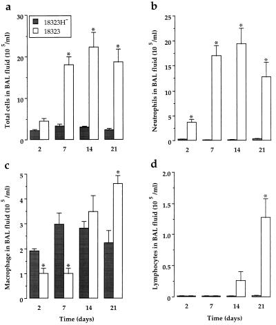
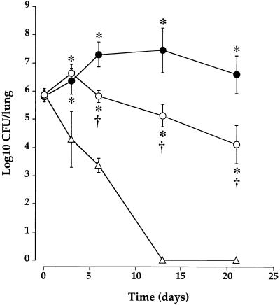
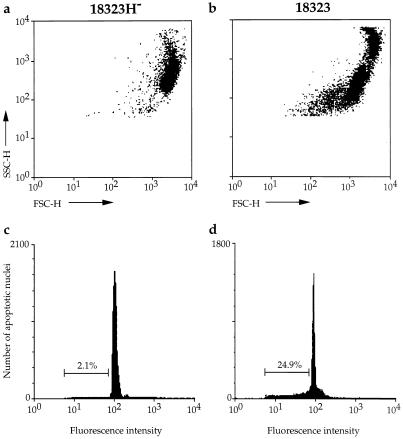
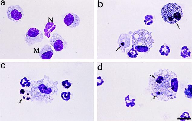
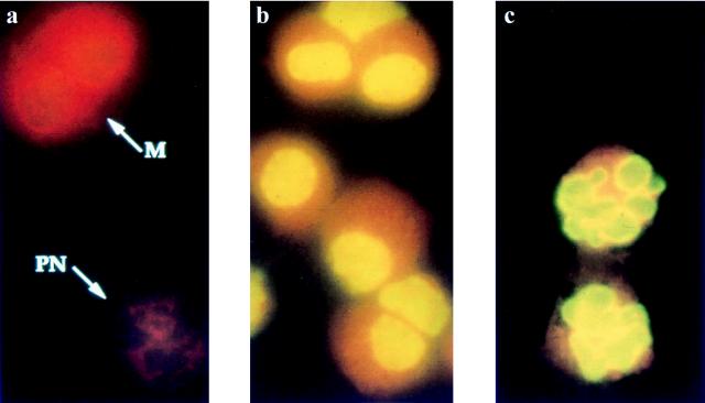
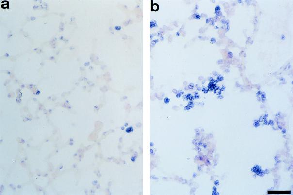
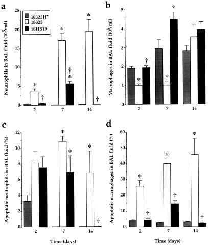
Bordetella pertussis induces in vitro apoptosis of murine alveolar macrophages by a mechanism that is dependent on expression of bacterial adenylate cyclase-hemolysin. Using a murine respiratory model, we found in this study that intranasal infection with a parental B. pertussis strain, but not with an isogenic variant deficient in the expression of all toxins and adhesins, induced a marked neutrophil accumulation in the bronchoalveolar lavage fluid and an early decrease in macrophage numbers. These phenomena paralleled a time-dependent rise in the proportion of apoptotic nuclei, as detected by flow cytometry, and of macrophages which had engulfed apoptotic bodies. Apoptotic death of bronchopulmonary cells was observed exclusively following intranasal infection with bacteria reisolated from lungs of infected animals and not with B. pertussis collected after in vitro subculture. Using the terminal deoxynucleotidyltransferase-mediated dUTP-biotin nick end labeling technique coupled to fluorescence microscopy and morphological analysis, we established that the apoptotic cells in bronchoalveolar lavage fluids were neutrophils and macrophages. Histological analysis of the lung tissues from B. pertussis-infected mice showed increased numbers of apoptotic cells in the alveolar compartments. Cellular accumulation in bronchoalveolar lavage fluids and apoptosis of alveolar macrophages were significantly attenuated in mice infected with a mutant deficient in the expression of adenylate cyclase-hemolysin, indicating a role of this enzyme in these processes.


Figures

FIG. 1
FIG. 1
Cellular distribution in BAL fluids from B. pertussis-infected mice. BAL fluids were collected from mice infected via the i.n. route with 1.5 × 10⁶ CFU of parental strain 18323 or variant 18323H and killed at different time points after the infection. Numbers of total cells (a), neutrophils (b), macrophages (c), and lymphocytes (d) were determined after cytocentrifugation and staining with Diff-Quick dye. Results are expressed as means ± SEM of six to nine experiments at each time point. ∗, P < 0.05 compared to variant 18323H-infected mice.
FIG. 2
FIG. 2
B. pertussis colonization of the lungs of mice. Mice were challenged i.n. with 1.5 × 10⁶ CFU of freshly isolated parental strain 18323 (•), variant 18323H (▵), and mutant strain 18HS19 (○). The plots show means ± SEM of 3 to 10 experiments at each time point. ∗, P < 0.05 compared to variant 18323H-infected mice. †, P < 0.05 compared to parental strain 18323-infected mice.
FIG. 3
FIG. 3
Flow cytometry analysis of apoptosis in BAL cells from B. pertussis-infected mice. Changes in light scattering properties (a and b) and DNA fluorescence flow cytometry profiles (c and d) of BAL cells collected 14 days after infection with variant 18323H (a and c) or with parental strain 18323 (b and d). Percentages values of apoptotic nuclei for each experimental condition are indicated in the corresponding panels.
FIG. 4
FIG. 4
Morphological analysis of BAL cells after B. pertussis infection. BAL fluids were collected from mice infected with the parental strain 18323 or with variant 18323H, and cytospin preparations were made. Slides were stained with Diff-Quick dye and examined in an oil immersion light microscope. (a) Normal morphology of macrophages (M) and a neutrophil (N) after infection with variant 18323H. Also shown are apoptotic macrophages (arrows; b) and neutrophil (c) after infection with the parental strain 18323 and macrophage engulfment of an apoptotic cell and of an apoptotic body (arrows) after infection with the parental strain 18323 (d). Scale bar, 12 μm.
FIG. 5
FIG. 5
Identification of apoptotic cells in BAL fluids from B. pertussis-infected mice. Cytospin preparations of BAL cells were stained by the TUNEL technique combined with fluorescence microscopy; fluorescein isothiocyanate was used for detection. (a) Neutrophils (PN) and macrophages (M) show normal red nuclei after infection with variant 18323H (arrows). Macrophages (b) and neutrophils (c) contain apoptotic green nuclei after infection with the parental strain 18323. Magnification, ×222.
FIG. 6
FIG. 6
Detection of apoptotic cells in lung sections from B. pertussis-infected mice. Cryostat lung sections were stained by the TUNEL technique followed by detection with alkaline phosphatase (fast blue substrate). Tissues were then counterstained with nuclear red dye. (a) Negative red cells after infection with variant 18323H (arrows). (b) Positive blue apoptotic cells in the alveolar spaces after infection with the parental strain 18323. Scale bar, 40 μm.
FIG. 7
FIG. 7
Cell numbers and apoptotic death in BAL fluids from mice infected with B. pertussis parental strain or mutants. BAL fluids were collected 2, 7, and 14 days after infection with variant 18323H, with parental strain 18323, or with mutant 18HS19. Neutrophils (a) and macrophages (b) were enumerated on cytospin preparations after Diff Quick staining. The proportions of apoptotic neutrophils (c) and macrophages (d) over the total number of each cell type were determined on cytospin preparations after TUNEL technique staining combined with morphological analysis. Results are expressed as means ± SEM of 6 to 12 experiments for each time point. ∗, P < 0.05 compared to variant 18323H-infected mice. †, P < 0.05 compared to parental strain 18323-infected mice.

Similar articles

Cited by

References

    1. Ameisen J C, Estaquier J, Idziorek T. From AIDS to parasite infection: pathogen-mediated subversion of programmed cell death as a mechanism for immune dysregulation. Immunol Rev. 1994;142:9–51. - PubMed
    1. Boschwitz J S, Batanghari J W, Kedem H, Relman D A. Bordetella pertussis infection of human monocytes inhibits antigen-dependent CD4 T cell proliferation. J Infect Dis. 1997;176:678–686. - PubMed
    1. Confer D L, Eaton J W. Phagocyte impotence caused by an invasive bacterial adenylate cyclase. Science. 1982;217:948–950. - PubMed
    1. Cox G, Crossley J, Xing Z. Macrophage engulfment of apoptotic neutrophils contributes to the resolution of acute pulmonary inflammation in vivo. Am J Respir Cell Mol Biol. 1995;12:232–237. - PubMed
    1. Douglas R S, Tarshis A D, Pletcher C H, Nowell P C, Moore J S. A simplified method for the coordinate examination of apoptosis and surface phenotype of murine lymphocytes. J Immunol Methods. 1995;188:219–228. - PubMed

Publication types