. 1998 Apr;66(4):1744-7.
doi: 10.1128/IAI.66.4.1744-1747.1998.

Enhancement of lipopolysaccharide-induced neutrophil oxygen radical production by tumor necrosis factor alpha

Affiliations

Enhancement of lipopolysaccharide-induced neutrophil oxygen radical production by tumor necrosis factor alpha

H P Jersmann et al. Infect Immun. 1998 Apr.

Abstract

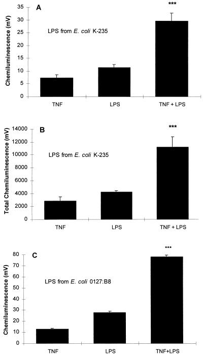
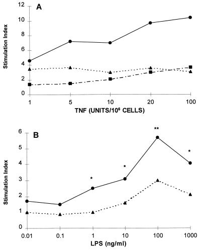
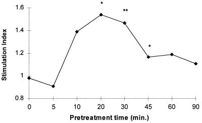
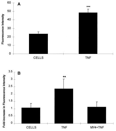
Although tissues become exposed to both exogenous and endogenous cell-activating mediators during infection, there is little appreciation of the effects of subjecting cells to multiple mediators. We examined the hypothesis that the response of neutrophils to bacterial lipopolysaccharide (LPS) is significantly altered in the presence of the endogenous mediator tumor necrosis factor alpha (TNF). The data showed that human neutrophils pretreated with TNF for 10 to 30 min, displayed significantly enhanced superoxide production in response to LPS (from either Escherichia coli K-235 or E. coli O127:B8), measured as lucigenin-dependent chemiluminescence (CL), seen as an increase in the initial peak rate as well as the total CL accumulated over the incubation period. TNF amplified the response to LPS at 1 to 100 U of TNF/10(6) neutrophils and was able to enhance the response to a wide range of concentrations of LPS (0.01 to 1,000 ng/ml). The TNF-induced increase in the LPS response was paralleled by an increase in LPS binding to the neutrophils, which could be abrogated by an anti-CD14 monoclonal antibody. The results demonstrate that TNF significantly increases the LPS-induced release of oxygen radicals in neutrophils through the upregulation of cell surface CD14.


Figures

FIG. 1
FIG. 1
(A) Effect of TNF on the LPS-induced neutrophil CL response. Neutrophils (106) were preincubated with 20 U of TNF for 20 min and then challenged with 100-ng/ml LPS (E. coli K-235), and CL was measured over 50 min of incubation. The values shown are the peak initial rate of CL and represent the means ± the standard error of the mean of eight experiments, each conducted in duplicate with neutrophils from eight different donors. The basal CL of 6.1 mV was deducted from each of the experimental values. Neutrophils treated with TNF and LPS showed a significantly increased response compared to responses induced by either of the agents alone (∗∗∗, P < 0.001 for TNF versus TNF plus LPS and LPS versus TNF plus LPS; P < 0.015 for the sum of values for the individual TNF and LPS treatments versus cotreatment with TNF plus LPS, [analysis of variance]). (B) Data expressed as a function of the total accumulated CL generated over the incubation period. The baseline value of 2528 mV was subtracted from each column. (∗∗∗, P < 0.001 for TNF versus TNF plus LPS and LPS versus TNF plus LPS; P < 0.015 for the sum of values for the individual TNF and LPS treatments versus cotreatment with TNF plus LPS [analysis of variance]). (C) Effect of LPS (E. coli 0127:B8) under the same conditions as in A. The basal CL of 13.3 mV was deducted from each of the experimental values. The increased response to stimulation with both TNF and LPS was highly significant (∗∗∗, P < 0.001 [analysis of variance]).
FIG. 2
FIG. 2
(A) Effects of varying TNF amounts on the LPS-induced CL response. Neutrophils were pretreated with the indicated concentrations of TNF or diluent for 20 min and then challenged with 1-μg/ml of LPS (E. coli K-235) or diluent for another 50 min. Neutrophils were treated with TNF only (▪), LPS only (▴), or TNF plus LPS (•). Results are the means of four duplicate experiments, expressed as stimulation indices. These were obtained by dividing the means of the treatments by the means of the baseline values. At all TNF doses, the effects of the combined action of TNF and LPS were significantly different from those of either agonist alone (P values of <0.0001 to <0.05 [analysis of variance]). (B) Response of TNF-primed neutrophils to varying concentrations of LPS (E. coli K-235). The cells were pretreated with 20 U of TNF/106 cells (•) or diluent (▴) for 20 min and then stimulated with the indicated concentrations of LPS. Values are stimulation indices. The data are the means of four experiments conducted in duplicate (∗, P < 0.05; ∗∗, P < 0.01 [analysis of variance]).
FIG. 3
FIG. 3
Effect of varying TNF pretreatment times on the response of neutrophils to LPS (E. coli K-235). Cells were pretreated with 20 U of TNF/106 cells for 20 min and then with LPS (100 ng/ml) for 50 min. The results are derived from eight experiments with different donor cells. The results are expressed as stimulation indices (∗, P, <0.035; ∗∗, P < 0.015 [analysis of variance]).
FIG. 4
FIG. 4
(A) Effect of TNF pretreatment on surface CD14 on neutrophils. Neutrophils 106 were incubated with either buffer or 20 U of TNF for 20 min. At 4°C the cells were incubated with anti-CD14 and then washed and incubated with goat anti-mouse phycoerythrin. The intensity of fluorescence was determined as described in the text. The data shown are the means and standard errors of five experiments. The increase in fluorescence was 2.1-fold (∗∗∗, P < 0.001 [Student t test]). (B) Effect of TNF on LPS binding. Neutrophils were stimulated as described above. The cells were washed and resuspended in serum (5%, autologous, heat inactivated for 30 min at 56°C) and incubated with FITC-labelled LPS (E. coli 0127:B8) at a final concentration of 16 μg/ml for 30 min on ice. After three washes, flow cytometry was performed as described in the text. The increase was 2.2-fold and could be abrogated by incubating the cells with MY4 (dilution, 1:10) for 30 min prior to adding serum and FITC-LPS. The data shown are the means and standard errors of five experiments (∗∗, P < 0.01 [analysis of variance]).

Similar articles

Cited by

References

    1. Beutler B, editor. Tumor necrosis factors, the molecules and their emerging role in medicine. New York, N.Y: Raven Press; 1992.
    1. Dahinden C A, Fehr J, Hugli T E. Role of cell surface contact in the kinetics of superoxide production by granulocytes. J Clin Invest. 1983;72:113–121. - PMC - PubMed
    1. Dziarski R. Cell-bound albumin is the 70-kDa peptidoglycan-, lipopolysaccharide-, and lipoteichoic acid-binding protein on lymphocytes and macrophages. J Biol Chem. 1994;269:20431–20436. - PubMed
    1. Fast D J, Schlievert P M, Nelson R D. Toxic shock syndrome-associated staphylococcal and streptococcal pyrogenic toxins are potent inducers of tumor necrosis factor production. Infect Immun. 1989;57:291–294. - PMC - PubMed
    1. Ferrante A, Kowanko I, Bates E J. Mechanisms of host tissue damage by cytokine-activated neutrophils. In: Coffey R G, editor. Granulocyte responses to cytokines: basic and clinical research. New York, N.Y: Marcel Dekker, Inc.; 1992. pp. 499–521. - PubMed

Publication types

MeSH terms