

doi: 10.1128/IAI.66.4.1755-1758.1998.
Agents that inhibit Rho, Rac, and Cdc42 do not block formation of actin pedestals in HeLa cells infected with enteropathogenic Escherichia coli
Affiliations
- PMID: 9529109
- PMCID: PMC108116
- DOI: 10.1128/IAI.66.4.1755-1758.1998
Item in Clipboard
Agents that inhibit Rho, Rac, and Cdc42 do not block formation of actin pedestals in HeLa cells infected with enteropathogenic Escherichia coli
Infect Immun.
1998 Apr.
Abstract
Enteropathogenic Escherichia coli (EPEC) induces formation of actin pedestals in infected host cells. Agents that inhibit the activity of Rho, Rac, and Cdc42, including Clostridium difficile toxin B (ToxB), compactin, and dominant negative Rho, Rac, and Cdc42, did not inhibit formation of actin pedestals. In contrast, treatment of HeLa cells with ToxB inhibited EPEC invasion. Thus, Rho, Rac, and Cdc42 are not required for assembly of actin pedestals; however, they may be involved in EPEC uptake by HeLa cells.
Figures
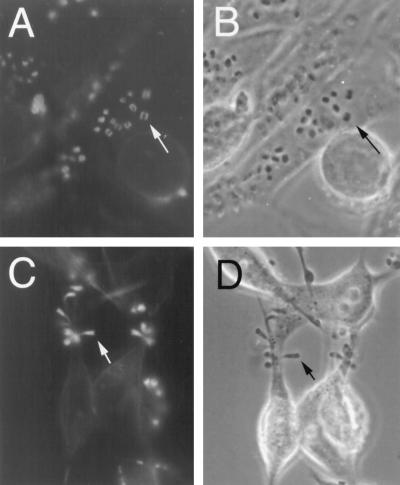
Formation of actin pedestals in HeLa cells treated with compactin. Untreated HeLa cells (A and B) or cells treated for 18 h with 50 μM compactin (C and D) were infected with JPN15 for 2 h. The infected cells were fixed and stained with phalloidin-rhodamine, and the fluorescent images (A and C), as well as the corresponding phase-contrast images (B and D), were photographed. Arrows indicate the EPEC-induced actin structures (A and C) and the corresponding bacteria above this structure (B) or at the tip of the actin pedestal (D). All images represent only one optical plane focusing on some of the EPEC-induced actin pedestals. The actin stress fibers in panel A are layered in a different optical plane and thus cannot be seen.
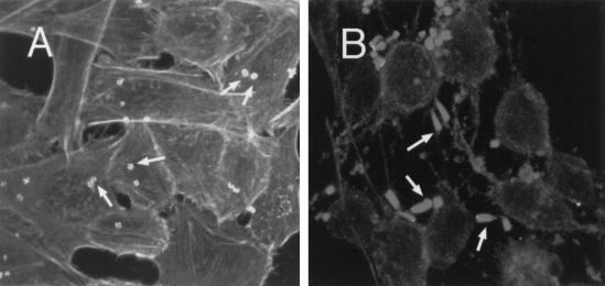
Formation of actin pedestals in HeLa cells treated with ToxB. Untreated HeLa cells (A) or cells treated with ToxB (10 ng/ml) for 3 h (B) were infected with JPN15 for 2 h. Then, cells were fixed and stained with phalloidin-rhodamine and analyzed by confocal microscopy. The cells were scanned at 0.5-μm intervals, and all optical sections were projected to form one image. The flat EPEC-induced actin structures in panel A and the elongated actin pedestals in panel B are indicated by arrows. The untreated cells are rich in stress fibers (A) that disappeared after treatment with ToxB (B).
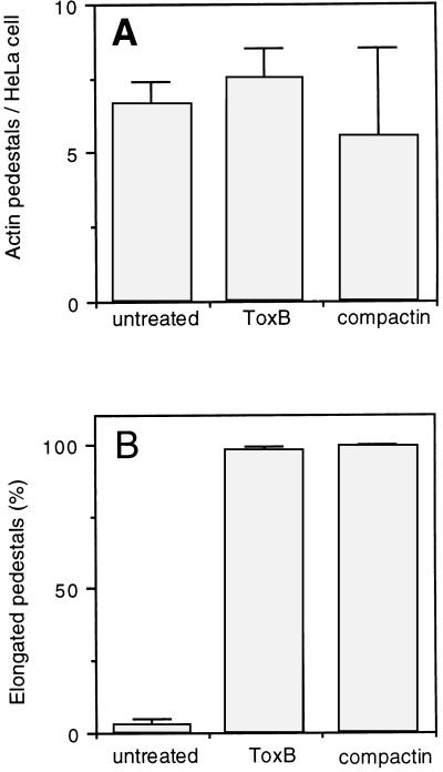
Formation of actin pedestals in HeLa cells treated with compactin or ToxB. The microscopic preparations described in the legends to Fig. 1 and 2 were used to quantify the formation of actin pedestals in infected HeLa cells (A), including untreated HeLa cells (n = 320), cells treated with compactin (n = 60), and cells treated with ToxB (n = 112). In addition, we determined the percentages of elongated pedestals (B) in untreated HeLa cells (n = 309), cells treated with compactin (n = 332), and cells treated with ToxB (n = 133). Standard deviations are indicated by error bars.
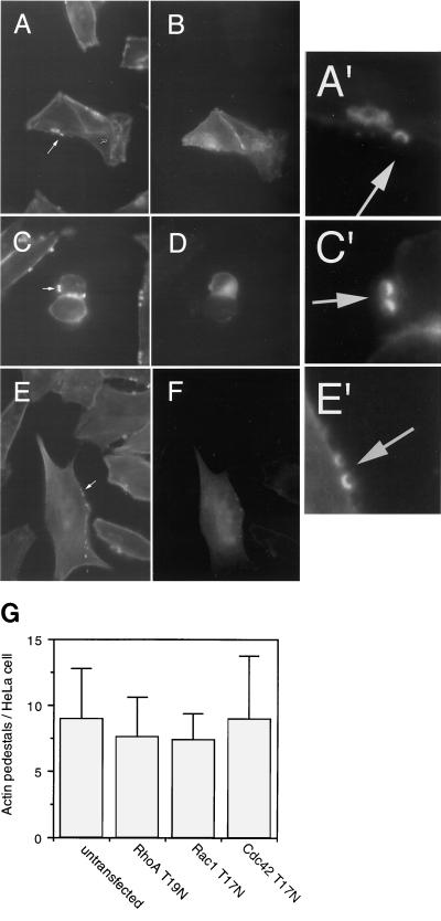
Formation of actin pedestals in HeLa cells expressing dominant negative Rho, Rac, and Cdc42. HeLa cells were transiently transfected with plasmids encoding dominant negative small GTP-binding proteins fused to GFP, including Rac1-T17N-GFP (A, A′, and B), Cdc42-T17N-GFP (C, C′, and D), and RhoA-T19N-GFP (E, E′, and F). The transfected cultures were infected with JPN15 and fixed, and the actin filaments were stained with phalloidin-rhodamine. Phalloidin stained all of the HeLa cells in the culture (A, C, and E). Among these, the transfected cells were identified as green fluorescing cells (B, D, and F). The arrows in panels A, C, and E indicate the localization of typical actin pedestals. These pedestals can be better seen in close-ups of the arrow-indicated regions (A′, C′, and E′). Only some of the actin pedestals associated with each cell could be visualized in a given optical plane, including those shown in panels A, C, and E. Additional actin pedestals were detected in other optical planes (not shown). (G) The average numbers of actin pedestals per HeLa cell were determined for untransfected cells (n = 128) and cells expressing either RhoA-T19N-GFP (n = 80), Rac1-T17N-GFP (n = 83), or Cdc42-T17N-GFP (n = 100). Standard deviations are indicated by error bars.
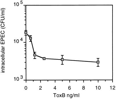
Inhibition of EPEC invasion by ToxB treatment of HeLa cells. HeLa cells, in a 24-well plate, were treated for 2 h with 500 μl of Dulbecco minimal essential medium containing different concentrations of ToxB and 10% fetal calf serum. Then, 5 μl of a fresh overnight bacterial culture was added, followed by incubation (37°C, 5% CO2) for an additional 3 h. The cells were washed twice and incubated for an additional 90 min in a medium containing 100 μg of gentamicin per ml; the monolayers were washed and then lysed with 1% Triton X-100, and appropriate dilutions were plated to determine the numbers of intracellular bacteria. The assays were carried out in duplicate.
References
-
- Chong L D, Kaplan T A, Bokoch G M, Schwartz M A. The small GTP-binding protein rho regulates a phosphatidylinositol 4-phosphate 5-kinase in mammalian cells. Cell. 1994;79:507–513. - PubMed
-
- Heim R, Cubitt A B, Tsien R Y. Improved green fluorescence protein. Nature. 1995;373:663–664. - PubMed
-
- Just I, Selzer J, Wilm M, Von Eichel-Streiber C, Mann M, Aktories K. Glucosylation of Rho proteins by Clostridium difficile toxin B. Nature. 1995;375:500–503. - PubMed
Publication types
MeSH terms
Substances
Grants and funding
LinkOut - more resources
Full Text Sources
Miscellaneous
