. 1998 Apr 6;187(7):1037-46.
doi: 10.1084/jem.187.7.1037.

Murine cytomegalovirus inhibits interferon gamma-induced antigen presentation to CD4 T cells by macrophages via regulation of expression of major histocompatibility complex class II-associated genes

Affiliations

Murine cytomegalovirus inhibits interferon gamma-induced antigen presentation to CD4 T cells by macrophages via regulation of expression of major histocompatibility complex class II-associated genes

M T Heise et al. J Exp Med. .

Abstract

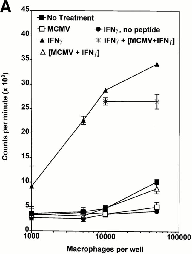
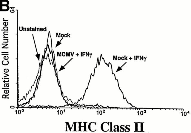
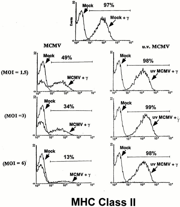
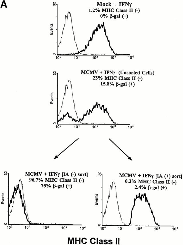
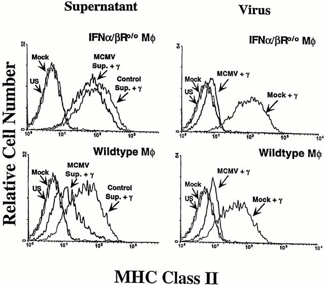
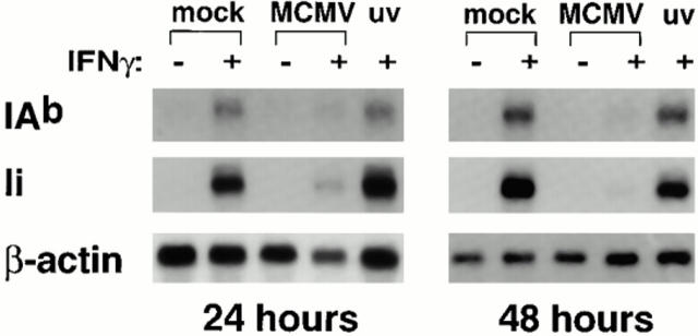
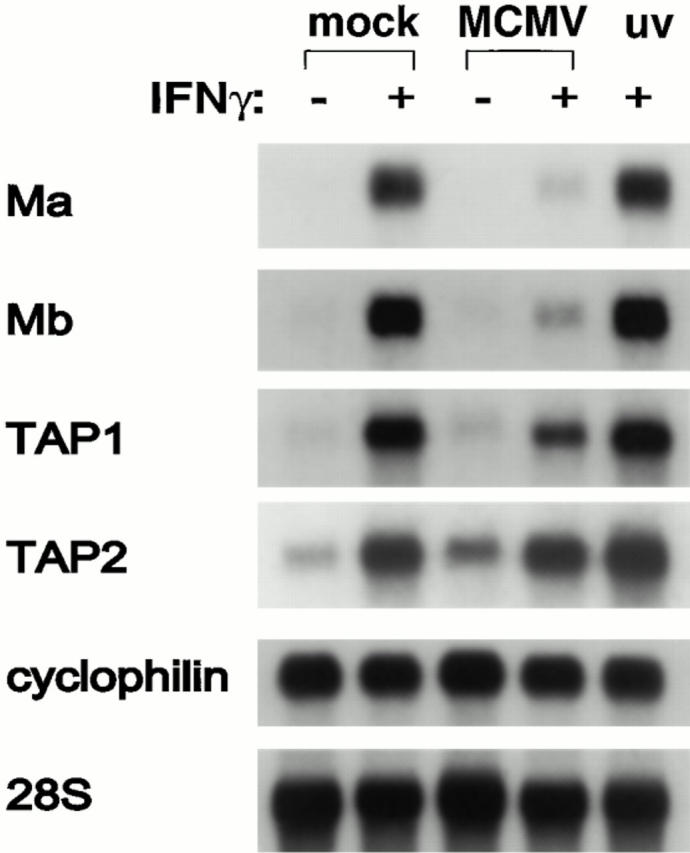
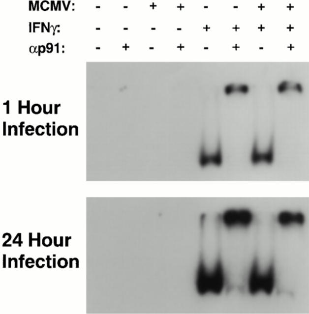
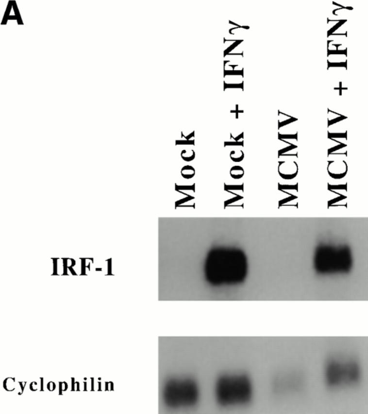
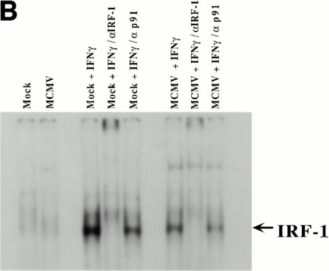
CD4 T cells and interferon gamma (IFN-gamma) are required for clearance of murine cytomegalovirus (MCMV) infection from the salivary gland in a process taking weeks to months. To explain the inefficiency of salivary gland clearance we hypothesized that MCMV interferes with IFN-gamma induced antigen presentation to CD4 T cells. MCMV infection inhibited IFN-gamma-induced presentation of major histocompatibility complex (MHC) class II associated peptide antigen by differentiated bone marrow macrophages (BMMphis) to a T cell hybridoma via impairment of MHC class II cell surface expression. This effect was independent of IFN-alpha/beta induction by MCMV infection, and required direct infection of the BMMphis with live virus. Inhibition of MHC class II cell surface expression was associated with a six- to eightfold reduction in IFN-gamma induced IAb mRNA levels, and comparable decreases in IFN-gamma induced expression of invariant chain (Ii), H-2Ma, and H-2Mb mRNAs. Steady state levels of several constitutive host mRNAs, including beta-actin, cyclophilin, and CD45 were not significantly decreased by MCMV infection, ruling out a general effect of MCMV infection on mRNA levels. MCMV effects were specific to certain MHC genes since IFN-gamma-induced transporter associated with antigen presentation (TAP)2 mRNA levels were minimally altered in infected cells. Analysis of early upstream events in the IFN-gamma signaling pathway revealed that MCMV did not affect activation and nuclear translocation of STAT1alpha, and had minor effects on the early induction of IRF-1 mRNA and protein. We conclude that MCMV infection interferes with IFN-gamma-mediated induction of specific MHC genes and the Ii at a stage subsequent to STAT1alpha activation and nuclear translocation. This impairs antigen presentation to CD4 T cells, and may contribute to the capacity of MCMV to spread and persist within the infected host.


Figures

Figure 1
Figure 1
MCMV infection impairs IFN-γ–enhanced peptide presentation by MHC class II. 129 mouse BMMφs were mock infected or infected with MCMV at an MOI of 5.0 for 1 h, and stimulated with IFN-γ (100 IU/ml) or medium alone. After 24 h BMMφs were assayed for ability to present peptide antigen, and analyzed for MHC class II expression by flow cytometry. (A) Mφs were plated at 103, 5 × 10³, 104, and 5 × 10⁴ cells per well, and incubated with the T cell hybridoma B11 (β-galactosidase 429-441, IAb-restricted) in the presence of β-galactosidase peptide (429–441) at a concentration of 10 μM. Supernatants were harvested and assayed for IL-2 content using [3H]thymidine incorporation by CTLL2 cells. Mock + IFN-γ and MCMV + IFN-γ BMMφs were mixed 1:1 and assayed as a control for potential toxic effects of MCMV-infected cells. Similar data was obtained at a peptide concentration of 1.0 μM. Data is from one of two similar experiments. All groups were plated in triplicate and are shown as mean cpm ± SEM. (B) Cell surface expression of the MHC class II molecule IAb on BMMφs infected with MCMV and stimulated with IFN-γ as above was measured by flow cytometry.
Figure 1
Figure 1
MCMV infection impairs IFN-γ–enhanced peptide presentation by MHC class II. 129 mouse BMMφs were mock infected or infected with MCMV at an MOI of 5.0 for 1 h, and stimulated with IFN-γ (100 IU/ml) or medium alone. After 24 h BMMφs were assayed for ability to present peptide antigen, and analyzed for MHC class II expression by flow cytometry. (A) Mφs were plated at 103, 5 × 10³, 104, and 5 × 10⁴ cells per well, and incubated with the T cell hybridoma B11 (β-galactosidase 429-441, IAb-restricted) in the presence of β-galactosidase peptide (429–441) at a concentration of 10 μM. Supernatants were harvested and assayed for IL-2 content using [3H]thymidine incorporation by CTLL2 cells. Mock + IFN-γ and MCMV + IFN-γ BMMφs were mixed 1:1 and assayed as a control for potential toxic effects of MCMV-infected cells. Similar data was obtained at a peptide concentration of 1.0 μM. Data is from one of two similar experiments. All groups were plated in triplicate and are shown as mean cpm ± SEM. (B) Cell surface expression of the MHC class II molecule IAb on BMMφs infected with MCMV and stimulated with IFN-γ as above was measured by flow cytometry.
Figure 2
Figure 2
Infectious MCMV impairs IFN-γ–induced MHC class II expression on Mφs in a dose-dependent manner. IFN-α/βR−/− BMMφs were mock infected or infected with live or UV-inactivated MCMV at an MOI of 1.5, 3.0, or 6.0 for 1 h before addition of 100 IU/ml IFN-γ. Mφs were harvested 48 h after infection and assayed for MHC class II expression (IAb) by flow cytometry. Shown is one of three experiments, each of which yielded similar results.
Figure 3
Figure 3
MCMV-infected BMMφs express low levels of MHC class II. IFN-α/βR−/− BMMφs were either mock infected or infected with recombinant MCMV expressing β-galactosidase (RM427) at an MOI of 0.6. 1 h after infection, cells were treated with IFN-γ (100 IU/ml) or medium alone for 48 h. Cells were harvested, stained for MHC class II (IAb) expression, and separated into MHC class II high and low populations by fluorescent activated cell sorting. The numbers of cells expressing β-galactosidase in total and sorted cell populations was determined by 5-bromo-4-chloro-3-indolyl-β-d-galactopyranoside (x-gal) staining. (A) Mock-infected or infected cells (top panels), and IAb high and low populations from infected cultures (bottom panels) were stained for IAb. The dotted line represents staining on unstimulated, mock-infected cells. The solid line depicts staining of IFN-γ– treated groups. (B) x-gal staining of mock- or MCMV-infected, and MHC class II high and low, populations is shown from one of three similar experiments.
Figure 3
Figure 3
MCMV-infected BMMφs express low levels of MHC class II. IFN-α/βR−/− BMMφs were either mock infected or infected with recombinant MCMV expressing β-galactosidase (RM427) at an MOI of 0.6. 1 h after infection, cells were treated with IFN-γ (100 IU/ml) or medium alone for 48 h. Cells were harvested, stained for MHC class II (IAb) expression, and separated into MHC class II high and low populations by fluorescent activated cell sorting. The numbers of cells expressing β-galactosidase in total and sorted cell populations was determined by 5-bromo-4-chloro-3-indolyl-β-d-galactopyranoside (x-gal) staining. (A) Mock-infected or infected cells (top panels), and IAb high and low populations from infected cultures (bottom panels) were stained for IAb. The dotted line represents staining on unstimulated, mock-infected cells. The solid line depicts staining of IFN-γ– treated groups. (B) x-gal staining of mock- or MCMV-infected, and MHC class II high and low, populations is shown from one of three similar experiments.
Figure 4
Figure 4
Supernatants from MCMV-infected Mφs inhibit IFN-γ induction of MHC class II on normal but not IFN-α/βR−/− BMMφs. Wild-type 129 BMMφs were either mock infected or infected with MCMV at an MOI of 5.0. 48 h after infection, supernatants were removed, ultracentrifuged to remove free virus, and placed on naive cultures of either 129 or IFN-α/βR−/− BMMφs. At the time of supernatant transfer, additional cultures were infected with MCMV (MOI = 5.0) as positive controls. After 1 h of incubation with the supernatants or MCMV, cells were treated with IFN-γ (100 IU/ml), incubated for 48 h, and harvested. MHC class II (IAb) expression was determined by flow cytometry. Shown is data from one of two experiments yielding similar results. (US, unstained).
Figure 5
Figure 5
MCMV infection reduces IFN-γ–induced MHC class II and Ii mRNA levels. IFN-α/βR−/− BMMφs were either mock infected or infected with MCMV or UV-inactivated MCMV at an MOI of 5.0. 1 h after infection, cells were treated with medium alone or with IFN-γ (100 IU/ml). Total cellular RNA was harvested at either 24 or 48 h after infection and analyzed by Northern blot hybridization for the IAb beta chain or Ii. β-actin hybridization is also shown. Similar results were obtained in three separate experiments.
Figure 8
Figure 8
MCMV infection inhibits H-2Ma, H-2Mb, and TAP1 mRNA expression, but not IFN-γ–induced TAP2 mRNA expression. IFN-α/ βR−/− BMMφ were either mock infected or infected with MCMV or UV-inactivated MCMV at an MOI of 5.0 in the presence or absence of IFN-γ (100 IU/ml) for 24 h. Total cellular RNA was harvested and analyzed by Northern blot. Blots were probed using radiolabeled probes for murine TAP1, TAP2, H-2Ma, H-2Mb, cyclophilin, and 28S ribosomal RNA. Shown is one of three experiments which yielded similar results.
Figure 6
Figure 6
STAT1α activation and nuclear translocation after IFN-γ stimulation is normal in MCMV-infected Mφs. IFN-α/ βR−/− BMMφs were infected with MCMV at an MOI of 5.0 or mock infected for 1 or 24 h. Harvested cells were incubated with IFN-γ for 10 min and nuclear extracts were prepared. Extracts were assayed for STAT1α activation by EMSA against a 32P-labeled IFN-γ activation sequence containing oligonucleotide derived from the murine FcγR1 promoter. The presence of STAT1α in the complex was determined by antibody super-shift using STAT1α-specific antiserum. Shown are STAT1α activation by IFN-γ at 1 h (100 IU/ml IFN-γ) or 24 h (5 IU/ml IFN-γ) after infection. Similar results were obtained in four separate experiments.
Figure 7
Figure 7
IRF-1 mRNA and DNA binding activity is inducible by IFN-γ in MCMV-infected Mφs. (A) IFN-α/βR−/− were either mock infected or infected with MCMV at an MOI of 5.0 for 3 h. Cultures were treated with IFN-γ for the final 2 h of infection and assayed for IRF-1 mRNA levels by Northern blot hybridization. Shown is one of two experiments yielding similar results. (B) IFN-α/βR−/− BMMφs were infected with MCMV at an MOI of 5.0 or mock infected for 5 h. Cells were treated with IFN-γ for the final 4 h of infection and nuclear extracts were prepared. IRF-1 DNA binding activity was assessed by incubating extract with a 32P-labeled IRF-E oligonucleotide probe, which was then analyzed by EMSA. The presence of IRF-1 within the complex was confirmed by incubation with antiserum against murine IRF-1 or STAT1α. Three separate experiments yielded similar results.
Figure 7
Figure 7
IRF-1 mRNA and DNA binding activity is inducible by IFN-γ in MCMV-infected Mφs. (A) IFN-α/βR−/− were either mock infected or infected with MCMV at an MOI of 5.0 for 3 h. Cultures were treated with IFN-γ for the final 2 h of infection and assayed for IRF-1 mRNA levels by Northern blot hybridization. Shown is one of two experiments yielding similar results. (B) IFN-α/βR−/− BMMφs were infected with MCMV at an MOI of 5.0 or mock infected for 5 h. Cells were treated with IFN-γ for the final 4 h of infection and nuclear extracts were prepared. IRF-1 DNA binding activity was assessed by incubating extract with a 32P-labeled IRF-E oligonucleotide probe, which was then analyzed by EMSA. The presence of IRF-1 within the complex was confirmed by incubation with antiserum against murine IRF-1 or STAT1α. Three separate experiments yielded similar results.

Similar articles

Cited by

References

    1. Lucin P, Pavic I, Polic B, Jonjic S, Koszinowski UH. Gamma interferon-dependent clearance of cytomegalovirus infection in salivary glands. J Virol. 1992;66:1977–1984. - PMC - PubMed
    1. Pavic I, Polic B, Crnkovic I, Lucin P, Jonjic S, Koszinowski UH. Participation of endogenous tumour necrosis factor-alpha in host resistance to cytomegalovirus infection. J Gen Virol. 1993;74:2215–2223. - PubMed
    1. Heise MT, Virgin HW. The T-cell-independent role of IFN-gamma and TNF-alpha in macrophage activation during murine cytomegalovirus and herpes simplex virus infection. J Virol. 1995;69:904–909. - PMC - PubMed
    1. Orange JS, Wang B, Terhorst C, Biron CA. Requirement for natural killer cell–produced interferon-γ in defense against murine cytomegalovirus infection and enhancement of this defense pathway by interleukin 12 administration. J Exp Med. 1995;182:1045–1056. - PMC - PubMed
    1. Orange JS, Biron CA. Characterization of early IL-12, IFN-alpha/beta, and TNF effects on antiviral state and NK cell responses during murine cytomegalovirus infection. J Immunol. 1996;156:4746–4756. - PubMed

Publication types

MeSH terms