. 1998 Apr 6;187(7):1057-67.
doi: 10.1084/jem.187.7.1057.

Ca2+ signaling modulates cytolytic T lymphocyte effector functions

Affiliations

Ca2+ signaling modulates cytolytic T lymphocyte effector functions

M T Esser et al. J Exp Med. .

Abstract

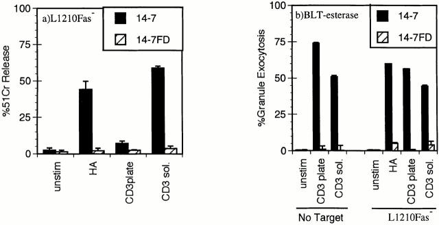
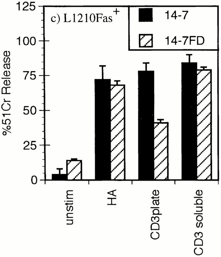
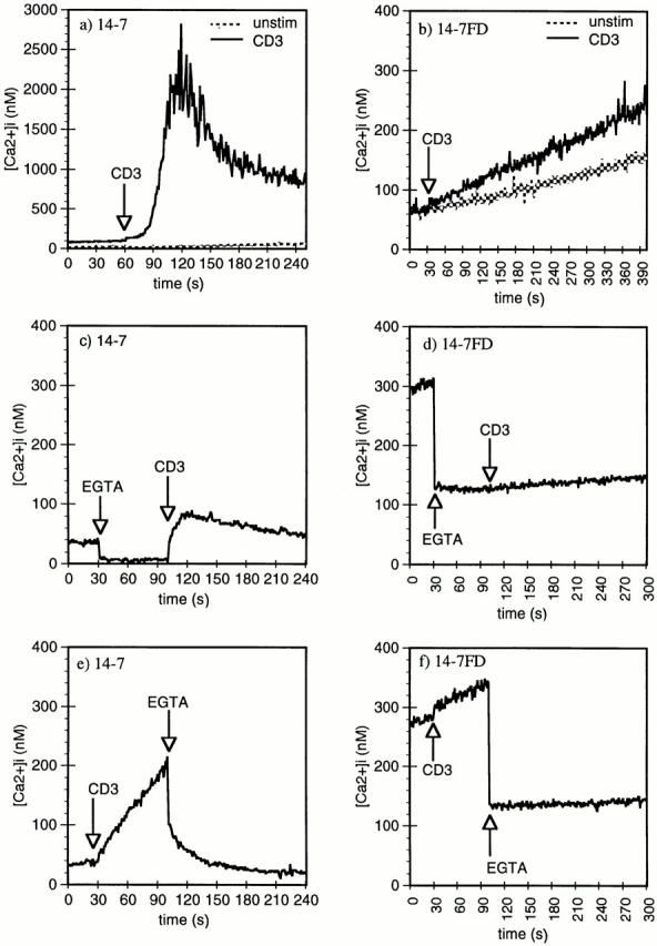
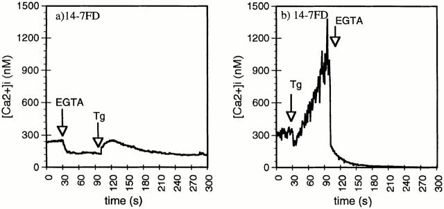
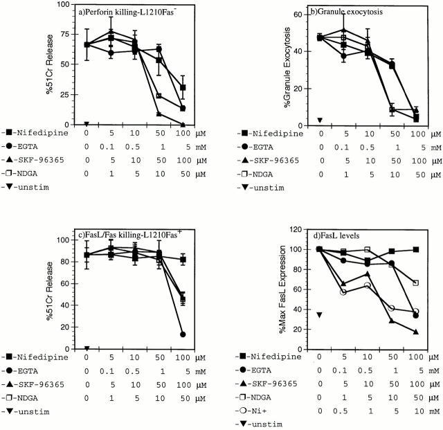
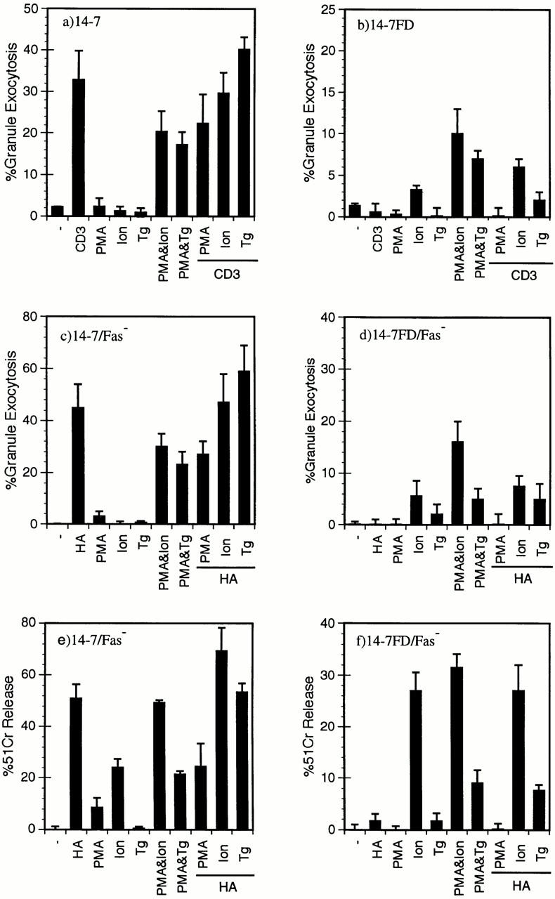
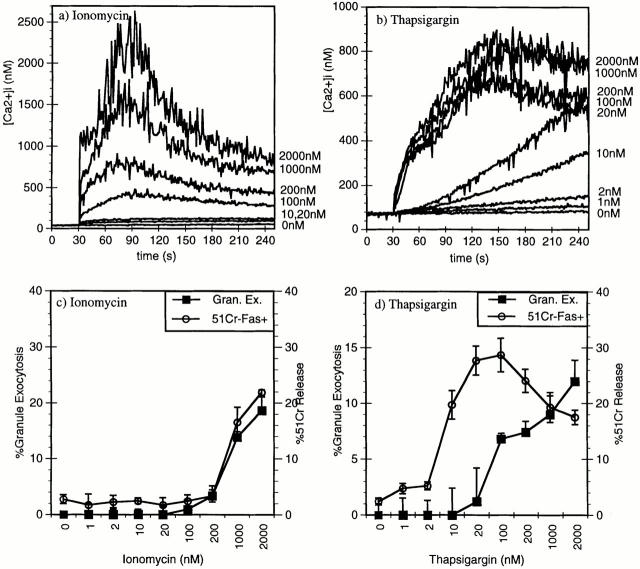
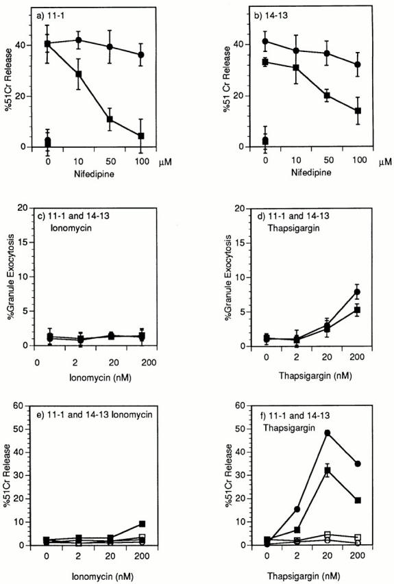
Cytolytic T cells use two mechanisms to kill virally infected cells, tumor cells, or other potentially autoreactive T cells in short-term in vitro assays. The perforin/granule exocytosis mechanism uses preformed cytolytic granules that are delivered to the target cell to induce apoptosis and eventual lysis. FasL/Fas (CD95 ligand/CD95)-mediated cytolysis requires de novo protein synthesis of FasL by the CTL and the presence of the death receptor Fas on the target cell to induce apoptosis. Using a CD8(+) CTL clone that kills via both the perforin/granule exocytosis and FasL/Fas mechanisms, and a clone that kills via the FasL/Fas mechanism only, we have examined the requirement of intra- and extracellular Ca2+ in TCR-triggered cytolytic effector function. These two clones, a panel of Ca2+ antagonists, and agonists were used to determine that a large biphasic increase in intracellular calcium concentration, characterized by release of Ca2+ from intracellular stores followed by a sustained influx of extracellular Ca2+, is required for perforin/granule exocytosis. Only the sustained influx of extracellular Ca2+ is required for FasL induction and killing. Thapsigargin, at low concentrations, induces this small but sustained increase in [Ca2+]i and selectively induces FasL/Fas-mediated cytolysis but not granule exocytosis. These results further define the role of Ca2+ in perforin and FasL/Fas killing and demonstrate that differential Ca2+ signaling can modulate T cell effector functions.


Figures

Figure 1
Figure 1
Anti-CD3ε stimulation of perforin/granule exocytosis and FasL/Fas killing. 14-7 and 14-7FD were incubated with either mock treated or peptide antigen HA529–537 (0.01 μM) pulsed target cells at an effector to target ratio of 5:1. For anti-CD3ε wells, soluble 145-2C11 (5 μg/ml) was added or the plate was coated with 5 μg/ ml 145-2C11 for 30 min at room temperature before addition of the CTL or the target. (a) 51Cr cytolysis assay with L1210Fas- target. (b) BLT-esterase assay with supernatants from a and from CTL stimulated in the absence of target cells. (c) 51Cr release assay with L1210Fas+ target.
Figure 1
Figure 1
Anti-CD3ε stimulation of perforin/granule exocytosis and FasL/Fas killing. 14-7 and 14-7FD were incubated with either mock treated or peptide antigen HA529–537 (0.01 μM) pulsed target cells at an effector to target ratio of 5:1. For anti-CD3ε wells, soluble 145-2C11 (5 μg/ml) was added or the plate was coated with 5 μg/ ml 145-2C11 for 30 min at room temperature before addition of the CTL or the target. (a) 51Cr cytolysis assay with L1210Fas- target. (b) BLT-esterase assay with supernatants from a and from CTL stimulated in the absence of target cells. (c) 51Cr release assay with L1210Fas+ target.
Figure 2
Figure 2
Anti-CD3ε induction of Ca2+ mobilization and influx in 14-7 and 14-7FD. Anti-CD3ε (145-2C11) (45 μg/ml) was used to trigger 14-7 and 14-7FD Ca2+ mobilization, and influx was monitored by indo-1 fluorescence. EGTA (5 mM) was used to examine the intra- and extracellular release components of the Ca2+ signaling pathways in 14-7 (c, e) and 14-7FD (d, f  ). EGTA was added before (c and e) or after (d and f  ) anti-CD3ε addition.
Figure 3
Figure 3
Thapsigargin (Tg) induction of intracellular Ca2+ release and Ca2+ influx in 14-7FD. 5 mM EGTA was added before (a) or after (b) stimulation of 14-7FD with 100 nM thapsigargin.
Figure 4
Figure 4
Extracellular Ca2+ is required for perforin/granule exocytosis and FasL/Fas-mediated cytotoxicity. 14-7 (a, b, d) and 14-7FD (c) were pretreated for 40 min with nifedipine, EGTA, SK&F-96365, NDGA, or Ni2+ at various concentrations before the start of a 51Cr cytolysis assay (a–c) or before anti-CD3ε stimulation (d). 14-7 was incubated with HA529–537 peptide (0.01 μM)–pulsed L1210Fas target in a 51Cr–release assay (a), and supernatants from a were used to measure granzyme A activity in a granule exocytosis assay (b). 14-7FD was incubated with HA529–537 peptide (0.01 μM)–pulsed L1210Fas+ targets in a 51Cr–release assay (c). 14-7 was stimulated with plate-bound anti-CD3ε for 6 h before staining for FasL with the Fas.Fc fusion protein by flow cytometry (d). All figures are representative of at least three separate experiments.
Figure 5
Figure 5
A large increase in [Ca2+]i is required for perforin/granule exocytosis killing. 14-7 (a) and 14-7FD (b) were stimulated with plate-bound anti-CD3ε, PMA (25 ng/ml), ionomycin (Ion; 1,000 nM), thapsigargin (Tg; 200 nM), or a combination of PMA and ionomycin, PMA and thapsigargin, or anti-CD3ε plus PMA, ionomycin, or thapsigargin, respectively, in the absence of target cells, and percentage of granule exocytosis was determined. 14-7 (c, e) and 14-7FD (d, f  ) were incubated with HA529–537 peptide (0.01 μM)–pulsed or mock-treated L1210Fas target cells. At the start of the assay PMA, ionomycin, or thapsigargin were added at the aforementioned concentrations. After 4 h, supernatants were collected and percentage of granule exocytosis (c and d) and of specific killing (e and f  ) was determined.
Figure 6
Figure 6
Thapsigargin or ionomycin induction of perforin versus FasL/Fas killing. 14-7 was stimulated with 2,000, 1,000, 200, 100, 20, or 10 nM of ionomycin (a) or thapsigargin (b) and [Ca2+]i was measured by indo-1 fluorescence. 14-7 was stimulated with PMA (25 ng/ml) plus different doses of ionomycin or thapsigargin, and granzyme A granule exocytosis was determined (c and d, ▪), or 14-7 was stimulated with ionomycin or thapsigargin in the absence of PMA and percentage of specific killing of the L1210Fas+ target was determined (c and d, ○).
Figure 7
Figure 7
Demonstration in two additional CD8+ CTL clones of nifidipene inhibition of perforin cytolysis and thapsigargin induction of cytolysis that is inhibited by anti-FasL. Clone 11-1 (a) and 14-13 (b) were pretreated for 40 min with nifedipine at various concentrations before the start of a 51Cr–release assay. The CTLs were incubated with 0.10 μM HA204–212 (a) or 0.01 μM nucleoprotein 147–155 (b) peptide–pulsed L1210Fas targets (▪) or L1210Fas+ targets (•) in a 51Cr–release assay (a and b). Clones 11-1(▪) and 14-13(•) were stimulated with PMA (25 ng/ ml) plus different doses of ionomycin (c) or thapsigargin (d) and granzyme A granule exocytosis was determined. Clones 11-1(▪) and 14-13(•) were stimulated with 200, 20, or 2 nM of ionomycin (e) or thapsigargin (f) and percentage of specific 51Cr–release of the L1210Fas+ target determined in the absence (▪, •) or presence (□, ○) of anti-FasL antibody.

References

    1. Kagi D, Vignaux F, Ledermann B, Burki K, Depraetere V, Nagata S, Hengartner H, Golstein P. Fas and perforin pathways as major mechanisms of T cell–mediated cytotoxicity. Science. 1994;265:528–530. - PubMed
    1. Zheng L, Fisher G, Miller RE, Peschon J, Lynch DH, Lenardo MJ. Induction of apoptosis in mature T cells by tumour necrosis factor. Nature. 1995;377:348–351. - PubMed
    1. Kagi D, Hengartner H. Different roles for cytotoxic T cells in the control of infections with cytopathic versus noncytopathic viruses. Curr Opin Immunol. 1996;8:472–477. - PubMed
    1. Podack ER. Execution and suicide: cytotoxic lymphocytes enforce Draconian laws through separate molecular pathways. Curr Opin Immunol. 1995;7:11–16. - PubMed
    1. Clark WR, Walsh CM, Glass AA, Huang MT, Ahmed R, Matloubian M. Cell-mediated cytotoxicity in perforin-less mice. Int Rev Immunol. 1995;13:1–14. - PubMed

Publication types

MeSH terms