. 1998 Apr;9(4):733-47.
doi: 10.1091/mbc.9.4.733.

Assembly of lampbrush chromosomes from sperm chromatin

Affiliations
Free PMC article

Assembly of lampbrush chromosomes from sperm chromatin

J G Gall et al. Mol Biol Cell. 1998 Apr.
Free PMC article

Abstract

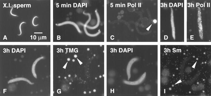
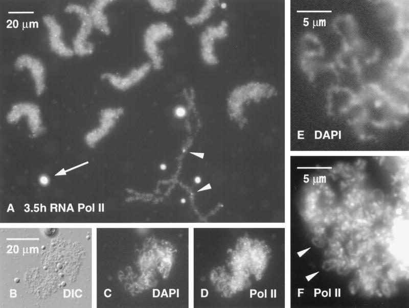
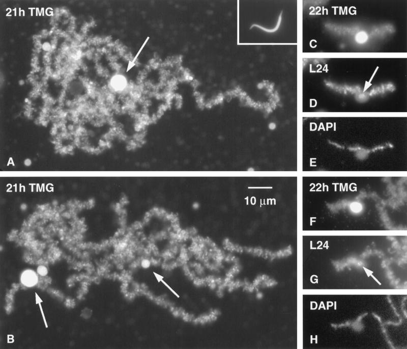
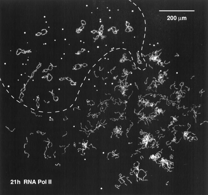
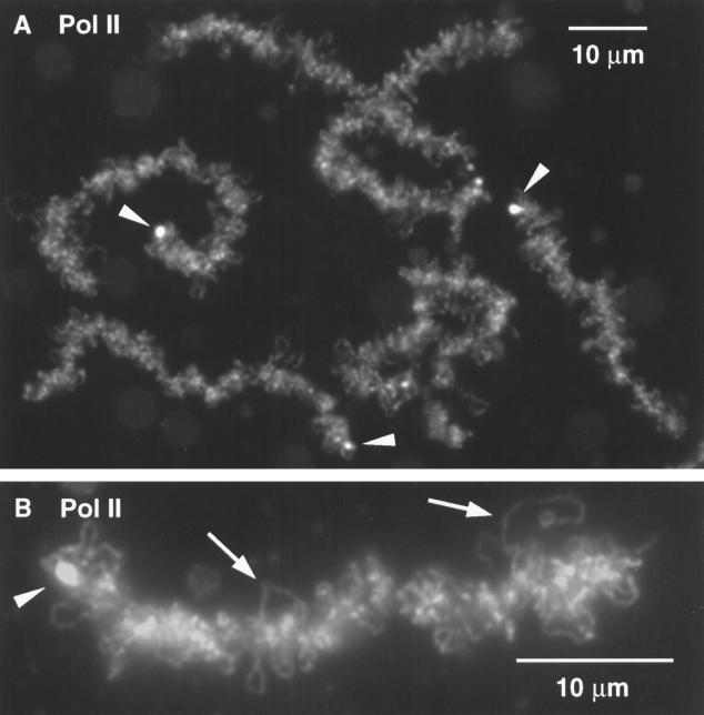
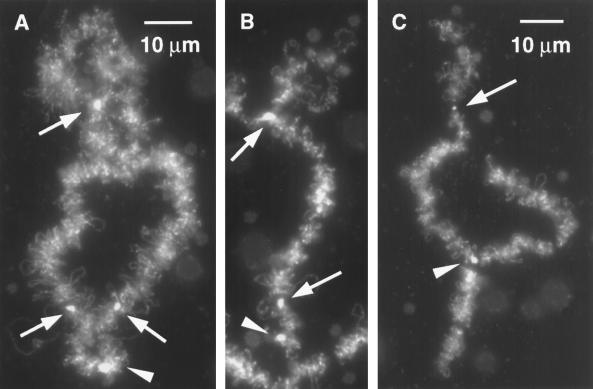
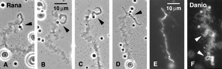
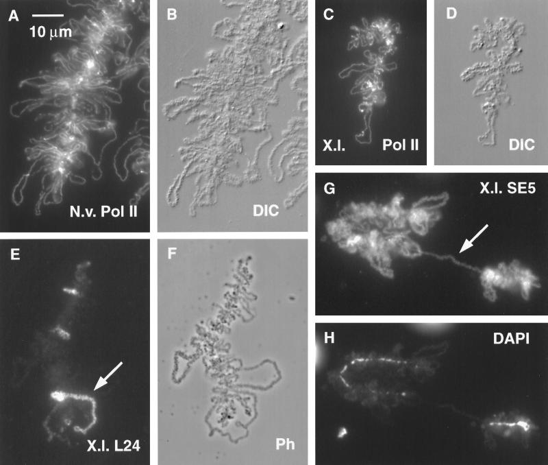
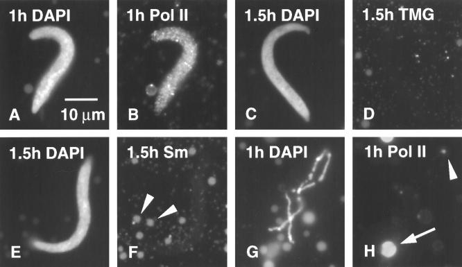
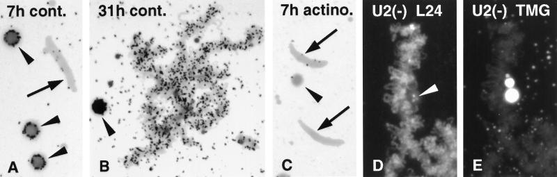
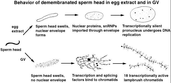
We have examined the behavior of demembranated sperm heads when injected into the germinal vesicle (GV) of amphibian oocytes. Xenopus sperm heads injected into Xenopus GVs swelled immediately and within hours began to stain with an antibody against RNA polymerase II (Pol II). Over time each sperm head became a loose mass of chromosome-like threads, which by 24-48 h resolved into individually recognizable lampbrush chromosomes (LBCs). Although LBCs derived from sperm are unreplicated single chromatids, their morphology and immunofluorescent staining properties were strikingly similar to those of the endogenous lampbrush bivalents. They displayed typical transcriptionally active loops extending from an axis of condensed chromomeres, as well as locus-specific "landmarks. " Experiments with [3H]GTP and actinomycin D demonstrated that transcription was not necessary for the initial swelling of the sperm heads and acquisition of Pol II but was required for maintenance of the lampbrush loops. Splicing was not required at any stage during formation of sperm LBCs. When Xenopus sperm heads were injected into GVs of the newt Notophthalmus, the resulting sperm LBCs displayed very long loops with pronounced Pol II axes, like those of the endogenous newt LBCs; as expected, they stained with antibodies against newt-specific proteins. Other heterologous injections, including sperm heads of the frog Rana pipiens and the zebrafish Danio rerio in Xenopus GVs, confirm that LBCs can be derived from taxonomically distant organisms. The GV system should help identify both cis- and trans-acting factors needed to convert condensed chromatin into transcriptionally active LBCs. It may also be useful in producing cytologically analyzable chromosomes from organisms whose oocytes do not go through a typical lampbrush phase or cannot be manipulated by current techniques.


Figures

Figure 1
Figure 1
(A) Demembranated sperm heads of X. laevis as injected (DAPI stain for DNA). (B) Swollen sperm heads 5 min after injection into a Xenopus GV (DAPI stain). (C) Same field stained with mAb H14 against RNA Pol II; sperm heads are negative, whereas a coiled body (sphere) stains brightly (arrowhead). (D) Sperm head 3 h after injection into a GV (DAPI stain). (E) Same sperm head shows strong staining with mAb H14 against RNA Pol II. (F) Sperm heads 3 h after injection into a GV (DAPI stain). (G) Same field stained with mAb K121 against the trimethylguanosine (TMG) cap of snRNAs. Sperm heads are negative; B-snurposomes are stained (arrowheads). (H) Sperm heads 3 h after injection into a GV (DAPI stain). (I) Same field stained with mAb Y12 against the Sm epitope of snRNPs. Sperm heads are negative; B-snurposomes are stained (arrowheads).
Figure 2
Figure 2
(A) Group of Xenopus sperm heads 3.5 h after injection into a Xenopus GV have swollen extensively and stain strongly with mAb H14 against RNA Pol II. An endogenous lampbrush chromosome is fused at its terminal granules to two other chromosomes (arrowheads). Several brightly staining coiled bodies (spheres) are also present (arrow). (B) DIC image of a single sperm head from the same GV. (C) DAPI stain reveals individual chromatids within the sperm head. (D) Staining with mAb H14 against RNA Pol II gives a strong reaction in the sperm head. (E) Higher magnification of C showing DAPI-positive chromatids. (F) Individual Pol II-positive loops (arrowheads) can be seen in this enlargement of the same region from D.
Figure 3
Figure 3
(A and B) Two Xenopus sperm heads 21 h after injection into a Xenopus GV. Each has resolved into a loose cluster of chromosomes with most of the features of the endogenous LBCs. Stained with mAb K121 against the TMG cap of the splicing snRNAs. The loops of the sperm LBCs stain strongly for snRNAs. Each cluster of chromosomes also displays one very large K121 (+) mass and several smaller masses (arrows). These almost certainly correspond to the similar masses that occur on chromosomes 6, 8, and 14. Inset in A shows sperm head at same magnification. (C) Single sperm LBC with a large K121 (+) mass attached near its center. The position of the mass and the relative size of this chromosome identify it as chromosome 14. (D) Same chromosome was double stained with serum L24 against protein xnf7 (Reddy et al., 1991). This antibody stains small inclusions in the mass (arrow) and most of the lampbrush loops, as it does on endogenous LBCs. (E) DAPI staining of the same chromosome to show the axis of condensed chromomeres. (F–H) K121, L24, and DAPI staining of another example of what is probably sperm LBC 14, attached at one end to another chromosome. Arrow in G points to L24 (+) granules inside the mass.
Figure 4
Figure 4
Overview of the contents of a single Xenopus GV 21 h after injection of Xenopus sperm heads. The sperm heads have been converted into loose clusters of chromatids or single chromatids that have the characteristic features of LBCs. This GV probably received 15–20 sperm heads. The 18 endogenous LBCs and most of the coiled bodies (spheres) occupy the area above the dashed line. This nucleus was stained with mAb H14 against RNA Pol II.
Figure 5
Figure 5
(A) Higher magnification of several sperm LBCs from the GV shown in Figure 4, stained with mAb H14 against RNA Pol II. The overall fuzzy appearance of the chromosomes is due to staining of the axes of the lateral loops. In addition, mAb H14 stains the terminal granules (arrowheads) found at the end of the long arm of 15 of the 18 Xenopus LBCs. (B) Single sperm LBC at still higher magnification, stained with mAb H14. The arrowhead points to the terminal granule, and arrows point to loop axes (DNA axes covered with Pol II).
Figure 6
Figure 6
(A) Bivalent 11 is identifiable by a terminal granule at the end of its long arm (arrowhead) and axial granules at positions 0.16 and 0.80 (arrows). In this example there is a single fused granule at position 0.80. The centromere lies immediately beyond the fused granule but is not visible with this stain. The short arm has a characteristic diffuse structure. Sample is stained with mAb H14 against RNA Pol II. (B) Chromosome 11 derived from a sperm. The terminal granule (arrowhead), two axial granules (arrows), and the diffuse short arm are recognizable. (C) Another example of chromosome 11 derived from a sperm. The axial granule at 0.16 is not evident in this image, but the chromosome is reliably identified by the axial granule at 0.80 (arrow), the diffuse short arm, and the terminal granule (arrowhead), which is fused to the terminal granules of two other chromosomes to give a triradiate figure.
Figure 7
Figure 7
(A–D) Phase-contrast images of four examples of the same sperm LBC derived from R. pipiens sperm heads injected 2 d previously into a Xenopus GV. Near one end of this chromosome is an unusually large loop with a prominent granule at its base (arrowheads). The loop is single, as expected for an unreplicated chromatid. (E) Same field as D, DAPI stain to show the prominent DNA axis. (F) Sperm LBC 2 d after injection of zebrafish D. rerio sperm heads into a Xenopus GV. Sample is stained with mAb K121 against TMG. Arrowheads point to two loops with unusually heavy matrix.
Figure 8
Figure 8
(A) Small portion of a typical LBC from a GV of the newt N. viridescens, stained with mAb H14 against RNA Pol II. (B) Same field by DIC. Compared with a Xenopus LBC, the newt LBC has longer loops, more heavily textured loop matrix, and unusually distinct Pol II (+) loop axes. In addition, newt LBCs are longer than Xenopus LBCs. (C) LBC derived from a Xenopus sperm head that had been injected 2 d previously into a newt GV. Sample was stained for Pol II. (D) Same field by DIC. Note how strongly this Xenopus LBC resembles a newt LBC except for length. (E) Xenopus sperm LBC 2 d after injection of Xenopus sperm into a newt GV. Sample was stained with serum L24 against xnf7 (Reddy et al., 1991). Only a few loops react strongly with this antibody. The large loop (arrow) near the chromosome end is single, not paired like the loops of an oocyte LBC. This loop consists of two tandem transcription units, only one of which stains. (F) Same field by phase contrast. (G) Xenopus sperm LBC 2 d after injection into a newt GV. Sample was stained with mAb SE5 (Roth and Gall, 1987), which normally stains newt but not Xenopus LBC loops. Staining demonstrates that newt proteins were used to assemble the Xenopus LBC. Arrow indicates a single-loop bridge described in the text. (H) Same field (DAPI stain) showing that the loop bridge spans a gap in the chromomere axis.
Figure 9
Figure 9
(A) Xenopus sperm head 1 h after injection into a Xenopus GV. The oocyte was incubated in actinomycin D (20 μg/ml) for 1 h before injection and was returned to the drug after injection (DAPI stain). (B) Same sperm head stained with mAb H14, showing uptake of Pol II by the sperm even though transcription was inhibited. (C) Sperm head 1.5 h after injection into an actinomycin-treated oocyte (DAPI stain). (D) Same sperm head stained with mAb K121 against TMG, showing the absence of splicing snRNAs. (E) Sperm head 1.5 h after injection into an actinomycin-treated oocyte (DAPI stain). (F) Same sperm head stained with mAb Y12, showing absence of Sm proteins. Arrowheads point to Y12 (+) B-snurposomes. (G) Highly contracted chromosome from the same GV that contained the sperm head shown in A and B (DAPI stain). (H) Same area showing absence of chromosomal stain with mAb H14, except for the terminal granule (arrowhead). A coiled body (sphere) shows typical staining (arrow).
Figure 10
Figure 10
(A) [3H]GTP was injected into the cytoplasm of a Xenopus oocyte and 1 h later Xenopus sperm heads were injected into the GV. An autoradiograph of the GV contents made 7 h later shows no label in a sperm head (arrow) but strong label in three nucleoli (arrowheads), indicative of rRNA transcription (1.5-d exposure). (B) Autoradiograph of labeled sperm LBCs 31 h after injection of [3H]GTP and sperm heads as in A. Active transcription takes place on the loops of the sperm LBCs. A nucleolus (arrowhead) is blackened by silver grains (1.5-d exposure). (C) Oocytes were preincubated for 1 h in actinomycin D (20 μg/ml), injected with [3H]GTP and sperm heads as in A, and then returned to the drug. An autoradiograph of GV contents made 7 h later shows no label in the sperm heads (arrows) or in the endogenous nucleoli (arrowhead; 1.5-d exposure). (D) Sperm LBC from an oocyte depleted for U2 snRNA and stained with serum L24 against xnf7 (Reddy et al., 1991). Loops are well stained, as are granules inside the large “mass” attached to the chromosome (arrowhead). (E) Same chromosome stained with mAb K121 against TMG. The loops show markedly reduced staining relative to untreated oocytes (compare Figure 3), but the K121 (+) masses are not affected.
Figure 11
Figure 11
Comparison of the behavior of demembranated sperm heads after incubation in egg extract (top row) or injection into the GV (bottom row).

References

    1. Almouzni G, Wolffe AP. Nuclear assembly, structure, and function: the use of Xenopus in vitro systems. Exp Cell Res. 1993;205:1–15. - PubMed
    1. Bellini M, Lacroix J-C, Gall JG. A zinc-binding domain is required for targeting the maternal nuclear protein PwA33 to lampbrush chromosome loops. J Cell Biol. 1995;131:563–570. - PMC - PubMed
    1. Birkenmeier EH, Brown DD, Jordan E. A nuclear extract of Xenopus laevis oocytes that accurately transcribes 5S RNA genes. Cell. 1978;15:1077–1086. - PubMed
    1. Bona M, Scheer U, Bautz EKF. Antibodies to RNA polymerase II (B) inhibit transcription in lampbrush chromosomes after microinjection into living amphibian oocytes. J Mol Biol. 1981;151:81–89. - PubMed
    1. Bregman DB, Du L, van der Zee S, Warren SL. Transcription-dependent redistribution of the large subunit of RNA polymerase II to discrete nuclear domains. J Cell Biol. 1995;129:287–298. - PMC - PubMed

Publication types

MeSH terms

LinkOut - more resources