. 1998 Apr;9(4):759-74.
doi: 10.1091/mbc.9.4.759.

New alleles of the yeast MPS1 gene reveal multiple requirements in spindle pole body duplication

Affiliations
Free PMC article

New alleles of the yeast MPS1 gene reveal multiple requirements in spindle pole body duplication

A R Schutz et al. Mol Biol Cell. 1998 Apr.
Free PMC article

Abstract

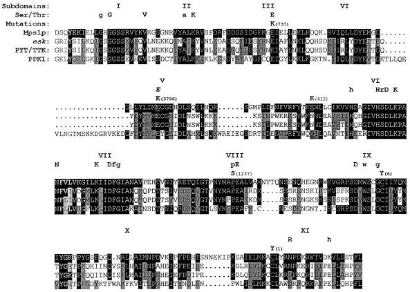
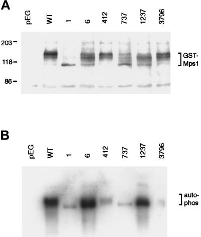
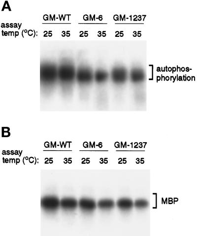
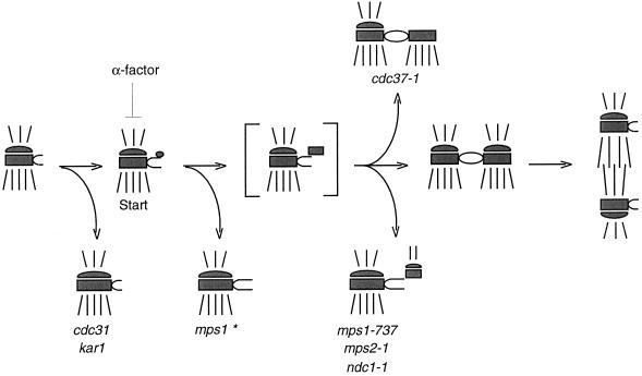
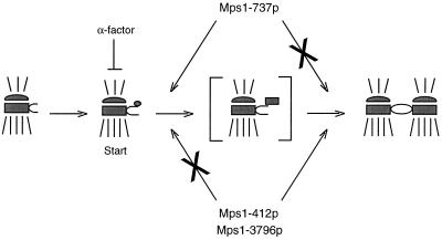
In Saccharomyces cerevisiae, the Mps1p protein kinase is critical for both spindle pole body (SPB) duplication and the mitotic spindle assembly checkpoint. The mps1-1 mutation causes failure early in SPB duplication, and because the spindle assembly checkpoint is also compromised, mps1-1 cells proceed with a monopolar mitosis and rapidly lose viability. Here we report the genetic and molecular characterization of mps1-1 and five new temperature-sensitive alleles of MPS1. Each of the six alleles contains a single point mutation in the region of the gene encoding the protein kinase domain. The mutations affect several residues conserved among protein kinases, most notably the invariant glutamate in subdomain III. In vivo and in vitro kinase activity of the six epitope-tagged mutant proteins varies widely. Only two display appreciable in vitro activity, and interestingly, this activity is not thermolabile under the assay conditions used. While five of the six alleles cause SPB duplication to fail early, yielding cells with a single SPB, mps1-737 cells proceed into SPB duplication and assemble a second SPB that is structurally defective. This phenotype, together with the observation of intragenic complementation between this unique allele and two others, suggests that Mps1p is required for multiple events in SPB duplication.


Figures

Figure 1
Figure 1
Strategy for identification of mutant lesions. The region of MPS1 that encodes the protein kinase consensus domain lies near the C terminus of the gene. Gapped plasmid repair data indicate that all mutations occur within this 800-bp region bounded by the BamHI and MroI restriction sites. The positions of unique BamHI, KpnI, and MroI restriction sites are shown. Arrows indicate the position and orientation of the oligonucleotide primers used for genomic PCR and for DNA sequence analysis (see MATERIALS AND METHODS). The location of each mutant lesion is marked with an X.
Figure 2
Figure 2
The six ts alleles each contain a single mutation in the kinase domain. The amino acid sequence of the Mps1p protein kinase domain is shown. Above the sequence, the 11 subdomains of the kinase domain and the residues conserved among the family of serine-threonine kinases are listed (Ser/Thr; see Hanks et al., 1988). Uppercase letters indicate highly conserved residues, and lowercase letters indicate less well conserved ones. The italicized E in subdomain V denotes a residue conserved in some dual-specificity kinases (Lindberg et al., 1992). The amino acid changes caused by each mps1 mutation and its corresponding allele number are noted above the Mps1p sequence, and the altered residue in Mps1p is underlined. Below the Mps1p sequence are those for the kinase domains of esk, PYT/TTK, and PPK1 (Douville et al., 1992; Lindberg et al., 1992; Kwart, personal communication). Identity between Mps1p and the other kinases is indicated by black boxes, and similarity is indicated by gray boxes. A homolog of MPS1 has also been found in Schizosaccharomyces pombe (He and Sazer, personal communication) but is not shown here. The sequences given here begin at the following amino acid numbers: Mps1p, position 437; esk, position 521; PYT/TTK, position 506; PPK1, position 144.
Figure 3
Figure 3
The mps1 mutant strains undergo aberrant mitosis, producing cells with increased ploidy. (A) mps1–6 cells (AS181–2b; Table 1) were exposed to the nonpermissive temperature and sampled for flow cytometry at 1-h intervals (2 h-4 h samples are shown). For comparison, a histogram for cells grown at the permissive temperature is shown (0 h). (B) The same experiment was performed with mps1–3796 cells (AS131–2d). Similar results were obtained for the remaining four alleles (our unpublished results). In these histograms, the x-axis is the relative DNA content determined by propidium iodide fluorescence, and the y-axis is the relative number of cells (see MATERIALS AND METHODS). The peaks corresponding to normal G1 and G2-M DNA content are indicated on the x-axis. Each sample represents 5,000 cells.
Figure 4
Figure 4
Most mps1 alleles share the mps1–1 SPB phenotype. (A) In mps1–1 cells, a single SPB with very prominent half-bridge (arrow) is seen at the nonpermissive temperature (Winey et al., 1991). (B and C) Strains with the mps1–6 (B; AS181–2b) or mps1–3796 (C; AS131–2d) mutations were shifted to the nonpermissive temperature for 4 h, and their SPB phenotypes were examined by electron microscopy. Both strains exhibited the same SPB morphology seen in panel A: a single enlarged SPB with prominent half-bridge (arrows). This was also observed in mps1–412 and mps1–1237 strains (our unpublished results). Bar, 0.1 μm.
Figure 5
Figure 5
Cells carrying the mps1–737 allele display an unusual SPB morphology. The mps1–737 strains AS132–3a or AS132–8c (Table 1) were exposed to the nonpermissive temperature for 3–4 h and examined by electron microscopy. In images A through D, the entire structure is seen in one thin section. The pairs of images in E and F represent two sections from the same nucleus, where relevant structures are seen in more than one section. A second SPB was found in four of 26 nuclei examined. (A and B) In most cells, a single SPB with a shorter and much less prominent half-bridge was found. (C and D) In each image, one complete SPB with enlarged half-bridge is present, and a second, structurally incomplete SPB (arrow) is perched at the end of the half-bridge on its cytoplasmic face. The second SPB has clear central and outer plaques. This morphology was observed in three cells. (E and E′) These images are two adjacent serial sections through the same nucleus. The intact SPB is seen in E, and the adjacent section (E′) reveals an aberrant SPB next to it (arrow). (F and F′) One normal SPB, which lies on an invagination of the nuclear envelope, is seen in F. In F′, the defective SPB is seen in another section from the same nucleus. This SPB has migrated away from its sibling to a distal location on the outer surface of the nuclear envelope, and microtubules can be seen emanating from its cytoplasmic face (arrowhead). This phenotype was observed once. Bar, 0.1 μm.
Figure 6
Figure 6
mps1–737 cells contain two dots of Spc42p-GFP fluorescence, indicating that two SPBs are present. Strains that carry various mps1 alleles and the GFP-tagged allele of SPC42 were shifted to 37°C for 4 h, briefly fixed, and stained with DAPI to visualize DNA. DAPI and GFP fluorescence were then observed. Only large budded cells with evidence of DNA missegregation were scored. The number of dots of GFP fluorescence in a given cell reflects the number of SPBs. (A) At 25°C, large-budded cells display normal DNA segregation patterns (DAPI), and one SPB dot (Spc42p-GFP) localizes with each region of DAPI staining. Shown here are mps1–3796 and mps1–737 strains (AS235-GFP and AS234-GFP, respectively; Table 1). (B) In the mps1–3796 strain at 37°C, aberrant DNA segregation becomes apparent in large-budded cells. Only one SPB dot can be found in most cells, and it is often located close to the bud neck. Similar results were observed for all alleles except mps1–737. (C) When shifted to 37°C, mps1–737 cells behave differently. DNA segregation patterns become aberrant, but most cells clearly contain two distinct SPB dots. Both dots are generally found in the same cell body, and one is often located in a region that does not overlap with DAPI staining (arrow). (D) Number of SPB dots observed in all six mutant backgrounds.
Figure 7
Figure 7
Autophosphorylation by GST-tagged mutant proteins varies greatly. Plasmids carrying the GST-tagged mps1 alleles were transformed into the wild-type strain FLY14A (Table 1), and expression of fusion proteins was induced as described (see MATERIALS AND METHODS). (A) GST fusions were detected on Western blots of whole cell lysates with anti-GST antibody. The control lane (pEG) shows cells carrying the GST vector without insert. The tagged wild-type protein (GM-WT) is phosphorylated, causing it to migrate above its predicted molecular weight of 112 kDa (Lauzéet al., 1995). (B) GST fusions were affinity-purified with glutathione-Sepharose and used for in vitro kinase assays at 25°C. Material was then separated on an SDS-PAGE gel and blotted. Proteins loadings are approximately equal across the gel, as determined by Western blot (our unpublished results).
Figure 8
Figure 8
In vitro activity of the GM-6 and GM-1237 mutant proteins is not thermolabile. Affinity-purified GM-WT, GM-6, and GM-1237 kinases were assayed in vitro at 25°C and 35°C, and then separated on an SDS-PAGE gel and blotted. This elevated assay temperature would be nonpermissive for growth of both mps1–6 and mps1–1237 mutant strains. (A) Autophosphorylation activity of the proteins at both assay temperatures. (B) Substrate-level phosphorylation of myelin basic protein (MBP). For each pair of samples (25°C and 35°C), protein loadings were approximately equal (determined by Western blot).
Figure 9
Figure 9
Proposed pathway for SPB duplication. In this schematic, the central and outer plaques of the SPB are depicted, with cytoplasmic and nuclear microtubules drawn above and below the SPB, respectively. The bracketed structure is a duplication intermediate that is inferred but has not been reported. Execution point experiments with mps1–1, mps2–1, and ndc1–1 mutants (Winey et al., 1991, 1993) indicate where in the pathway these mutations cause SPB duplication to fail. The mps1–1 mutation causes failure early on and is marked with an asterisk to indicate that four of the other five alleles share this SPB morphology. However, the mps1–737 mutation allows duplication to proceed farther before failure occurs and has been placed here at the same point in the pathway as mps2–1 and ndc1–1 because of their phenotypic similarity. Mutation of CDC37 also causes a later failure in SPB duplication, but generates a different, partially duplicated structure (Schutz et al., 1997). The two distinct mps1 mutant phenotypes suggest that this gene is required continuously or at multiple times during SPB duplication.
Figure 10
Figure 10
A model for intragenic complementation between mps1 alleles. The intragenic complementation observed between mps1–737 and either the mps1–412 or mps1–3796 alleles could be a result of multiple requirements for Mps1p during SPB duplication. The mps1–737 gene product, Mps1–737p, may be competent to meet the early requirement reported previously (Winey et al., 1991) but apparently cannot perform an important later function. Mps1–412p and Mps1–3796p clearly fail to meet the early requirement, but it is possible that they are competent to perform the later function that is lost in mps1–737 cells. If so, the combination of two partially functional kinases in mps1–737/mps1–412 or mps1–737/mps1–3796 diploid cells could meet both requirements, allowing the cell to grow at a higher temperature. According to this model, the three remaining mps1 alleles would be defective for both functions and thus would not display intragenic complementation. All six mps1 alleles are defective for the spindle assembly checkpoint function (Weiss and Winey, 1996).

References

    1. Anderson CW, Baum PR, Gesteland RF. Processing of adenovirus-2 induced proteins. J Virol. 1973;12:241–252. - PMC - PubMed
    1. Ausubel FM, Brent R, Kingston RE, Moore DD, Seidman JG, Smith JA, Struhl K. Current Protocols in Molecular Biology. New York: John Wiley & Sons; 1994.
    1. Byers B, Goetsch L. Duplication of spindle plaques and integration of the yeast cell cycle. Cold Spring Harbor Symp Quant Biol. 1974;38:123–131. - PubMed
    1. Byers B, Goetsch L. Behavior of the spindles and spindle plaques in the cell cycle and conjugation of Saccharomyces cerevisiae. J Bacteriol. 1975;124:511–523. - PMC - PubMed
    1. Carr AM, MacNeill SA, Hayles J, Nurse P. Molecular cloning and sequence analysis of mutant alleles of the fission yeast cdc2+ protein kinase gene: implications for cdc2 protein structure and function. Mol Gen Genetics. 1989;218:41–49. - PubMed

Publication types

MeSH terms

LinkOut - more resources