. 1998 Apr;9(4):865-74.
doi: 10.1091/mbc.9.4.865.

The thrombospondin receptor CD47 (IAP) modulates and associates with alpha2 beta1 integrin in vascular smooth muscle cells

Affiliations
Free PMC article

The thrombospondin receptor CD47 (IAP) modulates and associates with alpha2 beta1 integrin in vascular smooth muscle cells

X Q Wang et al. Mol Biol Cell. 1998 Apr.
Free PMC article

Abstract

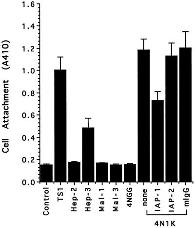
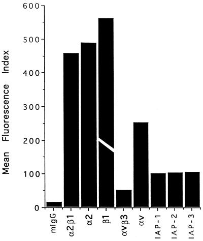
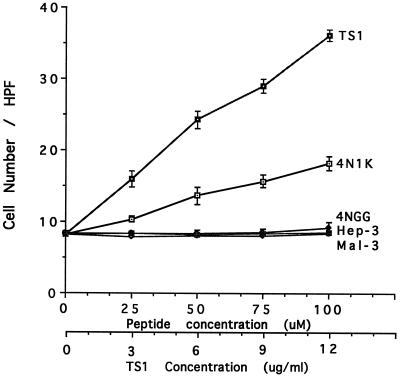
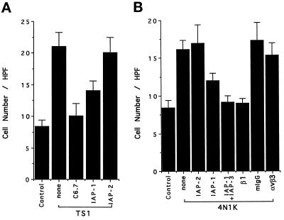
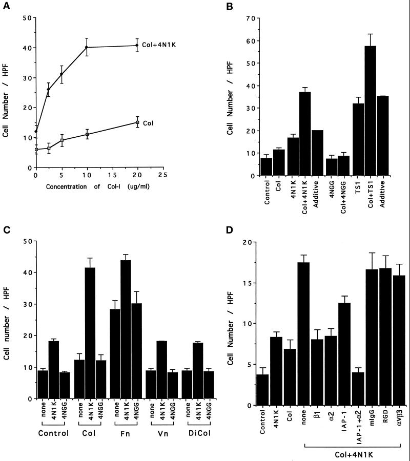
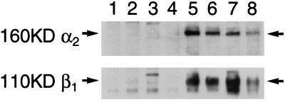
The carboxyl-terminal domain of thrombospondin-1 enhances the migration and proliferation of smooth muscle cells. Integrin-associated protein (IAP or CD47) is a receptor for the thrombospondin-1 carboxyl-terminal cell-binding domain and binds the agonist peptide 4N1K (kRFYVVMWKk) from this domain. 4N1K peptide stimulates chemotaxis of both human and rat aortic smooth muscle cells on gelatin-coated filters. The migration on gelatin is specifically blocked by monoclonal antibodies against IAP and a beta1 integrin, rather than alphav beta3 as found previously for 4N1K-stimulated chemotaxis of endothelial cells on gelatin. Both human and rat smooth muscle cells displayed a weak migratory response to soluble type I collagen; however, the presence of 4N1K peptide or intact thrombospondin-1 provoked a synergistic chemotactic response that was partially blocked by antibodies to alpha2 and beta1 integrin subunits and to IAP. A combination of antialpha2 and IAP monoclonal antibodies completely blocked chemotaxis. RGD peptide and antialphav beta3 mAb were without effect. 4N1K and thrombospondin-1 did not augment the chemotactic response of smooth muscle cells to fibronectin, vitronectin, or collagenase-digested type I collagen. Complex formation between alpha2 beta1 and IAP was detected by the coimmunoprecipitation of both alpha2 and beta1 integrin subunits with IAP. These data suggest that IAP can associate with alpha2 beta1 integrin and modulate its function.


Figures

Figure 1
Figure 1
Direct attachment of human smooth muscle cells to thrombospondin-1 and peptides from different domains of thrombospondin-1. Microtiter plates were coated with 50 μg/ml thrombospondin-1 or 50 μM peptides at 4°C overnight. Cells were suspended in 0.1% BSA in TBS or pretreated with 100 μg/ml antibodies in the same solution for 15 min at 37°C before being added to the wells. After 2 h at 37°C, the attached cells were quantified by the absorbance at 410 nm due to endogenous cellular phosphatase hydrolysis of p-nitrophenyl phosphate.
Figure 2
Figure 2
Expression of IAP and integrins on human smooth muscle cells. The bars represent the mean fluorescence index observed for each of the antibodies indicated. Mouse IgG was used as a control. IAP-1 is 1F7; IAP-2 is 2D3; IAP-3 is B6H12. The experiment was repeated twice. The actual value of the mean fluorescence index for the anti-β1 mAb P4C10 was 1298. The bar is truncated in the figure to emphasize differences among the values for the other mAbs.
Figure 3
Figure 3
Thrombospondin-1 and its peptides stimulate human smooth muscle cell chemotaxis on gelatin-coated filters. The indicated concentrations of thrombospondin-1, 4N1K, 4NGG, Hep-3, and Mal-3 peptides were added to the lower compartments of the Boyden chamber. Data are mean ± SE of the number of migrated cells counted per high-power field determined over five fields for each of triplicate wells.
Figure 4
Figure 4
(A) Human smooth muscle cell migration to thrombospondin-1 is inhibited by anti-IAP and thrombospondin-1 antibodies. Smooth muscle cells were preincubated with 100 μg/ml C6.7 or 1F7, 2D3 at 37°C for 15 min before being added to the upper compartment of the Boyden chamber. 4N1K (100 μM) was present in the lower compartment, and TBS was used as control. (B) Human smooth muscle cell migration to 4N1K is inhibited by antibodies against IAP and β1 integrin. Smooth muscle cells were preincubated in the presence or absence of β1 mAb (1:1500) or other mAbs as indicated (100 μg/ml) at 37°C for 15 min before being added to the upper compartment of the Boyden chamber.
Figure 5
Figure 5
(A) The synergistic effect of 4N1K and soluble collagen-I on rat smooth muscle cell chemotaxis. 4N1K (100 μM) and increasing concentrations of soluble collagen-I were used as chemoattractant. (B) Both 4N1K and thrombospondin-1 are synergistic with collagen-I. Collagen-I was present at 5 μg/ml along with 100 μM 4N1K or 4NGG, or 6 μg/ml thrombospondin-1 as chemoattractants. The experimentally observed values for the concerted effect of 4N1K, thrombospondin-1, and collagen (4N1K±Col, TS1±Col) were significantly greater than the calculated additive effect (additive). (C) Comparison of the effect of soluble collagen-I (Col), fibronectin (Fn), vitronectin (Vn), and digested collagen-I (DiCol) on 4N1K- induced rat smooth muscle cell chemotaxis. Collagen-I (5 μg/ml) was digested with 0.4 U/ml collagenase type I at 37°C overnight. 4N1K or 4NGG (100 μM) and 5 μg/ml of the different matrix proteins in MEM with 0.1% BSA were added to the lower compartment of Boyden chamber. (D) The effect of mAbs on collagen- and 4N1K- induced human smooth muscle cell chemotaxis. Smooth muscle cells were preincubated with mAbs at 37°C for 15 min before being added to the upper compartment of the Boyden chamber. The concentrations of mAbs were: anti-β1 mAb P4C10 (1:1500 dilution), anti-α2 mAb P1E6 (1:3000 dilution), anti-IAP-1 mAb 1F7 100 μg/ml, mIgG 100 μg/ml, RGD peptide 50 μM, anti-αvβ3 mAb 4C1 100 μg/ml.
Figure 6
Figure 6
Association of IAP and α2β1 integrin. Human smooth muscle cells were harvested by trypsin/EDTA and lysed in 30 mM n-octy-β-d-glucopyranoside as described in MATERIALS AND METHODS. Soluble material from equal numbers of cells was immunoprecipitated with the following mAbs: mouse IgG from Sigma (lane 1), anti-HLA (lane 2), anti-TNF receptor (lane 3), mouse IgG from Pierce (lane 4), anti-α2β1 BHA2.1 (lane 5), anti-IAP 2D3 and 1F7 (lanes 6 and 7). Lane 8 is a total cell lysate. α2 and β1 subunits were identified by Western blotting with polyclonal antibodies and ran at the expected sizes of 160 and 110 kDa, as indicated by the arrows.

Similar articles

Cited by

References

    1. Abedi H, Dawes KE, Zachary I. Differential effects of platelet-derived growth factor BB on p125 focal adhesion kinase and paxillin tyrosine phosphorylation and on cell migration in rabbit aortic vascular smooth muscle cells and Swiss 3T3 fibroblasts. J Biol Chem. 1995;270:11367–11376. - PubMed
    1. Adams JC, Tucker RP, Lawler J. The Thrombospondin Gene Family. Austin, TX: R.G. Landes Company; 1995.
    1. Asch AS, Barnwell J, Silverstein RL, Nachman RL. Isolation of the thrombospondin-1 membrane receptor. J Clin Invest. 1987;79:1054–1061. - PMC - PubMed
    1. Asch AS, Silbiger S, Heimer E, Nachman RL. Thrombospondin sequence motif (CSVTCG) is responsible for CD 36 binding. Biochem Biophys Res Commun. 1992;182:1208–1217. - PubMed
    1. Asch AS. The role of CD 36 as thrombospondin-1 receptor. In: Lahav J, editor. Thrombospondin. Boca Raton, FL: CRC Press; 1993. pp. 265–275.

Publication types

MeSH terms

LinkOut - more resources