. 1998 Apr;9(4):931-43.
doi: 10.1091/mbc.9.4.931.

Homology-dependent gene silencing in Paramecium

Affiliations
Free PMC article

Homology-dependent gene silencing in Paramecium

F Ruiz et al. Mol Biol Cell. 1998 Apr.
Free PMC article

Abstract

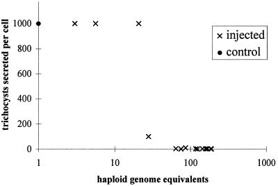
Microinjection at high copy number of plasmids containing only the coding region of a gene into the Paramecium somatic macronucleus led to a marked reduction in the expression of the corresponding endogenous gene(s). The silencing effect, which is stably maintained throughout vegetative growth, has been observed for all Paramecium genes examined so far: a single-copy gene (ND7), as well as members of multigene families (centrin genes and trichocyst matrix protein genes) in which all closely related paralogous genes appeared to be affected. This phenomenon may be related to posttranscriptional gene silencing in transgenic plants and quelling in Neurospora and allows the efficient creation of specific mutant phenotypes thus providing a potentially powerful tool to study gene function in Paramecium. For the two multigene families that encode proteins that coassemble to build up complex subcellular structures the analysis presented herein provides the first experimental evidence that the members of these gene families are not functionally redundant.


Figures

Figure 1
Figure 1
Maps of Paramecium gene sequences used in microinjection experiments. All sequences were cloned in plasmid vectors as described in MATERIALS AND METHODS. Open boxes, coding sequences; solid boxes, flanking or intron sequences; shaded boxes, coding sequences not included in the construct. Regions used as probes for hybridization are represented as a solid line; it is important to note that the ICL1b, T1b, and T4a probes are subfamily specific: they hybridize, respectively, with all members of the ICL1, T1, and T4 gene subfamilies on Southern blots (Madeddu et al., 1995, 1996). The pND7+ plasmid insert is derived from p201ND7 previously described by Skouri and Cohen (1997).
Figure 2
Figure 2
Maintenance of microinjected plasmid DNA. Total DNA was extracted from control uninjected nd7 cells (lane a), cells of a vegetative clone derived from a cell microinjected with pICL1b (lanes b and d), and cells of a postautogamous line derived from the pICL1b transformed clone (lane c). Undigested DNA was fractionated (4–8 μg/lane) on a 1% agarose gel by contour-clamped homogeneous electric field (CHEF) electrophoresis (9 V/cm; switch time, 0.1 s; angle, 120°; run time, 3 h), using a CHEF-DR III apparatus (Bio-Rad, Richmond, CA), and transferred to nitrocellulose. The same filter was sequentially hybridized with the ICL1 ( lanes a–c) and the pUC18 (lane d) specific probes. The 50- to 800-kb Paramecium macronuclear chromosomes are not resolved in these experimental conditions: the weak signals detected in control and postautogamous cells most likely arise from hybridization with the endogenous ICL1 genes.
Figure 3
Figure 3
Phenotypic characterization of cells transformed with ICL1 coding sequences. (a) Immunolabeling of an uninjected nd7 cell by the anti-centrin antibody 20H5. The ICL is formed of irregular polygonal meshes of various sizes; the regionalized differences in their arrangement reflect the polarized and asymmetrical organization of the Paramecium cortex. (b and c) Immunofluorescence images showing clonal descendants of pICL1b-microinjected cells, with total or partial disorganization of the ICL network. pICL1a-transformed cells presented similar phenotypes. Magnification, 700×. (d) Western blot of total cellular proteins from cells of three pICL1a- and three pICL1b-transformed clones, compared with uninjected (nd7) cells. The same blot was treated with the 20H5 antibody (bottom) and with an antibody directed against β-tubulin (top) as a control for the amount of total protein in each sample.
Figure 4
Figure 4
Northern blot analysis of pICL1b transformants. Total cellular RNA was prepared from uninjected nd7 cells (lane 1) and from two clones of pICL1b-transformed cells: clone 3a (lane 2) and clone 4a (lane 3). The same filter was hybridized sequentially with the ICL1b probe (a) and the T1b probe (b). The position of ICL1 mRNA (∼0.85 kb) is indicated. T1 mRNA migrates at ∼1.4 kb (Madeddu et al., 1995).
Figure 5
Figure 5
Phenotypic characterization of cells transformed with T1 and T4 coding sequences. Immunofluorescent images of trichocysts of a pT1b-transformed cell beneath an uninjected wild-type cell (a) and pT4a-transformed cells (20–25 divisions after microinjection) (b). Both pT1b- and pT4a-transformed cells presented undocked aberrantly shaped trichocysts, which tended to swell more readily than wild-type trichocysts under the fixation conditions used. Magnification, 500×.
Figure 6
Figure 6
Northern blot analysis of pT1b and pT4a transformants. (a) Total RNA was prepared from tam8 and wild-type cells (left), three clones of pT1b-transformed cells (middle), and two clones of pT4a-transformed cells (right). The blots were sequentially hybridized with the T1-specific probe (top) and with the T4-specific probe (bottom). Asterisks denote aberrantly migrating species that hybridized with a pUC18 vector-specific probe. (b) Histogram of the steady-state levels of T1 and T4 mRNAs in the various clones was established by quantification of the Northern blots shown in a, using hybridization to an ICL1-specific probe to normalize for the amount of total RNA in each sample.
Figure 7
Figure 7
Evaluation of exocytotic activity of cells transformed with ND7 sequences. The dark-field image shows cells treated with picric acid. Clonal descendants of cells injected with pND7−, which do not secrete any trichocysts (exo−), are shown above an nd7 cell whose exocytotic capacity has been restored (exo+) by transformation with a functional ND7 gene (pND7+). The latter, which is indistinguishable from uninjected wild-type cells, is surrounded by a dense halo of trichocysts. Magnification, ×200.
Figure 8
Figure 8
Evaluation of plasmid copy number in cells transformed with ND7 sequences. The number of clones as a function of copy number (in haploid genome equivalents) of pND7+ maintained after microinjection of either wild-type or nd7 mutant cells is given. The phenotypes of the clones are also indicated (white background, fewer than 10 trichocysts secreted per cell; lightly shaded background, approximately 100 trichocysts; darkly shaded background, indistinguishable from wild-type control cells; hatched background, no clones encountered). These data are compared with the number of copies of pND7− maintained in clones of wild-type cells. The pND7− data are the same as in Figure 9. In all cases, copy number was determined by a dot blot 10 fissions after injection.
Figure 9
Figure 9
Quantitative analysis of plasmid-induced silencing. The average number of trichocysts secreted per cell is plotted as a function of the copy number of pND7− in the macronucleus. Exocytotic capacity was determined by treatment of 10–20 cells with picric acid. Although it is possible to count up to 50 secreted trichocysts per cell, higher values represent an appreciation of the density of the halo, compared with that of a wild-type cell assumed to secrete all of its ∼1000 docked trichocysts. The copy number of plasmids, determined by a dot blot 10 fissions after injection, is expressed as the number of copies per haploid genome. ×, Each of the 15 transformed clones; •, clone of uninjected control cells.

References

    1. Adoutte A, Garreau de Loubresse N, Beisson J. Proteolytic cleavage and maturation of the crystalline secretion products of Paramecium. J Mol Biol. 1984;180:1065–1080. - PubMed
    1. Adoutte A. Exocytosis: biogenesis, transport and secretion of trichocysts. In: Görz H-D, editor. Paramecium. Heidelberg: Springer-Verlag; 1988. pp. 325–362.
    1. Baulcombe DC. RNA as a target and an initiator of post-transcriptional gene silencing in transgenic plants. Plant Mol Biol. 1996;32:79–88. - PubMed
    1. Beisson J, Rossignol M. Molecular Biology of Nucleocytoplasmic Relationships. The Netherlands: Elsevier; 1975. Movements and positioning of organelles in Paramecium aurelia; pp. 291–294.
    1. Bingham PM. Cosuppression comes to animals. Cell. 1997;90:385–387. - PubMed

Publication types

LinkOut - more resources