. 1998 Apr;9(4):945-56.
doi: 10.1091/mbc.9.4.945.

Swi5 controls a novel wave of cyclin synthesis in late mitosis

Affiliations
Free PMC article

Swi5 controls a novel wave of cyclin synthesis in late mitosis

B L Aerne et al. Mol Biol Cell. 1998 Apr.
Free PMC article

Abstract

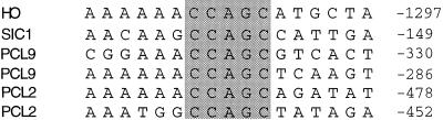
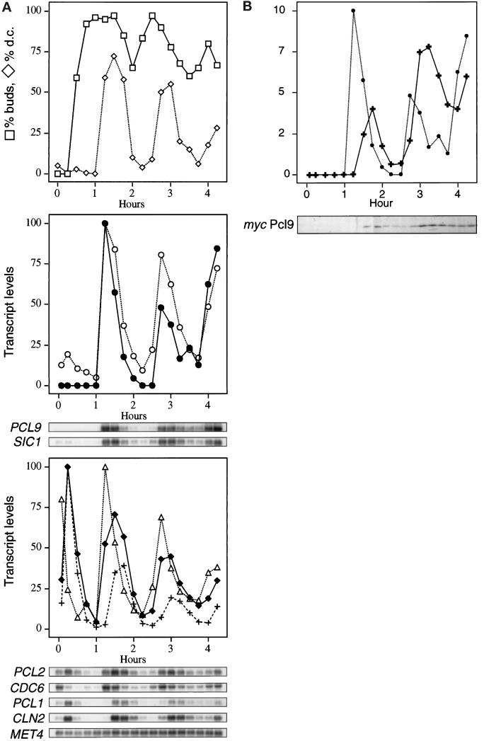
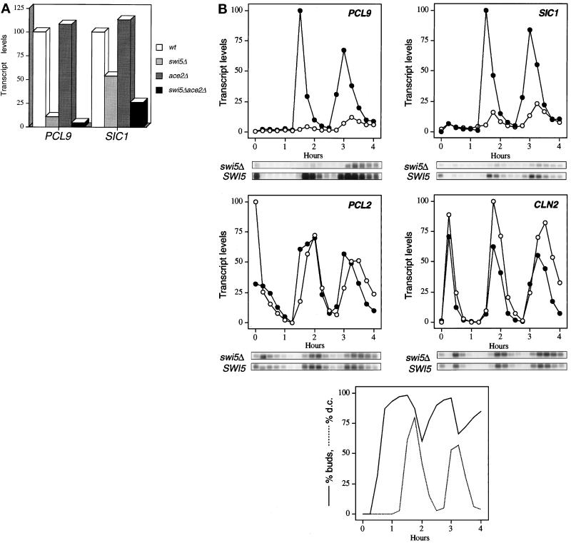
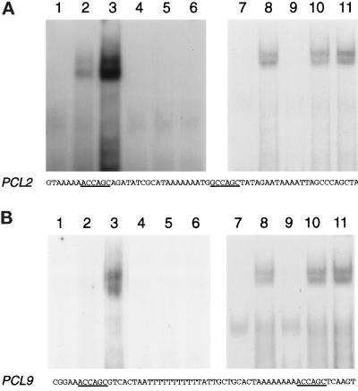
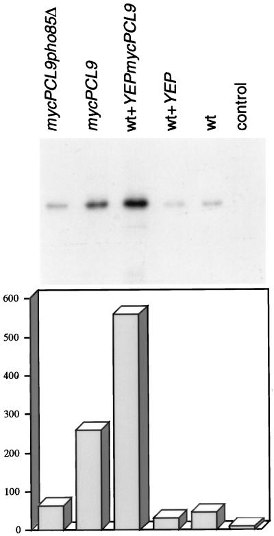
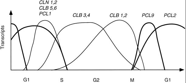
We have shown previously that the Swi5 transcription factor regulates the expression of the SIC1 Cdk inhibitor in late mitosis. This suggests that Swi5 might control other genes with roles in ending mitosis. We identified a gene with a Swi5-binding site in the promoter that encoded a protein with high homology to Pcl2, a cyclin-like protein that associates with the Cdk Pho85. This gene, PCL9, is indeed regulated by Swi5 in late M phase, the only cyclin known to be expressed at this point in the cell cycle. The Pcl9 protein is associated with a Pho85-dependent protein kinase activity, and the protein is unstable with peak levels occurring in late M phase. PCL2 is already known to be expressed in late G1 and we find that, in addition, it is also regulated by Swi5 in telophase. The expression of PCL2 and PCL9 at this stage of the cell cycle implies a role for the Pho85 Cdk at the end of mitosis. Consistent with this a synthetic interaction was observed between pho85delta and strains deleted for SIC1, SWI5, and SPO12. These and other studies support the notion that the M/G1 switch is a major cell cycle transition.


Figures

Figure 1
Figure 1
The PCL9 and PCL2 promoter contain putative Swi5 binding sites. Comparison of possible Swi5 binding sites found in the PCL9 and PCL2 promoter with those found in the promoters of SIC1 and HO. The conserved CCAGC motif is indicated.
Figure 2
Figure 2
Cell cycle regulation of PCL2 and PCL9 transcript levels and Pcl9 protein level. A wild-type strain containing a 3 myc epitope-tagged version of the PCL9 gene was arrested using α-factor and reinoculated into fresh medium. Samples were taken 5 min and 15 min after the release from the α-factor holding, and then at 15-min intervals. Samples were analyzed for (A) the percentage of budded cells and cells with divided chromatin; transcript levels of PCL9, SIC1, PCL2, CDC6, PCL1, CLN2, and MET4. Quantitation of transcript levels are relative to MET4; PCL9 (solid circles), SIC1 (open circles), PCL2 (solid diamonds), PCL1 (crosses), CDC6 (triangles). (B) mycPcl9 protein level. Solid circles, PCL9 transcript levels; crosses, Pcl9 protein levels.
Figure 3
Figure 3
A. SWI5 is required for PCL9 transcription. Densitometric quantitation of a Northern blot analysis using mRNA from exponentially growing wild-type (DTY7), swi5Δ (DTY87), ace2Δ (DTY91), and swi5Δace2Δ (DTY92) strains. Blots were hybridized with probes from PCL2 and SIC1 and normalized to ACT1 as a loading control. (B) Effect of deletion of SWI5 on the cell cycle regulation of PCL2 and PCL9 transcription. Cultures of swi5Δ strains containing either the plasmid YIpSWI5 or control plasmid YIplac211 were synchronized using α-factor, and samples were taken for Northern blot analysis (Toyn et al., 1997). The Northern blot was quantitated by densitometry and normalized to the level of actin mRNA detected on the same blot. The first sample on the autoradiograph is from log-phase cells before addition of α-factor and is not shown in the quantitations. Quantitations of transcript levels from the SWI5 (solid circles) and the swi5Δ experiment (open circles) are compared. The data points relating to the percentage buds and divided chromatin were described by Toyn et al. (1997).
Figure 4
Figure 4
PCL2 is expressed in late mitosis. The late mitotic cell cycle mutant dbf2 was grown at 25°C, sampled, and transferred to 37°C, and further samples were taken at 40-min intervals for 2 h. For the nocodazole-treated culture, the drug was added to midlog cell of strain CG378 to a final concentration of 25 μg/ml, sampled immediately, and then after 45-min intervals for 3 h. Samples were analyzed for PCL2, CDC6, and CLN2 transcript levels by Northern blot analysis. The Northern blot was quantitated by densitometry and normalized to the level of actin mRNA detected on the same blot. PCL2 (square), CDC6 (diamonds), CLN2 (circles).
Figure 5
Figure 5
Swi5 binds to the PCL2 and PCL9 promoters. Gel retardation assays were carried out using a DNA fragment consisting of the PCL2 and PCL9 promoter sequences shown in the figure; possible Swi5 binding sites are underlined. For the binding assay, bacterially produced Gst-Swi5 and Gst were used: lane 1, 10 ng Gst-Swi5; lane 2, 100 ng Gst-Swi5; lane 3, 500 ng Gst-Swi5; lane 4, 10 ng Gst; lane 5, 100 ng Gst; lane 6, 500 ng Gst; lane 7, no added protein; lane 8, 500 ng Gst-Swi5; lane 9, 500 ng Gst-Swi5 and unlabeled PCL2, respectively, PCL9 competitor DNA; lane 10, 500 ng Gst-Swi5 and unlabeled competitor DNA containing bp −634 to −496 of the CLN1 promoter region; lane 11, 500 ng Gst-Swi5 and unlabeled competitor DNA containing bp −261 to −130 of the YG100 promoter.
Figure 6
Figure 6
Pcl9 is associated with a Pho85-dependent kinase activity. mycPcl9 was immunoprecipitated from yeast extracts prepared from exponentially growing wild-type, mycPCL9, and mycPCL9pho85Δ cells, as well as from wild-type cells carrying either a YEpmycPCL9 plasmid or the empty YEp vector. In the control lane, the kinase reaction was carried out in the absence of substrate. Immunoprecipitates were coupled to protein G beads and analyzed for mycPcl9-associated kinase activity using Pho4 as a substrate. Bar diagram shows corresponding densitometric quantitation of the assay.
Figure 7
Figure 7
The cell cycle timing of cyclin mRNAs in Sacharomyces cerevisiae. A schematic diagram indicating the relative timing of PCL9 and PCL2 in comparison with CLN1, CLN2, CLB1, CLB2, CLB3, CLB4, CLB5, CLB6, and PCL1 mRNAs throughout the cell cycle.

References

    1. Amon A. Regulation of B-type cyclin proteolysis by Cdc28 associated kinases in Budding yeast. EMBO J. 1997;16:2693–2702. - PMC - PubMed
    1. Bobola N, Jansen R-P, Shin TH, Nasmyth K. Asymmetric accumulation of Ash1p in post anaphase nuclei depends on a myosin and restricts yeast mating-type switching to mother cells. Cell. 1996;84:699–709. - PubMed
    1. Cocker JH, Piatti S, Santocanale C, Nasmyth K, Diffley JF. An essential role for the Cdc6 protein in forming the pre-replicative complexes of budding yeast. Nature. 1996;379:180–182. - PubMed
    1. Cohen-Fix O, Peters JM, Kirschner MW, Koshland D. Anaphase initiation in Saccharomyces cerevisiae is controlled by the APC-dependent degradation of the anaphase inhibitor Pds1p. Genes Dev. 1996;10:3081–3093. - PubMed
    1. Dohrmann PR, Butler G, Tamai K, Dorland S, Greene JR. Parallel pathways of gene regulation: homologous regulators SWI5 and ACE2 differentially control transcription of HO and chitinase. Genes Dev. 1992;6:93–104. - PubMed

Publication types

MeSH terms