Review
. 1998 Mar;62(1):1-34.
doi: 10.1128/MMBR.62.1.1-34.1998.

Major facilitator superfamily

Affiliations
Review

Major facilitator superfamily

S S Pao et al. Microbiol Mol Biol Rev. 1998 Mar.

Abstract

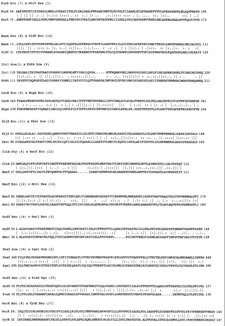
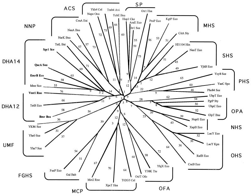
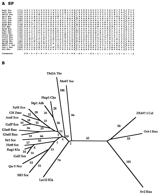
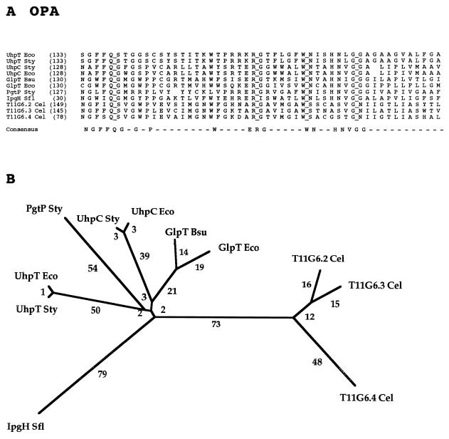
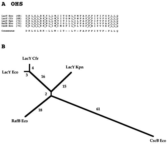
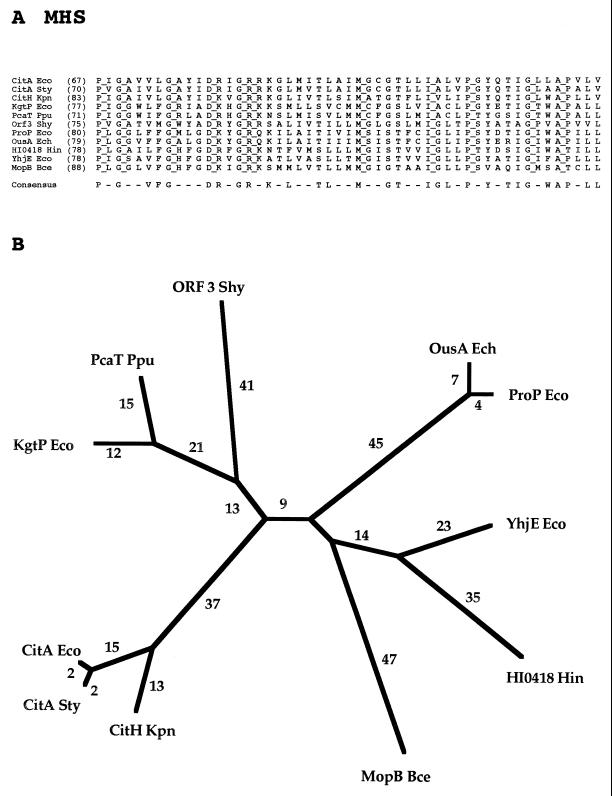
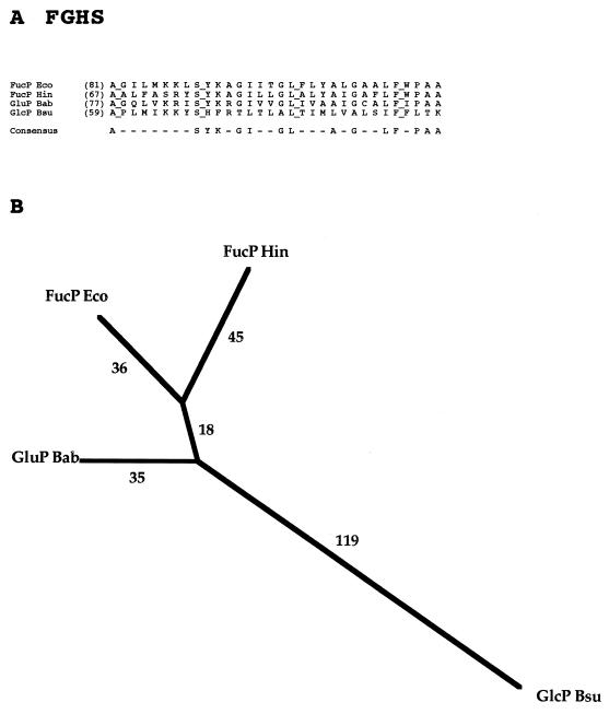
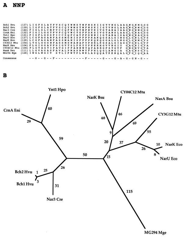
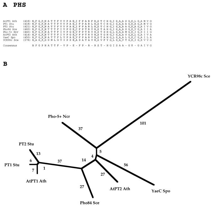
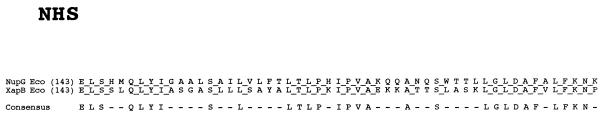
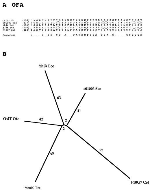
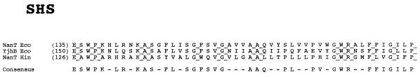
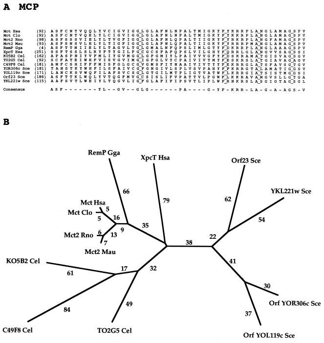
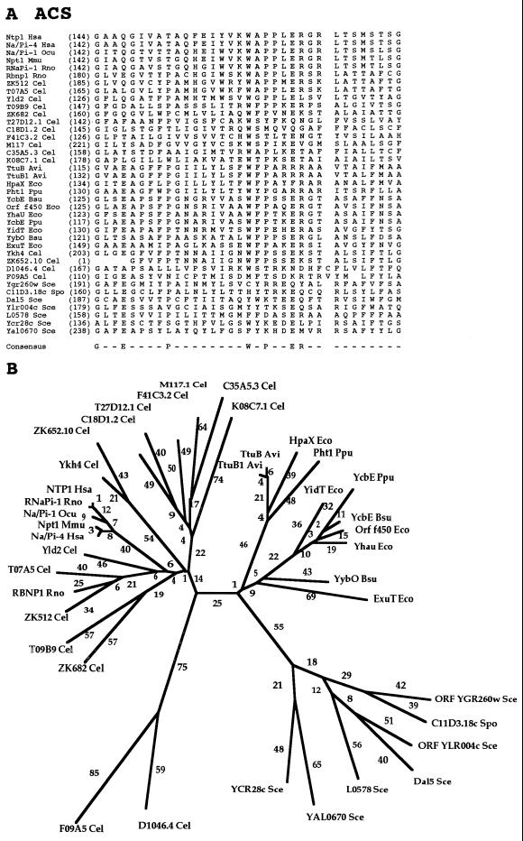
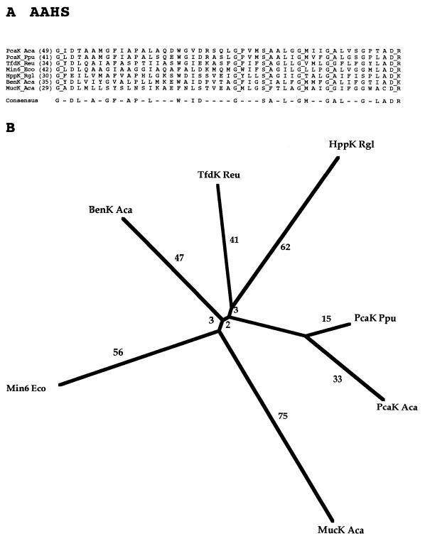
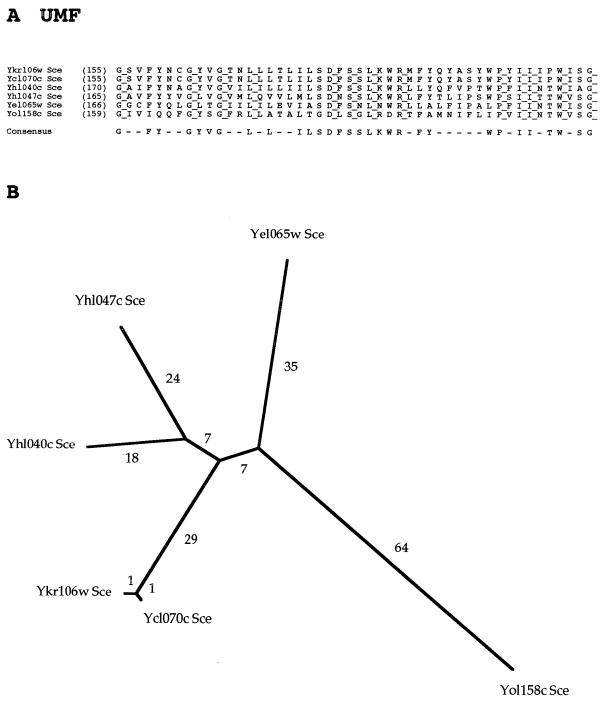
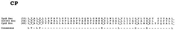
The major facilitator superfamily (MFS) is one of the two largest families of membrane transporters found on Earth. It is present ubiquitously in bacteria, archaea, and eukarya and includes members that can function by solute uniport, solute/cation symport, solute/cation antiport and/or solute/solute antiport with inwardly and/or outwardly directed polarity. All homologous MFS protein sequences in the public databases as of January 1997 were identified on the basis of sequence similarity and shown to be homologous. Phylogenetic analyses revealed the occurrence of 17 distinct families within the MFS, each of which generally transports a single class of compounds. Compounds transported by MFS permeases include simple sugars, oligosaccharides, inositols, drugs, amino acids, nucleosides, organophosphate esters, Krebs cycle metabolites, and a large variety of organic and inorganic anions and cations. Protein members of some MFS families are found exclusively in bacteria or in eukaryotes, but others are found in bacteria, archaea, and eukaryotes. All permeases of the MFS possess either 12 or 14 putative or established transmembrane alpha-helical spanners, and evidence is presented substantiating the proposal that an internal tandem gene duplication event gave rise to a primordial MFS protein prior to divergence of the family members. All 17 families are shown to exhibit the common feature of a well-conserved motif present between transmembrane spanners 2 and 3. The analyses reported serve to characterize one of the largest and most diverse families of transport proteins found in living organisms.


Figures

FIG. 1
FIG. 1
Interfamily binary alignments of representative regions within the larger alignments upon which the comparison scores recorded in Table 2 were based. The two proteins compared (families in parentheses) are presented above the alignment. Protein abbreviations are as indicated in Tables 3 to 17 for families 3 to 17, respectively. The number following the protein abbreviation and preceding its sequence is the residue number of the first residue shown. A vertical line specifies an identity, while double and single dots signify close and distant similarities, respectively.
FIG. 2
FIG. 2
Phylogenetic tree for the MFS including representative members of most of the currently recognized constituent families. All of the families listed in Table 1 are included except the AAHS family (family 15) and the putative POT family (putative family 18). The TREE program of Feng and Doolittle (22) was used to derive the tree.
FIG. 3
FIG. 3
Partial multiple alignment (A) and phylogenetic tree (B) for representative members of the SP family (family 1) of the MFS. The format of presentation for this figure is essentially the same as for subsequent family-specific figures (Fig. 4 to 17), as follows. (A) The abbreviation of each protein, provided in Table 3, is followed by the residue number in the protein, presented in parentheses, corresponding to the first residue in the alignment shown. Fully conserved residues are highlighted with a line to the right of that residue. A residue appears in the consensus sequence (bottom) if that residue occurs at the specified position in a majority of the aligned proteins. In the phylogenetic tree (B), the branch length (numerical values in arbitrary units) is a measure of sequence divergence and is assumed to be approximately proportional to the phylogenetic distance. The tree was based on the complete multiple-sequence alignment for the proteins included in the study. Both the multiple-sequence alignment and the phylogenetic tree were derived with the TREE program of Feng and Doolittle (22).
FIG. 4
FIG. 4
Partial multiple-sequence alignment (A) and phylogenetic tree (B) for the OPA family (family 4). The format of presentation for this figure and Fig. 5 to 17 is essentially as described in the legend to Fig. 3.
FIG. 5
FIG. 5
Partial multiple-sequence alignment (A) and phylogenetic tree (B) for the OHS family (family 5).
FIG. 6
FIG. 6
Partial multiple-sequence alignment (A) and phylogenetic tree (B) for the MHS family (family 6).
FIG. 7
FIG. 7
Partial multiple-sequence alignment (A) and phylogenetic tree (B) for the FGHS family (family 7).
FIG. 8
FIG. 8
Partial multiple-sequence alignment (A) and phylogenetic tree (B) for the NNP family (family 8).
FIG. 9
FIG. 9
Partial multiple-sequence alignment (A) and phylogenetic tree (B) for the PHS family (family 9).
FIG. 10
FIG. 10
Partial alignment of the sequences of the two currently sequenced proteins of the NHS family (family 10).
FIG. 11
FIG. 11
Partial multiple-sequence alignment (A) and phylogenetic tree (B) for the OFA family (family 11).
FIG. 12
FIG. 12
Partial alignment of the sequences of the three currently sequenced proteins of the SHS family (family 12).
FIG. 13
FIG. 13
Partial multiple-sequence alignment (A) and phylogenetic tree (B) for the MCP family (family 13).
FIG. 14
FIG. 14
Partial multiple-sequence alignment (A) and phylogenetic tree (B) for the ACS family (family 14).
FIG. 15
FIG. 15
Partial multiple-sequence alignment (A) and phylogenetic tree (B) for the AAHS family (family 15).
FIG. 16
FIG. 16
Partial multiple-sequence alignment (A) and phylogenetic tree (B) for the six currently sequenced proteins of the UMF family (family 16).
FIG. 17
FIG. 17
Partial multiple-sequence alignment for the three members of the CP family (family 17).

References

    1. Abe K, Ruan Z S, Maloney P C. Cloning, sequencing, and expression in Escherichia coli of OxlT, the oxalate:formate exchange protein of Oxalobacter formigenes. J Biol Chem. 1996;271:6789–6793. - PubMed
    1. Altschul S F, Gish W, Miller W, Myers E W, Lipman D J. Basic local alignment search tool. J Mol Biol. 1990;215:403–410. - PubMed
    1. Ambudkar S V, Maloney P C. Characterization of phosphate:hexose 6-phosphate antiport in membrane vesicles of Streptococcus lactis. J Biol Chem. 1984;259:12576–12585. - PubMed
    1. Anantharam V, Allison M J, Maloney P C. Oxalate:formate exchange: the basis for energy coupling in Oxalobacter. J Biol Chem. 1989;264:7244–7250. - PubMed
    1. Bailey T L, Elkan C. Proceedings of the Second International Conference on Intelligent Systems for Molecular Biology. Menlo Park, Calif: AAAI Press; 1994. Fitting a mixture model by expectation maximization to discover motifs in biopolymers; pp. 28–36. - PubMed

Publication types

Substances