Review
. 1998 Mar;62(1):35-54.
doi: 10.1128/MMBR.62.1.35-54.1998.

Asexual sporulation in Aspergillus nidulans

Affiliations
Review

Asexual sporulation in Aspergillus nidulans

T H Adams et al. Microbiol Mol Biol Rev. 1998 Mar.

Erratum in

  • Microbiol Mol Biol Rev 1998 Jun;62(2):545

Abstract

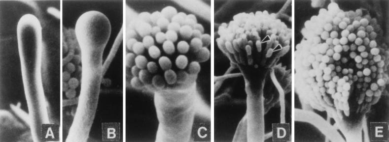
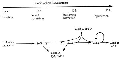
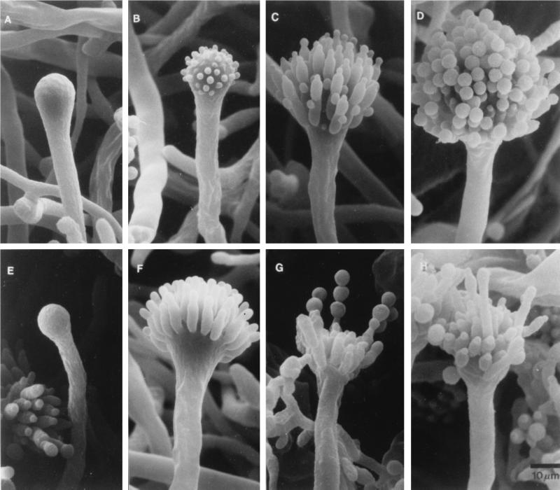
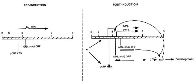
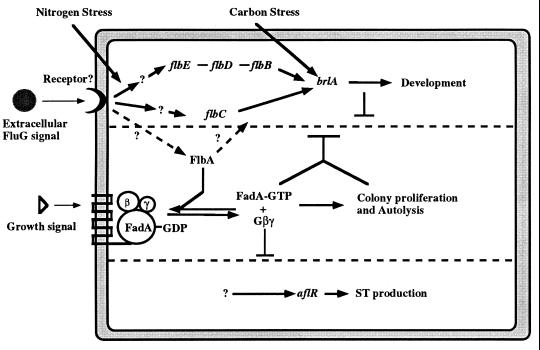
The formation of mitotically derived spores, called conidia, is a common reproductive mode in filamentous fungi, particularly among the large fungal class Ascomycetes. Asexual sporulation strategies are nearly as varied as fungal species; however, the formation of conidiophores, specialized multicellular reproductive structures, by the filamentous fungus Aspergillus nidulans has emerged as the leading model for understanding the mechanisms that control fungal sporulation. Initiation of A. nidulans conidiophore formation can occur either as a programmed event in the life cycle in response to intrinsic signals or to environmental stresses such as nutrient deprivation. In either case, a development-specific set of transcription factors is activated and these control the expression of each other as well as genes required for conidiophore morphogenesis. Recent progress has identified many of the earliest-acting genes needed for initiating conidiophore development and shown that there are at least two antagonistic signaling pathways that control this process. One pathway is modulated by a heterotrimeric G protein that when activated stimulates growth and represses both asexual and sexual sporulation as well as production of the toxic secondary metabolite, sterigmatocystin. The second pathway apparently requires an extracellular signal to induce sporulation-specific events and to direct the inactivation of the first pathway, removing developmental repression. A working model is presented in which the regulatory interactions between these two pathways during the fungal life cycle determine whether cells grow or develop.


Figures

FIG. 1
FIG. 1
Morphological changes during conidiophore formation. Shown are scanning electron micrographs of the stages of conidiation. (A) Early conidiophore stalk. (B) Vesicle formation from the tip of the stalk. (C) Developing metulae. (D) Developing phialides. (E) Mature conidiophores bearing chains of conidia. Reproduced from reference with permission of the publisher.
FIG. 2
FIG. 2
Experimental determination of the timing of developmental competence (Tc). Spores were inoculated into liquid medium at time zero and allowed to germinate in submerged culture. At the times indicated on the horizontal axis, mycelia were transferred to a solid substrate and observed to determine the earliest time of conidiophore formation. The delay time (d) is the interval between transfer to solid medium and the first appearance of conidiophores. The time Tc is the time after which d = D = constant. Reproduced from reference with permission of the publisher.
FIG. 3
FIG. 3
Starvation can induce conidiation in submerged culture. A wild-type strain was grown in glucose minimal medium for 18 h and then shifted to fresh minimal medium (A) or minimal medium lacking a carbon source (B). Conidiophores formed within 12 h after a shift to nitrogen starvation medium (development also occurred upon shifting to medium lacking a nitrogen source), but no conidiophores were seen in the culture shifted to minimal medium containing a usable carbon or nitrogen source. Reproduced from reference with permission of the publisher.
FIG. 4
FIG. 4
brlA mutants form indeterminate conidiophore stalks. Conidiophores from a wild-type strain (A) and a brlA mutant strain (B) are shown. In the wild-type strain, the stalks grow to a fairly uniform height and bear additional conidiophore-specific structures, while the stalks of a brlA mutant grow somewhat indeterminately and fail to elaborate other conidiophore-specific cells. Arrows indicate stalks. Reproduced from reference with permission of the publisher.
FIG. 5
FIG. 5
Overexpression of brlA activates sporulation in submerged culture. The brlA gene was placed under the control of the alcohol-inducible promoter alcA. The alcA(p)::brlA strain and a wild-type strain were grown for 12 h in liquid minimal medium containing glucose to repress brlA expression from the alcA promoter and then shifted to alcA(p)-inducing medium. (B) By 3 h after the medium shift, the alcA(p)::brlA strain produced spores from the tips of hyphae. (A) No conidiation was observed in the wild-type strain even 24 h after the medium shift.
FIG. 6
FIG. 6
Conidiophores from a wild-type strain (A) and an abaA mutant strain (B). The abaA mutant produces normal conidiophore stalks (ST) and vesicles (VS), but metulae (M) and phialides are abnormal and form abacus (AB) structures instead of conidia (C). Magnifications for panels A and B are equivalent. Reproduced from reference with permission of the publisher.
FIG. 7
FIG. 7
Electron micrographs of stuA and medA mutants. In the stuA mutant, abnormal conidia are formed either directly from the conidiophore vesicle (A) or from abnormal sterigmata (B and C). Normal metula and phialides are not produced. medA mutant conidiophores are initially nearly normal, but multiple layers of metulae are produced before phialides differentiate and begin to form conidia (D and E). Sometimes sterigmata redifferentiate to produce secondary conidiophores (F). Reproduced from reference with permission of the publisher.
FIG. 8
FIG. 8
Timeline of conidiation and the central regulatory pathway. The genes in the central regulatory pathway for conidiation are predicted to activate other genes responsible for the production of conidiophores. The activation of the class A, B, C, and D genes give rise to the formation of a conidiophore with the timing indicated in the upper part of the figure.
FIG. 9
FIG. 9
apsA mutants produce anucleate sterigmata. Electron micrographs showing conidiophore formation in a wild-type strain (A to D) are contrasted with micrographs of an apsA mutant (E to H). In apsA mutants, development of the conidiophore is normal up to the metula stage (E and F). Nuclei typically fail to enter the metulae, and development arrests with an anucleate metular bud (F). Occasionally, nuclei enter metulae, leading to production of functional phialides and normal chains of spores (G and H). Reproduced from reference with permission of the publisher.
FIG. 10
FIG. 10
Model for differential control of brlAα and brlAβ during conidiophore development. The brlAβ mRNA is transcribed in vegetative cells before developmental induction, but translation of the μORF represses translation of BrlA. Following induction, unknown regulatory factors activate BrlA translation from brlAβ by removing the translational block imposed by the μORF, increasing the transcription of brlAβ, or both. Activation of BrlA translation leads to transcription of abaA and other downstream regulatory proteins. This in turn activates a positive-feedback loop that leads to high levels of brlAα expression to cause further developmental changes. Reproduced from reference with permission of the publisher.
FIG. 11
FIG. 11
Phenotypic classes of fluffy mutants. Colonies were grown for 3 days on solid minimal medium. (A) Developmentally wild-type strain of A. nidulans. (B) fluG mutant. (C) flbA mutant. (D) Delayed conidiation mutant typical of flbB, flbC, flbD, and flbE mutants.
FIG. 12
FIG. 12
Overexpression of fluG, flbA, and flbD causes conidiophore production in submerged culture. The alcA promoter was used to drive the expression of fluG, flbA, and flbD. The alcA(p)::fluG, alcA(p)::flbA, and alcA(p)::flbD strains and a wild-type strain were grown for 14 h in liquid minimal medium containing glucose to repress alcA and then shifted to alcA-inducing medium. Overexpression of fluG (B), flbA (C), and flbD (D) led to production of conidiophores in ∼18 h, 9 h, and 9 h after the shift, respectively. The wild-type strain (A) never produced conidiophores or spores. Panel D is reproduced from reference with permission of the publisher.
FIG. 13
FIG. 13
Model describing fluffy gene interactions in controlling initiation of development. As described in the text, we have proposed that the activities of two antagonistic signaling pathways determine whether development and secondary metabolism occur (70, 86, 173). One pathway requires the product of FluG activity, which is proposed to work as an extracellular signal to activate a sporulation-specific pathway that requires flbB, flbC, flbD, and flbE. When the FadA Gα protein is GTP bound, it regulates downstream effectors to enhance proliferation and repress both sporulation and ST production. The FluG signal causes inactivation of FadA by activating FlbA, which functions as a GTPase-activating protein to turn off the FadA-dependent signaling pathway. This inactivation of FadA then allows both sporulation and ST biosynthesis to occur.

Similar articles

Cited by

References

    1. Adams T H, Boylan M T, Timberlake W E. brlA is necessary and sufficient to direct conidiophore development in Aspergillus nidulans. Cell. 1988;54:353–362. - PubMed
    1. Adams T H, Deising H, Timberlake W E. brlA requires both zinc fingers to induce development. Mol Cell Biol. 1990;10:1815–1817. - PMC - PubMed
    1. Adams T H, Hide W A, Yager L N, Lee B N. Isolation of a gene required for programmed initiation of development by Aspergillus nidulans. Mol Cell Biol. 1992;12:3827–3833. - PMC - PubMed
    1. Adams T H, Timberlake W E. Developmental repression of growth and gene expression in Aspergillus. Proc Natl Acad Sci USA. 1990;87:5405–5409. - PMC - PubMed
    1. Adams T H, Timberlake W E. Upstream elements repress premature expression of an Aspergillus developmental regulatory gene. Mol Cell Biol. 1990;10:4912–4919. - PMC - PubMed

Publication types

MeSH terms

LinkOut - more resources