Review
. 1998 Mar;62(1):181-203.
doi: 10.1128/MMBR.62.1.181-203.1998.

Growth of the stress-bearing and shape-maintaining murein sacculus of Escherichia coli

Affiliations
Review

Growth of the stress-bearing and shape-maintaining murein sacculus of Escherichia coli

J V Höltje. Microbiol Mol Biol Rev. 1998 Mar.

Abstract

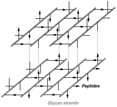
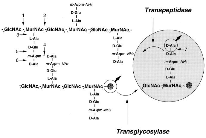
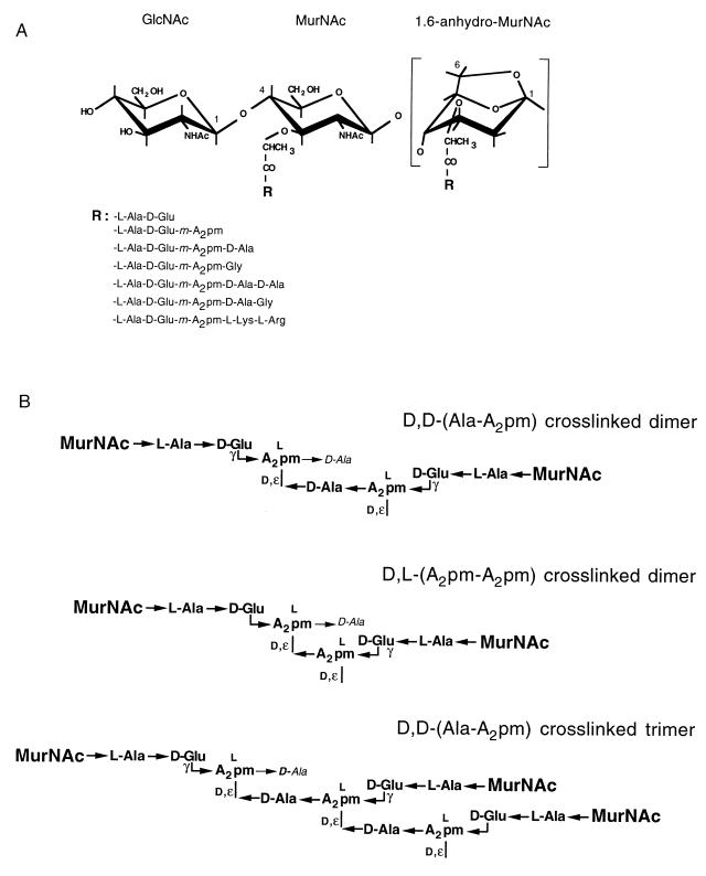
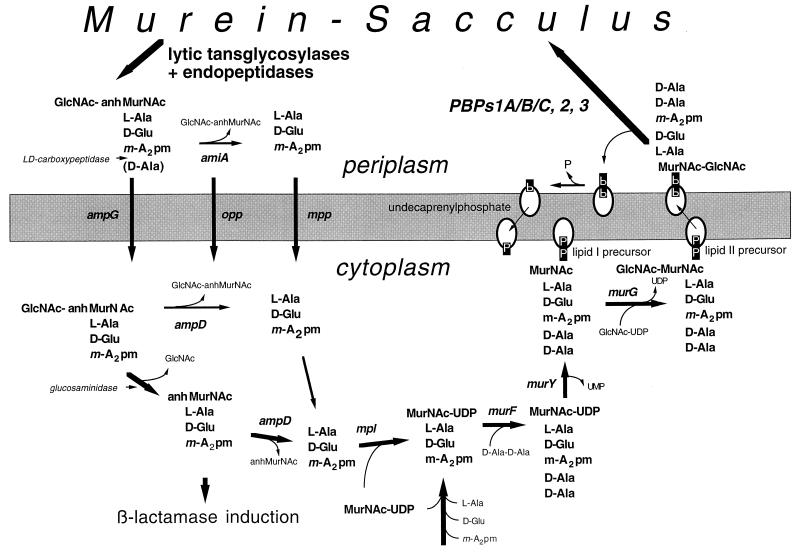
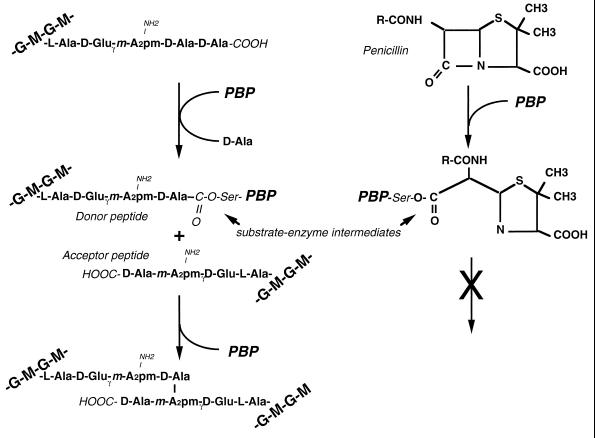
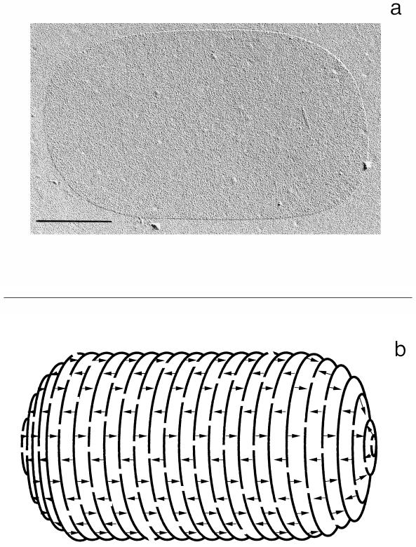
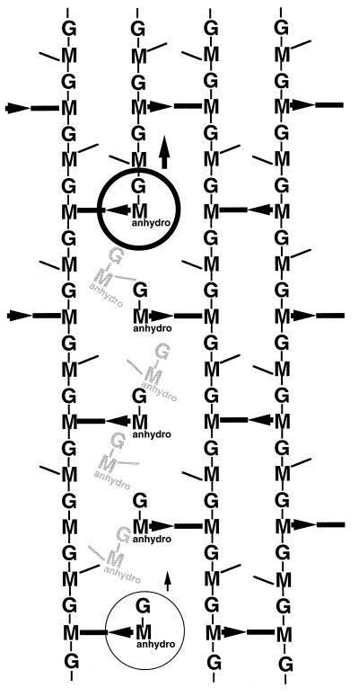
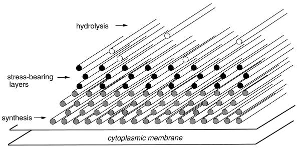
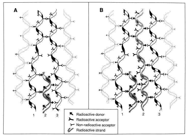
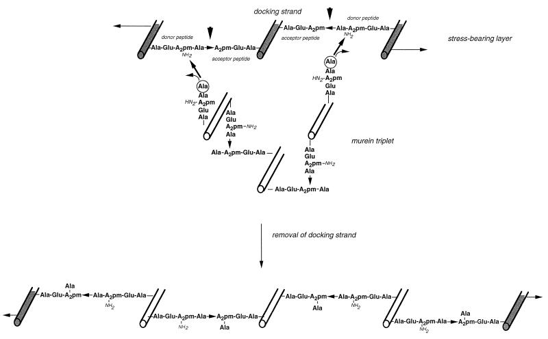
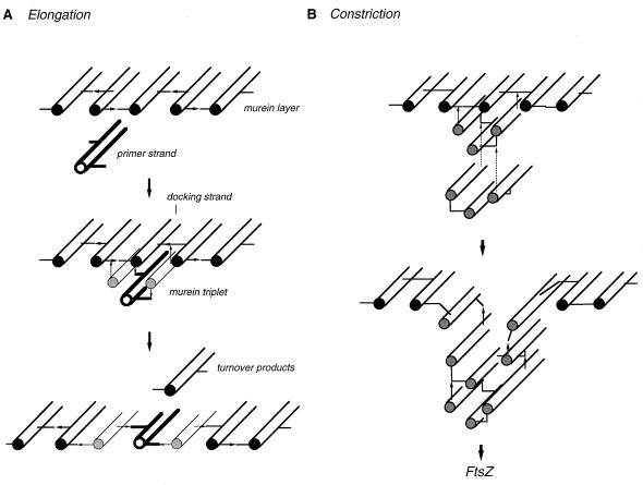
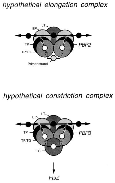
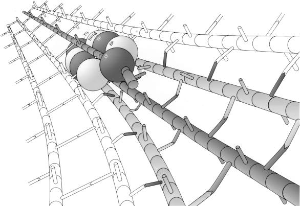
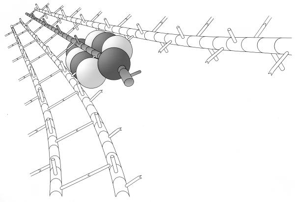
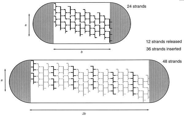
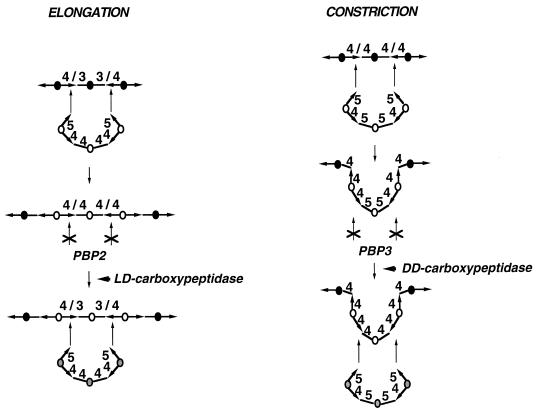
To withstand the high intracellular pressure, the cell wall of most bacteria is stabilized by a unique cross-linked biopolymer called murein or peptidoglycan. It is made of glycan strands [poly-(GlcNAc-MurNAc)], which are linked by short peptides to form a covalently closed net. Completely surrounding the cell, the murein represents a kind of bacterial exoskeleton known as the murein sacculus. Not only does the sacculus endow bacteria with mechanical stability, but in addition it maintains the specific shape of the cell. Enlargement and division of the murein sacculus is a prerequisite for growth of the bacterium. Two groups of enzymes, hydrolases and synthases, have to cooperate to allow the insertion of new subunits into the murein net. The action of these enzymes must be well coordinated to guarantee growth of the stress-bearing sacculus without risking bacteriolysis. Protein-protein interaction studies suggest that this is accomplished by the formation of a multienzyme complex, a murein-synthesizing machinery combining murein hydrolases and synthases. Enlargement of both the multilayered murein of gram-positive and the thin, single-layered murein of gram-negative bacteria seems to follow an inside-to-outside growth strategy. New material is hooked in a relaxed state underneath the stress-bearing sacculus before it becomes inserted upon cleavage of covalent bonds in the layer(s) under tension. A model is presented that postulates that maintenance of bacterial shape is achieved by the enzyme complex copying the preexisting murein sacculus that plays the role of a template.


Figures

FIG. 1
FIG. 1
Architecture of the murein layer. The drawing shows two murein layers and indicates (by dotted lines) how they can be stacked on one another to form a multilayered murein. Glycan strands are represented by solid bars. The peptide cross bridges are indicated by black lines (acceptor stem peptides) and black arrows (donor stem peptides).
FIG. 2
FIG. 2
Chemistry of the murein of E. coli. A section of the murein of E. coli is shown. On the right is shown how a new murein precursor linked to the undecaprenyl pyrophosphate group (represented by a spotted circle) is linked to the preexisting murein by the formation of two bonds. Concomitant with cleavage of the d,d-peptide bond between the two d-Ala residues of the pentapeptide precursor, a transpeptidase forms a d,d-peptide bond between the carboxyl group of the penultimate d-Ala of the precursor and the epsilon amino group of a diaminopimelic acid residue present in a peptide moiety of the growing murein sacculus. A transglycosylase splits the pyrophosphate bond between the undecaprenyl group and the MurNAc of a nascent glycan strand in the sacculus and forms a glycosidic bond to the hydroxyl group at carbon 4 of the GlcNAc of the precursor molecule. The numbers point to the bonds cleaved by specific murein hydrolases present in E. coli: 1, N-acetylglucosaminidase; 2, lytic transglycosylase; 3, N-acetylmuramyl-l-alanine amidase; 4, d,d-endopeptidase; 5, γ-d-glutamyl-l-diaminopimelic acid endopeptidase; 6, l,d-carboxypeptidase; 7, d,d-carboxypeptidase; m-A2pm, meso-diaminopimelic acid.
FIG. 3
FIG. 3
Fractionation of enzymatic degradation products of murein by HPLC. The upper panel shows the separation of sodium borohydride-reduced muropeptides obtained after complete digestion of isolated murein sacculi of E. coli with the muramidase Cellosyl. Chromatography was performed at 55°C on a 3-μm Hypersil ODS column with a linear gradient from 50 mM sodium phosphate (pH 4.32) to 15% methanol in 50 mM sodium phosphate (pH 4.95) (for details, see reference 48). The lower panel shows the separation of murein glycan strands obtained by digestion of isolated sacculi with human serum amidase. Chromatography was performed on a 5-μm Nucleosil ODS column. Elution was done at 50°C with a convex gradient of 100 mM sodium phosphate (pH 2.0) containing 5% acetonitrile to 100 mM sodium phosphate (pH 2.0) containing 11% acetonitrile (for details, see reference 62). This was followed by elution with 100% methanol.
FIG. 4
FIG. 4
Muropeptide structures. (A) Monomeric muropeptide structures identified from the murein of E. coli. The different peptide moieties (R) that can substitute the lactyl group of MurNAc are listed. (B) Chemistry of the major cross-linked dimers and trimers found in the murein of E. coli. A2pm, diaminopimelic acid.
FIG. 5
FIG. 5
Recycling pathway of murein turnover products in E. coli. During growth, lytic transglycosylases together with d,d-endopeptidases release monomeric 1,6-anhydromuropeptides in the periplasm, which are mostly tripeptide derivatives due to the presence of a l,d-carboxypeptidase. The turnover products can enter two different metabolic pathways. In one case, they are first degraded by an amidase (AmiA) (161) in the periplasm to the disaccharide and the peptide, which can be transported into the cell by the general oligopeptide transport system (Opp) (53) or the specific murein peptide permease (Mpp) (118). The fate of the sugars is not well established. Alternatively, the turnover products can be taken up by the cell through the AmpG transporter, which accepts intact muropeptides (99, 114). In the cytoplasm, the muropeptides are further degraded by an amidase (AmpD) with a specificity for 1,6-anhydromuropeptides (72, 81) and an N-acetylglucosaminidase (176), yielding free tripeptides, anhydro-MurNAc, and GlcNAc. The intermediate 1,6-anhydro-N-acetylmuramyltripeptide functions as an inducer of AmpC β-lactamases by interacting with the AmpR regulator (79). The tripeptide can be reused directly for the synthesis of murein precursor molecules. A specific tripeptide ligase (Mpl) (103) forms UDPMurNAc tripeptide, which is a normal intermediate of the de novo murein biosynthetic pathway. Accordingly, a d-Ala–d-Ala dipeptide is linked to the tripeptide intermediate. The resulting UDPMurNAc-pentapeptide is then translocated to undecaprenyl phosphate, yielding the lipid I precursor that is supplemented by the addition of GlcNAc from UDPGlcNAc to the final lipid II precursor, the bactoprenol-linked disaccharide-pentapeptide. After its translocation to the periplasmic side of the membrane, the precursor is inserted into the murein sacculus by transpeptidation and transglycosylation reactions catalyzed by the high-molecular-weight PBP1A, PBP1B, PBP1C, PBP2, and PBP3. The released undecaprenyl pyrophosphate is hydrolyzed to undecaprenyl phosphate before it can enter a new cycle.
FIG. 6
FIG. 6
d,d-Transpeptidation reaction. The general scheme of the two-step transpeptidation reaction, resulting in the formation of a cross-link between two glycan strands, is shown on the left. In the first step, the d-Ala–d-Ala peptide bond of a donor peptide is cleaved by a PBP enzyme, and a covalent bond between the carboxyl group of the penultimate d-Ala and the hydroxyl group of a serine in the active center of the enzyme is formed, giving rise to a mureinyl enzyme intermediate. In a second reaction step, the mureinyl moiety is transferred to a free epsilon amino group of a diaminopimelic acid residue in another peptide moiety (acceptor peptide). Concomitant with the formation of the cross-link, the PBP enzyme is released. The interaction of the PBP enzyme with penicillin is indicated on the right. By analogy to the cleavage of the d-Ala–d-Ala bond during the transpeptidation, the β-lactam ring is cleaved and a penicilloyl enzyme intermediate is formed. In contrast to the mureinyl enzyme intermediate, the penicilloyl enzyme intermediate is rather inert, and thus the enzyme is blocked by a covalent bond to the antibiotic, a kind of suicide substrate for PBPs. G, GlcNAc; M, MurNAc; A2pm, diaminopimelic acid; Ser is from the catalytic center of the PBP.
FIG. 7
FIG. 7
Murein sacculus of E. coli. (a) Electron micrograph (agar filtration) (prepared by H. Frank) of an isolated murein sacculus. Bar, 0.5 μm. (b) Idealized schematic drawing of the architecture of the murein sacculus. The parallel lines represent a few of the vast number of glycan strands, and the arrows indicate the peptide bridges.
FIG. 8
FIG. 8
Maturation of murein. Changes in the relative amounts of defined muropeptide structures in the murein of E. coli were analyzed by pulse-chase labelling experiments. Exponentially growing A2pm auxotrophic E. coli W7 (with a generation time of 30 min) was pulse-labelled with [3H]diaminopimelic acid for 2 min and chased for different times as indicated. Experimental details are given in reference . (A) Changes of the major monomeric muropeptides. Solid circles, LysArg-modified disaccharide tripeptides; open circles, disaccharide tripeptides; triangles, disaccharide pentapeptides. (B) Alteration in time of the anhydromuropeptides representing the chain ends. Solid circles, monomers; open circles, cross-linked muropeptides; dotted line, total amount of anhydromuropeptides. (C) Changes in the cross-linkages. Solid squares, cross-linkage by d,d-A2pm-Ala; open squares, cross-linkage by l,d-A2pm-A2pm. (D) Distribution of labelled diaminopimelic acid in donor and acceptor peptides of cross-linked muropeptides. Solid circles, muropeptides cross-linked by two tetra peptides; open triangles, muropeptides cross-linked by one tri- and one tetrapeptide; solid squares, trimeric muropeptides cross-linked by three tetra-peptides.
FIG. 9
FIG. 9
Mode of action of exoglycosylases. The processive degradation of murein strands by a lytic transglycosylase is shown. Structures shown in light grey depict released (soluble) muropeptides. The circle represents the enzyme molecule, with the thin circle marking the site from where the exoenzyme started its action. G, GlcNAc; M, MurNAc; bars and arrows indicate acceptor and donor peptides, respectively.
FIG. 10
FIG. 10
Inside-to-outside growth of the peptidoglycan of gram-positive bacteria. The addition of newly synthesized murein layers in a relaxed state underneath the stress-bearing layers with concomitant degradation of the outermost layers is schematically illustrated. The rods represent the murein strands. For the sake of simplicity, the cross-links between the glycans have been omitted.
FIG. 11
FIG. 11
Model for the growth of murein following a cut-and-insertion strategy as proposed by Park (116). The structure of the murein at a growth site for elongation of the murein sacculus as it may exist during a pulse-labelling experiment is shown. The stippled, helical strand represents a new radioactive strand of murein inserted between preexisting strands initially (A) and later when, as a result of continued insertion along a helical path, a radioactive strand contacts the radioactive strand formed a few minutes earlier (B). The arrows between strands indicate the direction of cross-links in dimers from donor to acceptor strands. Reprinted from reference with permission.
FIG. 12
FIG. 12
Model for the growth of murein following a make-before-break strategy (three-for-one growth model). Three cross-linked glycan strands from the monolayered, stress-bearing murein are shown in grey. A triple pack of newly synthesized, cross-linked glycans still in a relaxed state is shown in outline. The murein triplet is covalently attached to the free amino groups present in the donor peptides of the cross-links on both sides of a strand, called the docking strand, that is substituted by acceptor peptides. Specific cleavage of the preexisting cross-links (arrowheads) results in the replacement of the docking strand by the murein triplet. The rods represent the glycan strands. A2pm, diaminopimelic acid.
FIG. 13
FIG. 13
Cell elongation and cell constriction according to the three-for-one growth mechanism. (A) Murein synthesis during cell elongation is proposed to use preexisting free glycan strands (primer strands) that are supplemented to form murein triplets by the synthesis and cross-linkage of two strands on both sides of the primer strand. The triplet is then attached to the peptide bridges on the right and left of the docking strand in the stress-bearing murein layer. Removal of the docking strand by murein hydrolases results in the insertion of the murein triplet into the murein sacculus. (B) During cell constriction, three glycan strands are simultaneously synthesized and cross-linked to one another to form a murein triplet that is attached underneath a docking strand located at the site of cell division. Formation of a constriction is the outcome of the repeated addition (dotted lines) of murein triplets, each followed by the release of the middle strand and an inward pull of the membrane-anchored enzymes resulting from the contracting FtsZ ring (12, 107, 122, 130). The glycan strands are represented by rods, and the peptide bridges are shown by lines (acceptor peptides) and arrows (donor peptides).
FIG. 14
FIG. 14
Hypothetical murein-synthesizing enzyme complexes. Two similar multienzyme complexes may exist that would differ in their specificities mainly as a result of the presence of either PBP2 (elongation complex) or PBP3 (constriction complex). The large circles represent the enzymes (LT, lytic transglycosylase; EP, endopeptidase; TP, transpeptidase; TP/TG, bifunctional transpeptidase-transglycosylase; TG, transglycosylase). The small circles indicate the glycan strands running perpendicular to the plane of the drawing (solid circles represent preexisting strands; open circles represent the newly synthesized murein triplet). Bars and arrows indicate the cross-linking donor and acceptor peptides.
FIG. 15
FIG. 15
Proposed murein replicase holoenzyme in action. It is proposed that the multienzyme complex (Fig. 14) synthesizes three new murein strands (shown in grey), attaches these strands to the cross bridges on both sides of the docking strand (single grey strand), and at the same time degrades this strand by the concerted action of a processive lytic transglycosylase and a dimer of an endopeptidase. The complex slides along the docking strand with the murein synthases in front of the hydrolases, thereby acting according to the make-before-break strategy. LT, lytic transglycosylase; EP, endopeptidase; TP, transpeptidase; TP/TG, bifunctional transpeptidase-transglycosylase; TG, transglycosylase.
FIG. 16
FIG. 16
Idling of the murein replicase in the presence of murein synthesis inhibitors. It is assumed that in the presence of murein synthesis inhibitors, the multienzyme complex is still pushed along the murein strands by the action of the murein hydrolases processively degrading the docking strand while the synthases idle due to a block in the supply or utilization of the murein precursors (see the legend to Fig. 15).
FIG. 17
FIG. 17
Theoretical doubling of the cylindrical middle part of the murein sacculus by a three-for-one growth modus. The murein of the cylindrical middle part of a rod-shaped sacculus is schematically spread out inside the outlines of a cell, with the metabolically inert polar caps indicated by striped semicircles. Lines running perpendicular to the long axis of the rod-shaped cell indicate glycan strands. The docking strands are shown as fat lines. Arrows and lines arranged parallel to the long axis of the cell represent acceptor and donor peptides.
FIG. 18
FIG. 18
Proposed changes in the cross-linkages during elongation and constriction of the murein sacculus. It is proposed that during cell elongation, murein triplets are exclusively hooked to tetra-tri peptide bridges due to the specificity of the PBP2-containing elongation machinery (Fig. 14). The newly inserted murein triplet is assumed to be cross-linked by tetra-tetra cross-links. Only after the action of an l,d-carboxypeptidase are the peptide bridges converted to tetra-tri cross-links that can function as acceptors for the addition of new murein triplets by the elongation complex. During cell constriction, the newly synthesized murein triplet is assumed to be hooked to tetra-tetra cross-links due to the specificity of the PBP3-containing cell division machinery (Fig. 14). The high rate of localized murein synthesis during cell division is likely to result in the presence of tetra-penta cross-links in the constriction site that may not be accepted by PBP3. Thus, the action of a d,d-carboxypeptidase converting the tetra-penta cross-links to tetra-tetra bridges may be needed for cell constriction. The circles represent the glycan strands, and the arrows and lines represent the donor and acceptor peptides of the cross-links. The numbers above the cross-links indicate whether a tri-, tetra-, or pentapeptide is present.

Similar articles

Cited by

References

    1. Asoh S, Matsuzawa H, Ishino F, Strominger J L, Matsuhashi M, Ohta T. Nucleotide sequence of the pbpA gene and characteristics of the deduced amino acid sequence of penicillin-binding protein 2 of Escherichia coli K12. Eur J Biochem. 1986;160:231–238. - PubMed
    1. Baquero M-R, Bouzon M, Quintela J C, Ayala J A, Moreno F. dacD, an Escherichia coli gene encoding a novel penicillin-binding protein (PBP6b) with d,d-carboxypeptidase activity. J Bacteriol. 1996;178:7106–7111. - PMC - PubMed
    1. Barbas J A, Diaz J, Rodriguez-Tebar A, Vazquez D. Specific location of penicillin-binding proteins within the cell envelope of Escherichia coli. J Bacteriol. 1986;165:269–275. - PMC - PubMed
    1. Bayer M E. Areas of adhesion between wall and membrane of Escherichia coli. J Gen Microbiol. 1968;53:395–404. - PubMed
    1. Bayer M H, Keck W, Bayer M E. Localization of penicillin-binding protein 1b in Escherichia coli: immunoelectron microscopy and immunotransfer studies. J Bacteriol. 1990;172:125–135. - PMC - PubMed

LinkOut - more resources