. 1998 Apr 1;12(7):927-42.
doi: 10.1101/gad.12.7.927.

The fission yeast SPB component Cut12 links bipolar spindle formation to mitotic control

Affiliations

The fission yeast SPB component Cut12 links bipolar spindle formation to mitotic control

A J Bridge et al. Genes Dev. .

Abstract

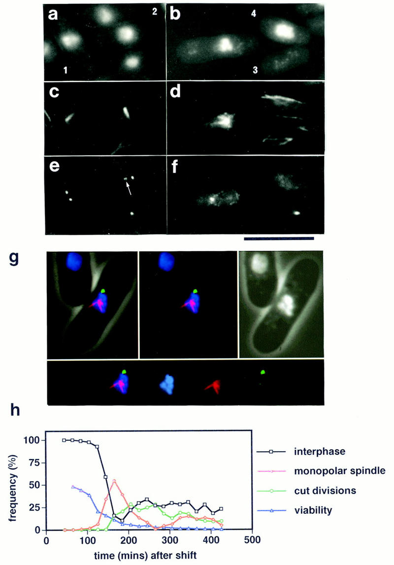
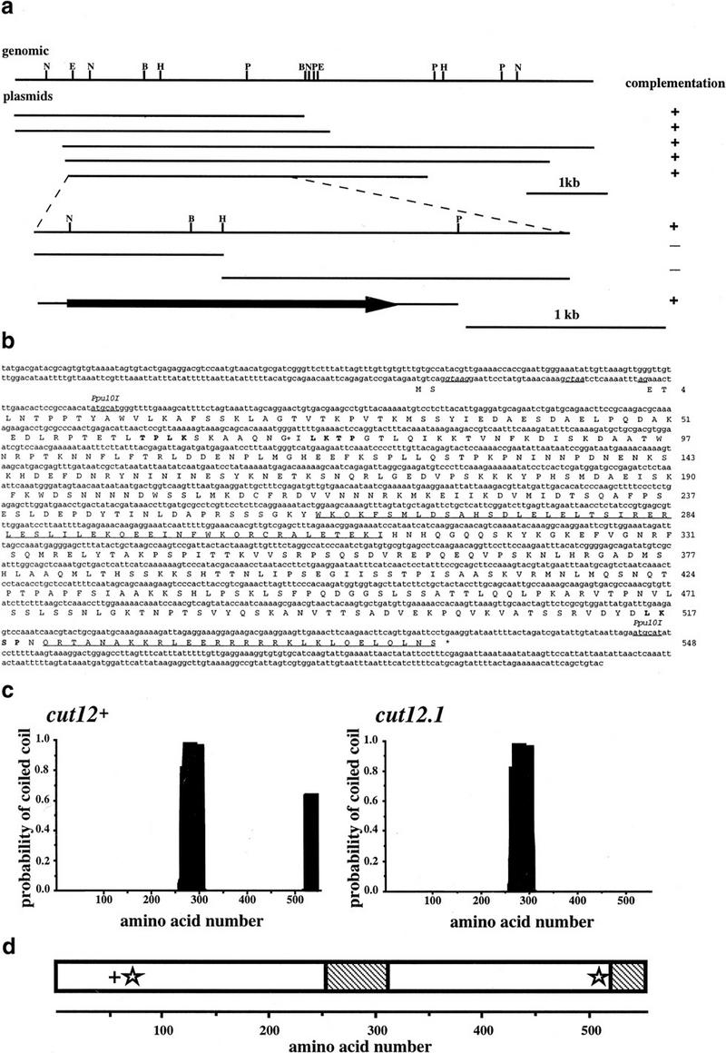
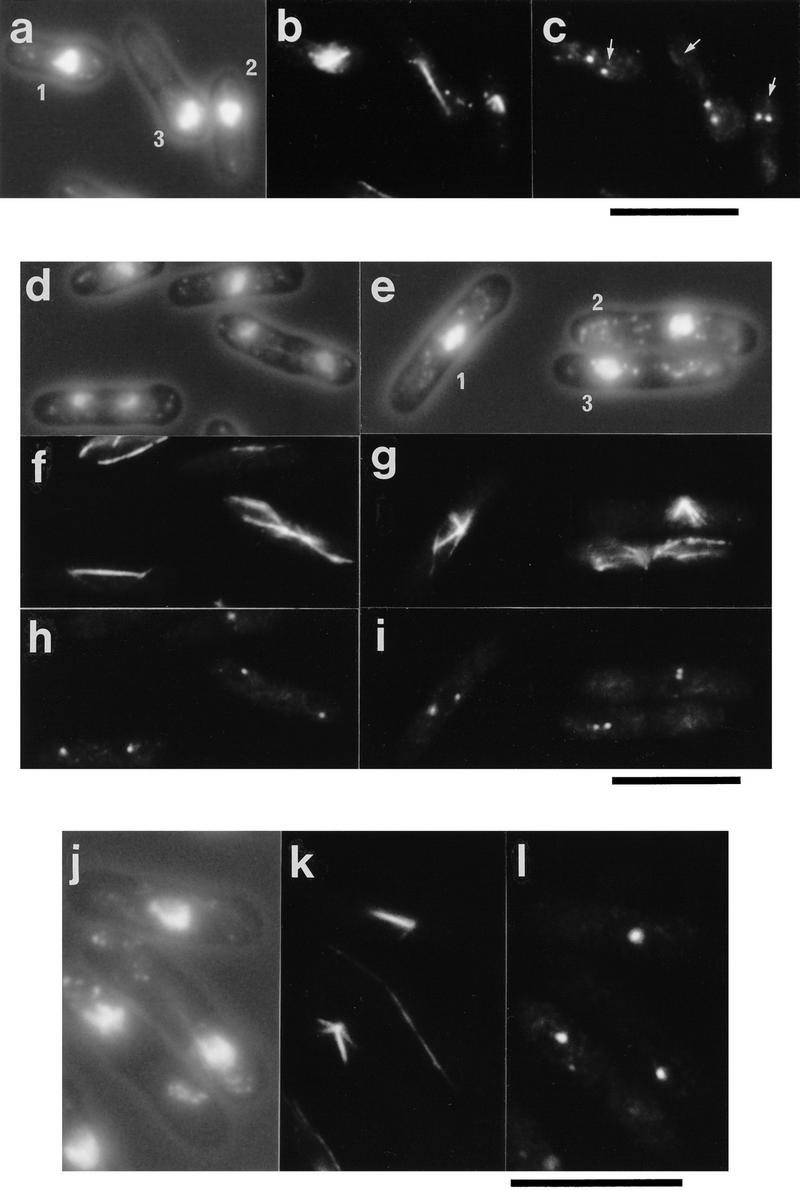
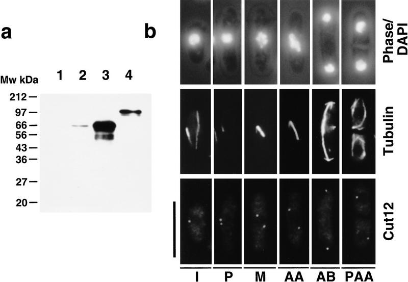
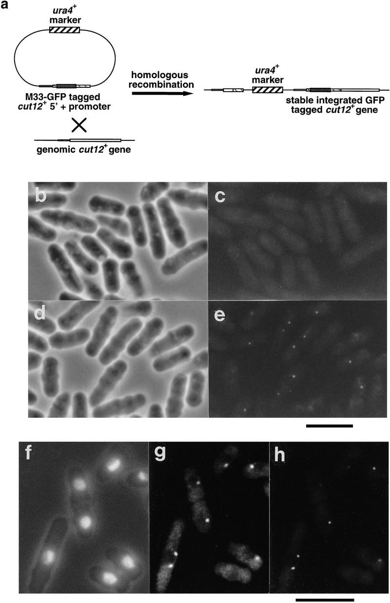
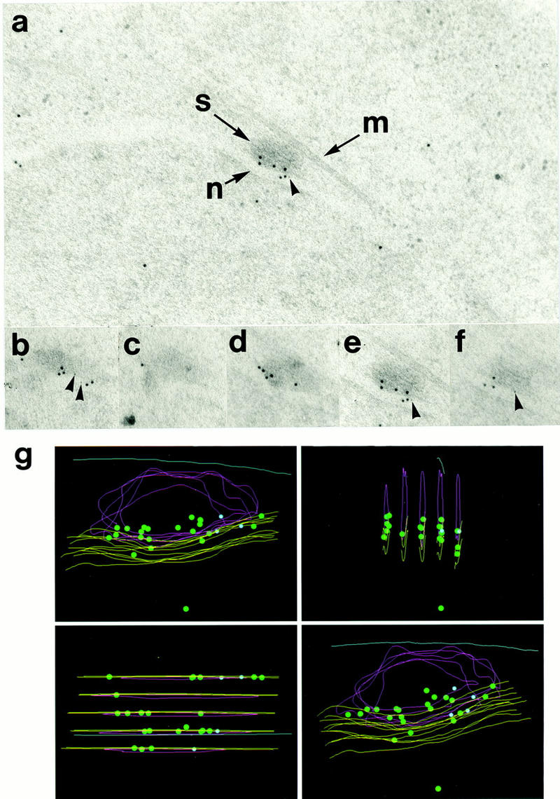
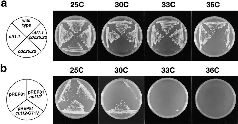
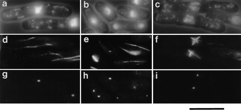
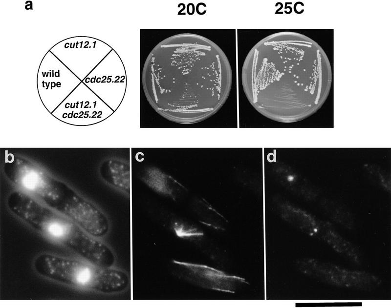
During fission yeast mitosis, the duplicated spindle pole bodies (SPBs) nucleate microtubule arrays that interdigitate to form the mitotic spindle. cut12.1 mutants form a monopolar mitotic spindle, chromosome segregation fails, and the mutant undergoes a lethal cytokinesis. The cut12(+) gene encodes a novel 62-kD protein with two predicted coiled coil regions, and one consensus phosphorylation site for p34(cdc2) and two for MAP kinase. Cut12 is localized to the SPB throughout the cell cycle, predominantly around the inner face of the interphase SPB, adjacent to the nucleus. cut12(+) is allelic to stf1(+); stf1.1 is a gain-of-function mutation bypassing the requirement for the Cdc25 tyrosine phosphatase, which normally dephosphorylates and activates the p34(cdc2)/cyclin B kinase to promote the onset of mitosis. Expressing a cut12(+) cDNA carrying the stf1.1 mutation also suppressed cdc25.22. The spindle defect in cut12.1 is exacerbated by the cdc25.22 mutation, and stf1.1 cells formed defective spindles in a cdc25.22 background at high temperatures. We propose that Cut12 may be a regulator or substrate of the p34(cdc2) mitotic kinase.


Figures

Figure 1
Figure 1
Monopolar mitotic spindle formation in the cut12.1 mutant. (a–f) Immunofluorescence analysis of an asynchronous culture of the cut12.1 mutant at restrictive temperature of 36°C. (a,b) phase/DAPI reveals cell outline and chromatin; (c,d) anti-tubulin staining; (e,f) anti-Sad1 staining. Monopolar mitotic spindles arose from one of the two foci of Sad1 staining (cells 1 and 2). The microtubules normally arose from the less bright staining of the two Sad1 foci (indicated by an arrow in cell 2). Asymmetric segregation of the chromatin and Sad1 staining material led to the formation of aneuploid cells containing Sad1 (cell 3) and diploid cells that lack any detectable Sad1 staining (cell 4). Scale bar, 10 μm. (g) False color immunofluorescence image of a defective monopolar spindle in the cut12.1 mutant. Microtubules are shown in red, Sad1 staining in green, and chromatin in blue. (Top left) Merged image reveals cell outline and spindle structures; (top middle) spindle structure alone; (top right) phase/DAPI staining; (bottom) individual spindle components. The microtubules arose from the less bright staining focus of Sad1 staining. (h) Time course of the loss of viability and the appearance of monopolar spindles and cut divisions in a population of the cut12.1 mutant grown at 25°C, synchronized in early G2 by lactose gradient centrifugation, and then shifted to 36°C. Note that the loss of viability precedes the appearance of defective spindles and cut divisions.
Figure 2
Figure 2
Cloning and molecular analysis of the cut12+ gene. (a) Restriction mapping and complementation analysis of the cut12+ containing clones isolated by complementation of the cut12.1 mutant, and of subclones generated from the overlapping 2.8-kb region found in all cut12.1 complementing plasmids. (b) Nucleotide and predicted amino acid sequence of the cut12+ gene. The cut12+ gene contained 2 exons of 7 and 1637 nucleotides, which together encode a predicted 548 amino acid protein of 62 kD. The two exons are separated by an intron of 42 bp; the consensus intron splice and branch sequences are shown underlined and in italics. The two regions of the Cut12 protein predicted to form coiled coils are underlined. The potential p34cdc2 and MAP kinase consensus sites are indicated in boldface type. (G+) The site of the stf1.1 mutation at glycine 71. The GenBank accession no. for the cut12+ sequence is Y16837. (c) Predicted coiled–coil regions in the Cut12 protein were determined by applying the algorithm of Lupas et al. (1991) with a 28 amino acid window. (d) Schematic representation of the main features of the Cut12 protein. (+) p34cdc2 kinase consensus site; (star) MAP kinase consensus site; (hatched region) predicted region of coiled coil.
Figure 3
Figure 3
Defective spindle formation in cells deleted for the cut12+ gene and cells overexpressing cut12+. (a–c) Defective spindle formation and cut divisions in germinating cut12.d1 spores. Haploid spores prepared from a cut12+/cut12.d1 diploid were germinated in minimal medium lacking uracil at 30°C and processed for immunofluorescence from 12 to 22 hr after inoculation. Shown are three cells fixed 16 hr after inoculation. (A) Phase/DAPI reveals cell outline and chromatin; (B) anti-tubulin staining; (C) anti-Sad1 staining. Germinating cut12.d1 spores appeared to contain two foci of Sad1 staining, although the microtubules arose from a position distal to both foci, indicated by an arrow for each of the three cells. Cells also underwent cut divisions (cell 3). Scale bar, 10 μm. (d–i) Monopolar spindle formation and cut divisions following the switch off of plasmid borne cut12+ expression in cut12.d1 haploid cells. A culture of the cut12.d1 haploid maintained by the expression of a plasmid borne copy of the cut12+ cDNA from the attenuated thiamine repressible nmt1+ promoter was grown to log phase in minimal medium in the absence of thiamine at 30°C. Aliquots were then inoculated into thiamine-free media to allow continued expression of cut12+, and a second aliquot inoculated into medium containing thiamine to repress cut12+ expression. Samples were processed for immunofluorescence from 14 to 28 hr later. (d,f,h) cut12+ expressed; (e,g,i) cut12+ repressed. (d,e) Phase/DAPI reveals cell outline and chromatin staining; (f,g) anti-tubulin staining; (h,i) anti-Sad1 staining. Defective spindles formed in cells ∼14 hr after repression of cut12+. Monopolar spindles arose from only one of the two Sad1 foci (cell 1 and 2), similar to the ts cut12.1 mutant, and Cut divisions occurred (cell 3). Scale bar, 10 μm. (j–l) Immunofluorescence analysis of cells overexpressing cut12+. Cells carrying the cut12+ cDNA linked to the wild-type nmt1+ promoter of the pREP1 plasmid were grown to mid log phase, the culture was divided in two, and cut12+ expression was derepressed in one half. Samples were taken every hour from 12 to 20 hr following induction. Shown are cells in which cut12+ was derepressed for 16 hr. (j) Phase contrast/DAPI stain; (k) anti-tubulin staining; (l) anti-Sad1 staining. Cells overexpressing the cut12+ gene formed monopolar spindles that seemed to arise from the single focus of Sad1 staining visible within the cell. Cells in which cut12+ expression was repressed formed normal bipolar spindles (not shown). Scale bar, 10 μm.
Figure 4
Figure 4
Characterization of anti-Cut12 antibodies and immunofluorescence localization of Cut12. (a) Affinity-purified rabbit antibodies against amino acids 33–548 of the Cut12 protein fused to GST were used to probe Western-blotted extracts from cut12.d1 cells (1), wild-type cells (2), cells overexpressing a full length cut12+ cDNA from the nmt1+ promoter (3), or cells in which the wild-type cut12+ gene had been replaced with a version bearing the GFP tag inserted at methionine 33 of the cut12+ sequence (4), thereby causing a shift in predicted molecular weight of the Cut12 protein from 62 to 89 kD. (b) Immunolocalization of the Cut12 protein through the cell cycle. Wild-type fission yeast cells were grown to mid log phase, processed for immunofluorescence, and stained with anti-Cut12 and anti-tubulin antibodies. (Top) Cell outline and position of chromatin as revealed by DAPI; (middle) anti-tubulin staining; (bottom) anti-Cut12 staining. (I) interphase; (P) prophase; (M) metaphase; (AA) anaphase A; (AB) anaphase B; (PAA) postanaphase array. Interphase cells were found to contain a single dot of anti-Cut12 staining associated with the interphase chromatin, whereas in mitotic cells anti-Cut12 staining was localized to the spindle poles. Scale bar, 10 μm.
Figure 5
Figure 5
Localization of the Cut12 protein by use of GFP. (a). The wild-type cut12+ gene was replaced with a version bearing the GFP protein by homologous recombination with a plasmid containing the 5′ region of the cut12+ gene tagged at methionine 33 with the GFP cDNA. (b–e). Wild-type cells and cells bearing the GFP-tagged cut12+ gene were removed from culture and examined by phase microscopy (b,d) and fluorescence microscopy with standard FITC filters to reveal GFP fluorescence (c,e). (b,c) Wild-type cells; (d,e) Cut12-GFP tag cells. Cells carrying the integrated GFP-tagged cut12+ gene contained one or two dots of GFP fluorescence that were absent from wild-type cells. Scale bar, 10 μm. (f–h). Cells carrying integrated GFP-tagged cut12+ were processed for immunofluorescence and stained with anti-Sad1 antibodies. (f) Cell outline and chromatin revealed by DAPI; (g) Sad1 localization; (h) GFP-Cut12 localization. The GFP–Cut12 and Sad1 signals were found to colocalize in both interphase and mitotic cells. Scale bar, 10 μm.
Figure 6
Figure 6
Electron microscopic localization of Cut12 protein within the SPB. (a) Immunogold staining of a section through an SPB by use of antibodies to the Pk epitope tag and a polyclonal anti-Cut12 antibody. Secondary antibodies were conjugated to gold particles of 5 nm (Pk) and 10 nm (Cut12). (S) Spindle pole body; (M) cytoplasmic microtubule laterally associated with the SPB; (N) nuclear envelope. The Cut12 epitopes were localized specifically to the inner face of the main body of the cytoplasmic SPB, adjacent to the nuclear envelope. The Pk epitope tag appeared localized more to one side of the inner face of the SPB. (b–f) Five serial sections through the single SPB shown in a were stained with antibodies to the Pk epitope tag and polyclonal antibodies to the Cut12 protein. Secondary antibodies were conjugated to gold particles of 5 nm (Pk) and 10 nm (Cut12). (g) Immunogold localization of Cut12 epitopes in serial sections through a single SPB in the Pk epitope tagged cut12+ strain. The five serial sections shown in b–f were superimposed. (Green dots) 10 nm-gold particles (anti-Cut12); (blue dots) 5 nm gold particles (anti-Pk); (yellow lines) nuclear envelope; (red lines) SPB; (blue line) cytoplasmic microtubule.
Figure 7
Figure 7
The cut12+ and stf1+ genes are identical. (a) Temperature sensitivity of the cdc25.22 and stf1.1 single mutants and a cdc25.22 stf1.1 double mutant. Cells were streaked onto complete medium at 25°C, 30°C, 33°C, and 36°C. The stf1.1 mutation allowed growth of a cdc25.22 mutant on complete medium at all four temperatures tested, although at 36°C growth was slow relative to wild-type cells, and colony size is irregular. (b) Suppression of cdc25.22 by a point mutation in the cut12+ gene. cdc25.22 mutant cells carrying a pREP81 plasmid containing no insert, a full-length wild-type cut12+ cDNA, or the mutant cut12.G71V cDNA, were streaked onto minimal medium lacking thiamine at 25°C, 30°C , 33°C, and 36°C. Although cdc25.22 cells carrying a blank plasmid or the wild-type cut12+ gene were unable to form colonies at 30°C, cdc25.22 cells carrying the cut12.G71V mutant cDNA were able to grow at 30°C but not 33°C. This is consistent with the ability of a cdc25.22/cdc25.22 stf1.1/stf1+ diploid to grow at temperatures below 33°C (Hudson et al. 1990).
Figure 8
Figure 8
The stf1.1 mutant has a temperature-sensitive spindle defect in a cdc25.22 background. (a–i) Immunofluorescence analysis of the cdc25.22 and stf1.1 single mutants and the double mutant cdc25.22 stf1.1. Cells were cultured in complete medium to mid-log phase at 25°C and then shifted to 37°C, and processed for immunofluorescence analysis. (a–c) Phase/DAPI; (d–f) anti-tubulin staining; (g–i) anti-Sad1 staining. (a,d,g) cdc25.22; (e,f,h) stf1.1; (c,f,i) cdc25.22 stf1.1. The single cdc25.22 mutant arrested with an interphase cytoskeleton characteristic of a G2 cell, whereas the stf1.1 mutant continued to form normal spindles and divide. The double-mutant cdc25.22 stf1.1 formed aberrant star-shaped spindles arising from a single large Sad1 focus within the cell. Scale bar, 10 μm.
Figure 9
Figure 9
Synthetic lethality between cut12.1 and cdc25.22. (a) Temperature sensitivity of cdc25.22, cut12.1, and cdc25.22 cut12.1 mutants. Cells were streaked onto complete medium at 20°C and 25°C. Although the single mutants cdc25.22 and cut12.1 both grew at 25°C, the double mutant cdc25.22 cut12.1 was inviable at this temperature. (b–d) Immunofluorescence analysis of phenotype of the cdc25.22 cut12.1 mutants. Cells were cultured in complete medium at 20°C and then shifted to 25°C and processed for immunofluorescence. (b) Cell outline and chromatin; (c) anti-tubulin staining; (d) anti-Sad1 staining. On shift to 25°C, the cdc25.22 cut12.1 mutant formed defective spindles. Although the cdc25.22 cut12.1 cells appeared to contain two foci of Sad1 staining, cells formed defective monopolar spindles that arose from the less bright staining Sad1 focus, as in the cut12.1 mutant at 36°C. Scale bar, 10 μm.

References

    1. Alfa CE, Ducommun B, Beach D, Hyams JS. Distinct nuclear and spindle pole body populations of cyclin-cdc2 in fission yeast. Nature. 1990;347:680–682. - PubMed
    1. Bailly E, Doree M, Nurse P, Bornens M. p34cdc2 is located both in nucleus and cytoplasm; part is centrosomally associated at G2/M and enters vesicles at anaphase. EMBO J. 1989;8:3985–3995. - PMC - PubMed
    1. Bailly E, Pines J, Hunter T, Bornens M. Cytoplasmic accumulation of cyclin B1 in human cells: Association with a detergent resistant compartment and with the centrosome. J Cell Sci. 1992;101:529–545. - PubMed
    1. Balczon R. The centrosome in animal cells and its functional homologs in plant and yeast cells. Int Rev Cytol. 1996;169:25–82. - PubMed
    1. Barbet N, Muriel WJ, Carr AM. Versatile shuttle vectors and genomic libraries for use with Schizosaccharomyces pombe. Gene. 1992;114:59–66. - PubMed

Publication types

Substances

Associated data