. 1998 Apr 1;12(7):956-67.
doi: 10.1101/gad.12.7.956.

Translational readthrough in the hdc mRNA generates a novel branching inhibitor in the drosophila trachea

Affiliations

Translational readthrough in the hdc mRNA generates a novel branching inhibitor in the drosophila trachea

P Steneberg et al. Genes Dev. .

Abstract

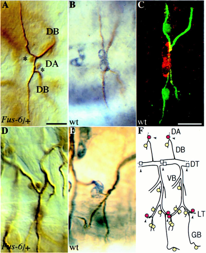
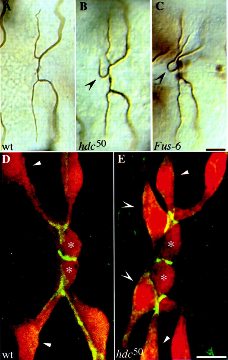
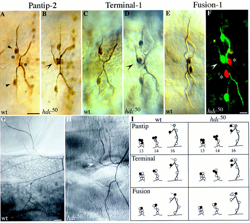
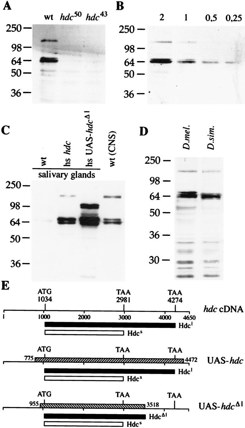
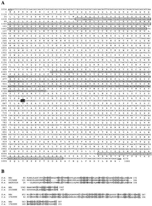
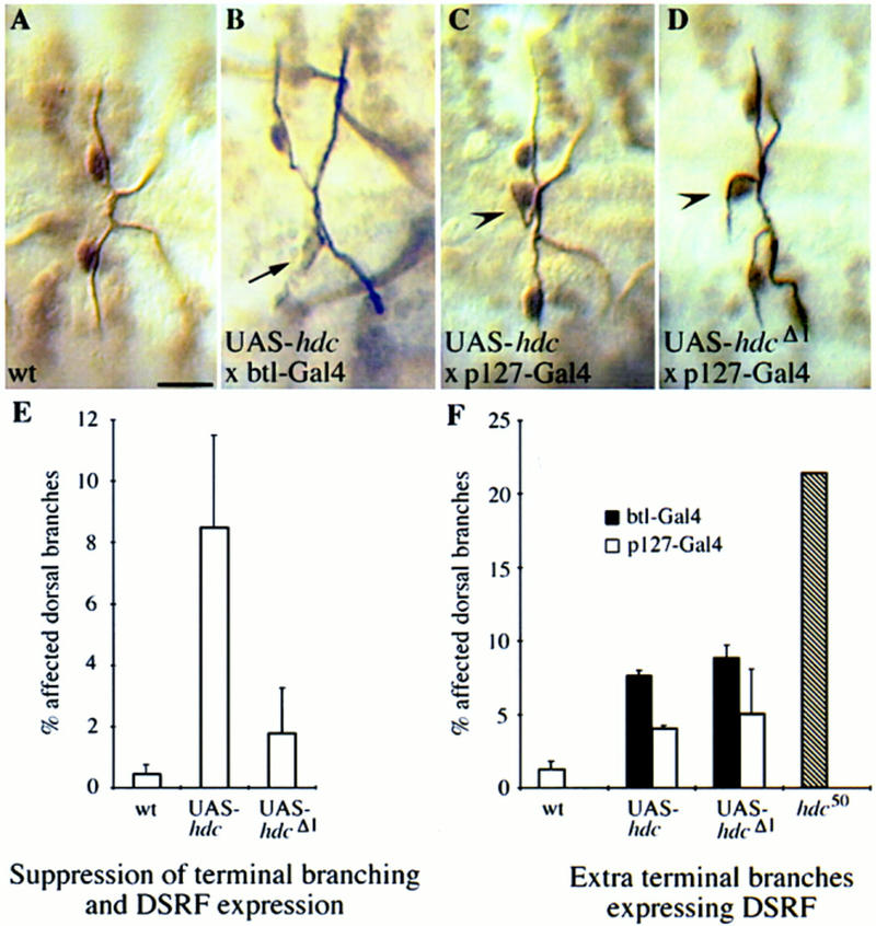
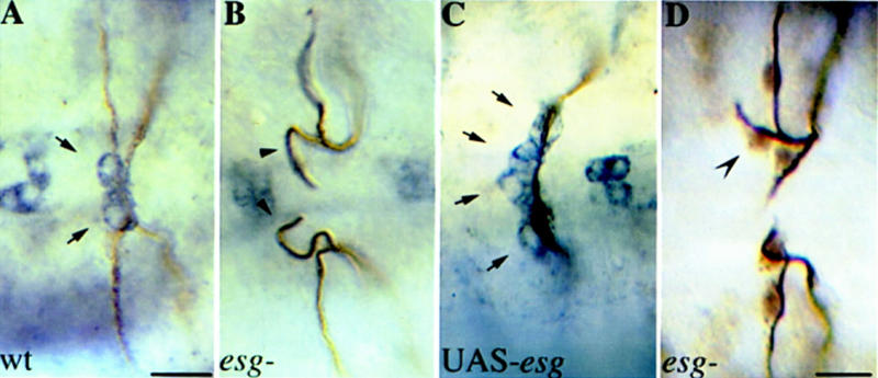
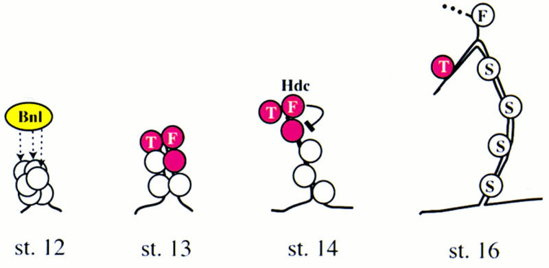
A central question in the development of many branched tubular organs, including the Drosophila trachea, concerns the mechanisms and molecules that control the number and pattern of new branches arising from preexisting vessels. We report on a branching inhibitor, Fusion-6 (Fus-6) produced by specialized tracheal cells to prevent neighboring cells from branching. In Fus-6 mutants, cells that are normally quiescent acquire the branching fate and form an increased number of sprouts emanating from the primary branches. Fus-6 is identified as the headcase (hdc) gene and is expressed in a subset of the cells that extend fusion sprouts to interconnect the tracheal network. hdc expression is regulated by the transcription factor escargot (esg) because it is not expressed in the fusion cells of esg mutants and is ectopically activated in the trachea in response to esg misexpression. We show that the hdc mRNA encodes two overlapping protein products by an unusual suppression of translational termination mechanism. Translational readthrough is necessary for hdc function because rescue of the tracheal mutant phenotype requires the full-length hdc mRNA. In ectopic expression experiments with full-length and truncated hdc constructs, only the full-length cDNA encoding both proteins could inhibit terminal branching. We propose that hdc acts non-autonomously in an inhibitory signaling mechanism to determine the number of cells that will form unicellular sprouts in the trachea.


Figures

Figure 1
Figure 1
Expression of Fus-6 (hdc) in a subset of the tracheal fusion cells. The tracheal lacZ expression in the Fus-6 enhancer trap strain is identical to the expression of hdc. (A,D) Fus-6 embryos have been stained with antibodies against β-galactosidase and a tracheal lumenal antigen, (B,E) wild-type embryos were stained with antibodies against Hdc in purple and the tracheal lumen in brown. (A) Dorsal view (anterior left) focused at the dorsal anastomosis (DA). The nuclear lacZ marker is selectively expressed in the two fusion cells (indicated by asterisks) of the dorsal branches (DB) connecting two contralateral metameres. (B) hdc is expressed in the fusion cells forming the DA. lacZ (D) and hdc (E) expression in the fusion cells connecting the lateral trunk (lateral view, anterior left). (C) Hdc is cytoplasmic in the fusion cells of the dorsal anastomosis in contrast to the nuclear localization of DSRF in the adjacent terminal cells (dorsal view, anterior left). Embryos have been stained with antibodies against Hdc (red), DSRF (green), and tracheal lumen (green). (F) Schematic diagram of two central abdominal tracheal metameres (lateral view, anterior left) showing the cells expressing hdc as darker (red) circles and the neighboring cells expressing terminal markers as lighter (yellow) circles. Fusion points at the dorsal anastomosis (DA), lateral trunk (LT), and dorsal trunk (DT) are indicated by arrowheads. hdc is not expressed in the fusion cells of the DT (squares) that do not contact terminal cells. Bar in A and D, 10 μm; in B, C, and E, 10 μm.
Figure 2
Figure 2
Tracheal phenotypes in Fus-6 (hdc) mutants. Additional sprouts emanate from the DBs in hdc and Fus-6 mutants. Dorsal views, anterior to the left, of wild-type and mutant embryos at stage 16. Wild-type (A), hdc50 homozygous (B), and Fus-6 homozygous (C) embryos stained with antibodies against the lumenal antigen and β-galactosidase. Extra branches are marked by concave arrowheads. Bar, 10 μm. Extra cells sprout off the DBs in hdc50 embryos. (D,E) Confocal images of the DB in wild-type (1-eve-1/+) and hdc50 (1-eve-1, hdc50/hdc50) embryos showing tracheal cells (anti-β-galactosidase in red) and cell junctions (anti-Coracle antiserum in green). The fusion cells are marked by asterisks, the terminal sprouts in the wild type by arrowheads, and the extra sprouting cells in the mutant by concave arrowheads. Bar, 5 μm.
Figure 3
Figure 3
Expression of branching markers in hdc mutants. The extra sprouting cells express pantip and terminal but not fusion cell markers. Dorsal views, anterior to the left, of wild-type and hdc mutant embryos at stage 16. For the pantip and fusion markers, wild-type and hdc50 mutant embryos carrying one copy of the marker gene were double stained for β-galactosidase and the tracheal lumen. For the terminal marker panels, wild-type and hdc mutant embryos were triple stained with antibodies against the lumen, β-galactosidase, and DSRF. (A) A pair of contralateral DBs in a wild-type embryo, each expressing the pantip-2 marker strongly in the terminal (arrowheads) and weaker in the fusion (asterisks) cells. (B) Similar view of a hdc50 mutant embryo showing that the extra sprouting cell strongly expresses the pantip marker (concave arrowhead). (C,D) DBs of wild-type and hdc50 embryos stained for the DSRF terminal marker. The extra sprouting cell expressing DSRF is indicated by a concave arrowhead. (E,F) Expression of the Fus-1 marker in the fusion cells of wild-type DBs in E and hdc50 mutant in F. In the confocal micrograph in F, hdc50 embryos were stained both for the Fus-1 marker in red, and the terminal DSRF marker and tracheal lumen in green to show that the extra sprouting cell (concave arrowhead) expressed terminal but not fusion markers. Fusion cells are indicated by asterisks. Bar in A–E, 10 μm; in F, 5 μm. (G,H) Extra sprouts in hdc mutants ramify in to tracheoles during larval life. (G) Pair of DBs in a wild-type 3rd instar larva viewed by Nomarski optics, showing the extensive fine tracheoles emanating from each terminal branching cell. (H) Same view of a hdc50 mutant larva with an additional terminal branch extending tracheoles (concave arrowhead). Bar, 50 μm. (I) Schematic summary of pantip, terminal, and fusion marker expression in the wild-type and hdc mutants. The extra sprouting cells express pantip and terminal markers at stage 14.
Figure 4
Figure 4
Translational readthrough generates two hdc products. Western blots probed with the antibody against the hdc products (A). The antibody recognizes two proteins in CNS and imaginal disk extracts from wild-type larvae. Both bands are absent in extracts from larvae homozygous for hdc50 and hdc43. (B) Quantitation of the amount of the two products relative to each other. Serial dilutions of a CNS and imaginal disk extract were loaded. The shorter product is ∼fourfold more abundant than the longer one. (C) Full-length cDNA is necessary to generate a product with the same size as the longer endogenous protein. Extracts of salivary glands from transgenic larvae carrying the full-length or a truncated version of the hdc cDNA under heat shock control were compared with CNS and imaginal disk extracts from wild type. (D) Two Hdc proteins are detected in extracts from D. simulans larval CNS and disks. (E) Drawings of the hdc cDNA clone and the fragments used to generate the transgenic strains shown as hatched bars. Start and stop codons are indicated and the predicted products are shown as black or white bars under each construct. Hdcl designates the longer product, and Hdcs the shorter one.
Figure 5
Figure 5
(A) Deduced amino acid sequence of the hdc longer product. The nucleotide sequence derives from the cDNA sequence in Weaver and White (1995) and our longest, partially sequenced, cDNA clone. Start and stop codons are underlined and X is used as a symbol for the unknown amino acid incorporated at the internal UAA stop codon. The different regions homologous to the two C. elegans ORFs are highlighted. (B) BLASTP amino acid sequence comparisons of the hdc products to the two C. elegans ORFs and the human EST sequence. The C. elegans ORF 1763999 is homologous (34% identity, 60% similarity) to the region between amino acids 453–587 in the Hdc sequence shared by both products (highlighted in gray in A). The C. elegans ORF 2315488 and the ORF deriving from the human EST are homologous to a region between amino acids 85 and 172 (38% identity, 63% homology) in the Hdc sequence shared by both products (highlighted in a gray box in A). The same C. elegans ORF is also homologous (36% identity, 56% homology) to a region between amino acids 1042 and 1067 unique to the carboxyl terminus of the longer product (highlighted in a gray box in A).
Figure 6
Figure 6
Misexpression of hdc in all tracheal cells can suppress terminal branching. The GAL4 expression system was used to express wild-type and mutant Hdc proteins throughout the developing tracheal system (B,E,F) and selectively in the fusion cells (C,D,F) of hdc+ embryos. Expressing UAS–hdc in all tracheal cells with the pantracheal btl–GAL4 driver results in suppression of terminal branching and expression of the terminal marker DSRF (arrow in B). (E) Comparison of the terminal branching suppression phenotype of UAS–hdc and UAS–hdcΔl. The full-length construct is necessary for suppression. (C) Expression of UAS–hdc with the p127-GAL4 fusion cell-specific driver or the pantracheal btl–GAL4 driver causes extra branching phenotypes similar to the hdc50 loss-of-function mutant (arrowheads). Expression of the UAS–hdcΔ1 construct with the same drivers results in a similar phenotype (D,F). Bar in A–D, 10 μm.
Figure 7
Figure 7
hdc expression is regulated by esg. (A–C) Embryos double stained with antibodies against the tracheal lumen in brown and Hdc in blue. (A) Dorsal view focusing on hdc expression in the dorsal branches (DBs) of wild type, (B) esg mutant, and (C) C38–GAL4/UAS-esg embryos. (Arrows in A) hdc expression in the two fusion cells of wild-type embryos. hdc expression was not detectable in esg mutants (Arrowheads in B), whereas in UAS-esg embryos, hdc is expressed in four cells at the tip of the DBs (Arrows in C). Bar in A–C, 10 μm. (D) Dorsal view of an esg mutant embryo stained with antibodies against tracheal lumen and DSRF. A third branching cell expressing DSRF is indicated by a concave arrowhead. Bar, 10 μm.
Figure 8
Figure 8
Model of the inhibitory action of hdc on sprouting. Secreted Bnl protein (dashed arrows) activates the expression of pantip marker genes (red) in the tip cells of the dorsal primary branch closest to the source of the signal. Single cells of the pantip group are selected to express terminal (T) or fusion (F) cell-specific markers. The fusion cell expresses hdc to inhibit the Bnl mediated induction of terminal branching in the remaining cell of the pantip group, which later becomes a stalk cell (S).

References

    1. Ashburner M, Thompson P, Roote J, Lasko PF, Grau Y, El Messal M, Roth S, Simpson P. The genetics of a small autosomal region of Drosophila melanogaster containing the structural gene for alcohol dehydrogenase. VII. Characterization of the region around the snail and cactus loci. Genetics. 1990;126:679–694. - PMC - PubMed
    1. Bergstrom D, Merli C, Cygan J, Shelby R, Blackman R. Regulatory autonomy and molecular characterization of the Drosophila out at first gene. Genetics. 1995;139:1331–1346. - PMC - PubMed
    1. Böck A, Forchhammer K, Heider J, Baron C. Selenoprotein synthesis: An expansion of the genetic code. Trends Biochem Sci. 1991;16:463–467. - PubMed
    1. Bodmer R, Carretto R, Jan Y. Neurogenesis of the peripheral nervous system in Drosophila embryos: DNA replication patterns and cell lineages. Neuron. 1989;3:21–32. - PubMed
    1. Brand AH, Perrimon N. Targeted gene expression as a means of altering cell fates and generating dominant phenotypes. Development. 1993;118:401–415. - PubMed

Publication types