. 1998 Apr 1;12(7):996-1009.
doi: 10.1101/gad.12.7.996.

A coactivator of pre-mRNA splicing

Affiliations

A coactivator of pre-mRNA splicing

B J Blencowe et al. Genes Dev. .

Abstract

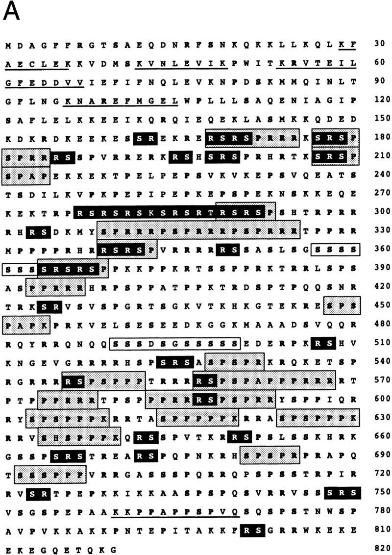
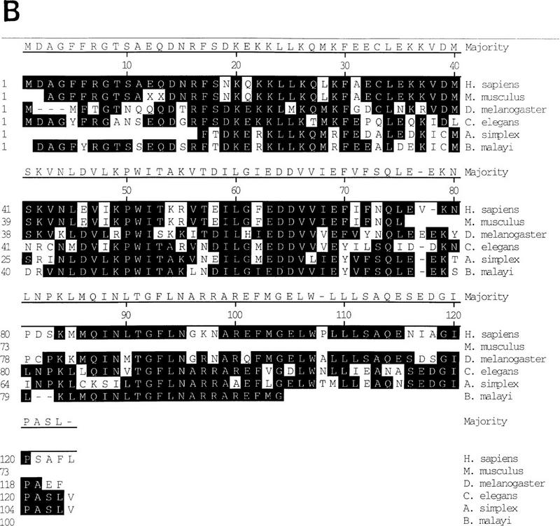
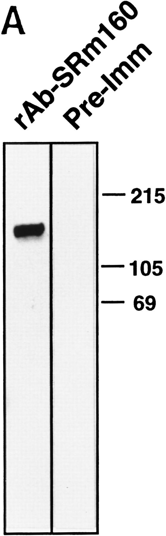
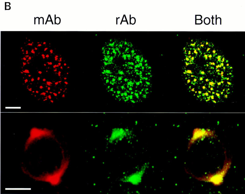
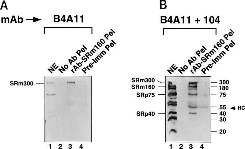
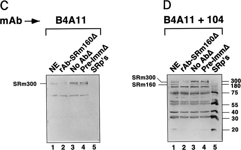
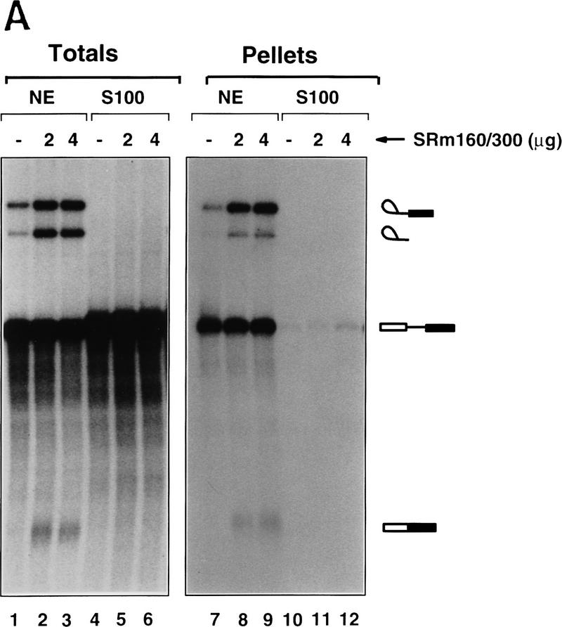
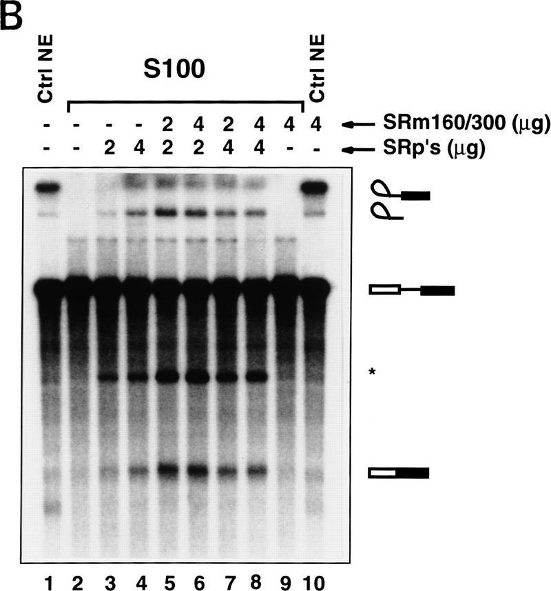
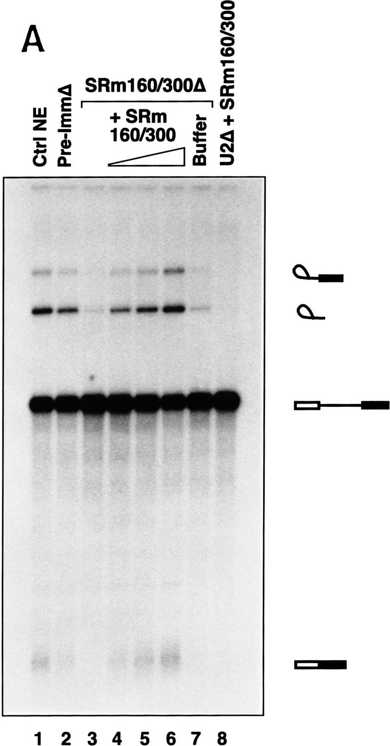
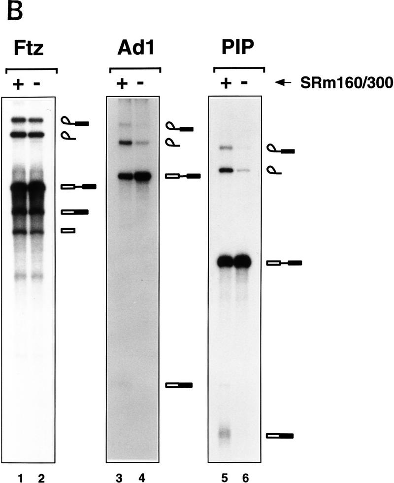
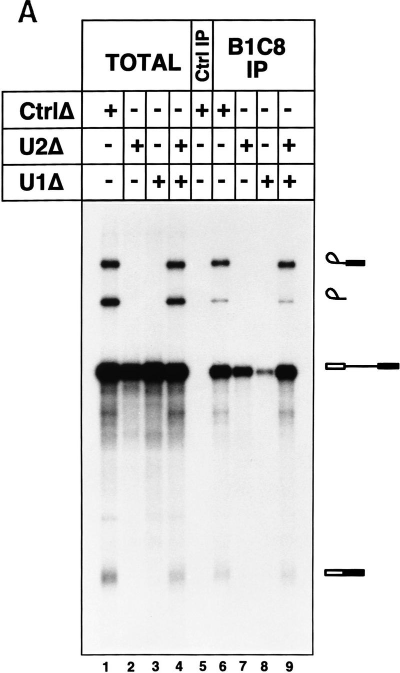
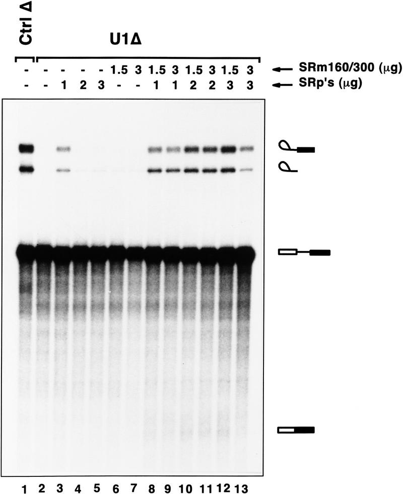
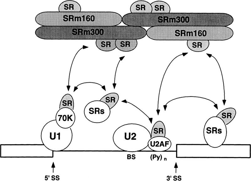
The nuclear matrix antigen recognized by the monoclonal antibody (mAb) B1C8 is a novel serine (S) and arginine (R)-rich protein associated with splicing complexes and is named here SRm160 (SR-related matrix protein of 160 kD). SRm160 contains multiple SR repeats, but unlike proteins of the SR family of splicing factors, lacks an RNA recognition motif. SRm160 and a related protein SRm300 (the 300-kD nuclear matrix antigen recognized by mAb B4A11) form a complex that is required for the splicing of specific pre-mRNAs. The SRm160/300 complex associates with splicing complexes and promotes splicing through interactions with SR family proteins. Binding of SRm160/300 to pre-mRNA is normally also dependent on U1 snRNP and is stabilized by U2 snRNP. Thus, SRm160/300 forms multiple interactions with components bound directly to important sites within pre-mRNA. The results suggest that a complex of the nuclear matrix proteins SRm160 and SRm300 functions as a coactivator of pre-mRNA splicing.


Figures

Figure 1
Figure 1
The SRm160 ORF and related sequences. (A) Amino acid sequence of the SRm160 ORF. Arginine (R), serine (S), and proline (P) amino acid residues are highlighted in bold-face type. SR/RS dipeptides are highlighted in black boxes. Serine-rich domains are highlighted in white boxes. Other repeat motifs containing P, R, or S residues are highlighted in gray boxes (see Table 1). The latter represent only the most frequently occurring repeats and are a subset of the total repeats identified as statistically significant by the statistical analysis of protein sequence (SAPS) algorithm (Brendel et al. 1992). Microsequences of lys-C peptides obtained from purified SRm160 protein are underlined. The SRm160 cDNA sequence has been deposited in GenBank (accession no. AF048977). (B) Multiple alignment of sequences homologous to the amino-terminal 150 amino acids of human SRm160. Partial cDNA ORF sequences from mouse (Mus musculus), fly (D. melanogaster), and three namatode species (C. elegans, A. simplex, and B. malayi) identified by BLAST searches were aligned using the Clustal algorithm. Sequences included in the alignment correspond to the following GenBank accession numbers: M. musculus, W67104; D. melanogaster, AA567455, AA440655, AA539347; C. elegans, D76066, C43686; A. simplex, S51495; B. malayi, AA089428. Residues identical to the majority sequence (top line) are shaded in black. Amino-terminal residues not present in the EST cDNAs are indicated by a blank space; gaps are indicated by a dash, and undefined residues are indicated by an X.
Figure 1
Figure 1
The SRm160 ORF and related sequences. (A) Amino acid sequence of the SRm160 ORF. Arginine (R), serine (S), and proline (P) amino acid residues are highlighted in bold-face type. SR/RS dipeptides are highlighted in black boxes. Serine-rich domains are highlighted in white boxes. Other repeat motifs containing P, R, or S residues are highlighted in gray boxes (see Table 1). The latter represent only the most frequently occurring repeats and are a subset of the total repeats identified as statistically significant by the statistical analysis of protein sequence (SAPS) algorithm (Brendel et al. 1992). Microsequences of lys-C peptides obtained from purified SRm160 protein are underlined. The SRm160 cDNA sequence has been deposited in GenBank (accession no. AF048977). (B) Multiple alignment of sequences homologous to the amino-terminal 150 amino acids of human SRm160. Partial cDNA ORF sequences from mouse (Mus musculus), fly (D. melanogaster), and three namatode species (C. elegans, A. simplex, and B. malayi) identified by BLAST searches were aligned using the Clustal algorithm. Sequences included in the alignment correspond to the following GenBank accession numbers: M. musculus, W67104; D. melanogaster, AA567455, AA440655, AA539347; C. elegans, D76066, C43686; A. simplex, S51495; B. malayi, AA089428. Residues identical to the majority sequence (top line) are shaded in black. Amino-terminal residues not present in the EST cDNAs are indicated by a blank space; gaps are indicated by a dash, and undefined residues are indicated by an X.
Figure 2
Figure 2
Association of SRm160 with interphase nucleoplasmic speckles, foci, and the mitotic spindle apparatus. (A) Total HeLa nuclear extract was separated on an SDS–polyacrylamide gel and immunoblotted with an affinity-purified polyclonal antiserum raised to a GST–SRm160 fusion protein containing SRm160 ORF amino acids 7–160 (rAb–SRm160, lane 1), and the corresponding preimmune serum (lane 2). (B) Interphase (top row) and metaphase (bottom row) human CaSki cells were double-immunolabeled with mAb B1C8 (red) and rAb–SRm160 (green) and visualized by confocal microscopy. The images were superimposed to reveal sites of overlap (yellow). The interphase image corresponds to a single confocal section proximal to the midline of the nucleus. The metaphase image corresponds to a stack of merged sections through the whole cell. Bar, 5 μm.
Figure 2
Figure 2
Association of SRm160 with interphase nucleoplasmic speckles, foci, and the mitotic spindle apparatus. (A) Total HeLa nuclear extract was separated on an SDS–polyacrylamide gel and immunoblotted with an affinity-purified polyclonal antiserum raised to a GST–SRm160 fusion protein containing SRm160 ORF amino acids 7–160 (rAb–SRm160, lane 1), and the corresponding preimmune serum (lane 2). (B) Interphase (top row) and metaphase (bottom row) human CaSki cells were double-immunolabeled with mAb B1C8 (red) and rAb–SRm160 (green) and visualized by confocal microscopy. The images were superimposed to reveal sites of overlap (yellow). The interphase image corresponds to a single confocal section proximal to the midline of the nucleus. The metaphase image corresponds to a stack of merged sections through the whole cell. Bar, 5 μm.
Figure 3
Figure 3
A complex of the SRm160 and SRm300 nuclear matrix proteins (SRm160/300) associates with SR family proteins comigrating with SRp40 and SRp75. (A,B) Proteins immunoprecipitated from HeLa nuclear extract by rAb–SRm160 were separated by SDS-PAGE, blotted, and probed with mAb B4A11 (A), followed by mAb104 (B). Protein immunoprecipitated from ∼0.5 mg of nuclear extract was loaded in lanes 2–4 and ∼0.15 mg of total nuclear extract was loaded in lane 1. Immunoprecipitations were performed with protein A beads alone (lane 2), rAb–SRm160 (lane 3), and preimmune serum (lane 4). Sizes of SR proteins SRm160 and SRm300 are indicated in kD. (HC) immunoglobulin heavy chain. (C,D) Supernatant fractions from the immunoprecipitations in A and B were separated and immunoblotted with mAbs B4A11 and 104, as in A and B. Total nuclear extract proteins are shown in lanes 1, and proteins recovered from the immunoprecipitation supernatant fractions prepared with rAb–SRm160, protein A beads alone and preimmune serum, are shown in lanes 2, 3, and 4, respectively. Approximately 0.15 mg of nuclear extract and of each supernatant fraction was loaded per lane. A sample of purified SR family proteins (∼4 μg) was separated as a marker in lane 5.
Figure 3
Figure 3
A complex of the SRm160 and SRm300 nuclear matrix proteins (SRm160/300) associates with SR family proteins comigrating with SRp40 and SRp75. (A,B) Proteins immunoprecipitated from HeLa nuclear extract by rAb–SRm160 were separated by SDS-PAGE, blotted, and probed with mAb B4A11 (A), followed by mAb104 (B). Protein immunoprecipitated from ∼0.5 mg of nuclear extract was loaded in lanes 2–4 and ∼0.15 mg of total nuclear extract was loaded in lane 1. Immunoprecipitations were performed with protein A beads alone (lane 2), rAb–SRm160 (lane 3), and preimmune serum (lane 4). Sizes of SR proteins SRm160 and SRm300 are indicated in kD. (HC) immunoglobulin heavy chain. (C,D) Supernatant fractions from the immunoprecipitations in A and B were separated and immunoblotted with mAbs B4A11 and 104, as in A and B. Total nuclear extract proteins are shown in lanes 1, and proteins recovered from the immunoprecipitation supernatant fractions prepared with rAb–SRm160, protein A beads alone and preimmune serum, are shown in lanes 2, 3, and 4, respectively. Approximately 0.15 mg of nuclear extract and of each supernatant fraction was loaded per lane. A sample of purified SR family proteins (∼4 μg) was separated as a marker in lane 5.
Figure 4
Figure 4
Pre-mRNA binding and promotion of splicing by SRm160/300 requires SR family proteins. (A) Immunoprecipitations were performed from splicing reactions incubated with PIP85A pre-mRNA for 40 min. The splicing reactions contained nuclear (lanes 1–3,7–9) or S100 cytoplasmic (lanes 4–6,10–12) extract, with (lanes 2,3,5,6,8,9,11,12) or without (lanes 1,4,7,10) purified SRm160/300 proteins added; the preparation of SRm160/300 is as described in Fig. 4, Blencowe et al. (1995). Fifty percent of the total RNA recovered directly from each splicing reaction (Totals) was loaded in lanes 1–6), whereas all of the RNA recovered after immunoprecipitation with mAb B1C8 (Pellets) was loaded in lanes 7–12. (B) S100 splicing reactions (lanes 2–9) containing β-globin pre-mRNA and varying amounts of SRm160/300 and SR family proteins, as indicated, were incubated for 1 hr. Control splicing reactions incubated in parallel contained nuclear extract (lane 1) or S100 extract (lane 2), without added proteins, and a reaction containing nuclear extract and 4 μg SRm160/300 (lane 10). The asterisk (*) indicates an RNA fragment protected from endogenous nuclease activity.
Figure 4
Figure 4
Pre-mRNA binding and promotion of splicing by SRm160/300 requires SR family proteins. (A) Immunoprecipitations were performed from splicing reactions incubated with PIP85A pre-mRNA for 40 min. The splicing reactions contained nuclear (lanes 1–3,7–9) or S100 cytoplasmic (lanes 4–6,10–12) extract, with (lanes 2,3,5,6,8,9,11,12) or without (lanes 1,4,7,10) purified SRm160/300 proteins added; the preparation of SRm160/300 is as described in Fig. 4, Blencowe et al. (1995). Fifty percent of the total RNA recovered directly from each splicing reaction (Totals) was loaded in lanes 1–6), whereas all of the RNA recovered after immunoprecipitation with mAb B1C8 (Pellets) was loaded in lanes 7–12. (B) S100 splicing reactions (lanes 2–9) containing β-globin pre-mRNA and varying amounts of SRm160/300 and SR family proteins, as indicated, were incubated for 1 hr. Control splicing reactions incubated in parallel contained nuclear extract (lane 1) or S100 extract (lane 2), without added proteins, and a reaction containing nuclear extract and 4 μg SRm160/300 (lane 10). The asterisk (*) indicates an RNA fragment protected from endogenous nuclease activity.
Figure 5
Figure 5
SRm160/300 is required for splicing of specific pre-mRNAs. (A) Depletion of SRm160/300 blocks the first step of splicing of PIP85A pre-mRNA. Splicing reactions containing nuclear extract depleted of SRm160/300 proteins (see Fig. 3C,D, lane 2) were incubated for 1 hr with PIP85A pre-mRNA in the presence (lanes 4–6), or absence (lanes 3,7), of purified SRm160/300 proteins. Control splicing reactions contained regular nuclear extract (lane 1), nuclear extract mock-depleted with preimmune serum (lane 2; see Fig. 3C,D, lane 4), SRm160/300-depleted extract plus protein buffer (lane 7), and U2 snRNP-depleted nuclear extract plus SRm160/300 proteins (lanes 8). Splicing reactions were performed in 30-μl reactions containing a range of 2–8 μg of SRm160/300 proteins in lanes 4–6 and 8 μg of SRm160/300 proteins in lane 8. (B) Comparision of the splicing activity of different pre-mRNAs in mock depleted (lanes 1,3,5) versus SRm160/300-depleted (lanes 2,4,6) nuclear extracts. Reactions were incubated for 1 hr with the Drosophila fushi tarazu (ftz, lanes 1,2), adenovirus major late (Ad1; lanes 3,4), and PIP85A (PIP; lanes 5,6) pre-mRNAs.
Figure 5
Figure 5
SRm160/300 is required for splicing of specific pre-mRNAs. (A) Depletion of SRm160/300 blocks the first step of splicing of PIP85A pre-mRNA. Splicing reactions containing nuclear extract depleted of SRm160/300 proteins (see Fig. 3C,D, lane 2) were incubated for 1 hr with PIP85A pre-mRNA in the presence (lanes 4–6), or absence (lanes 3,7), of purified SRm160/300 proteins. Control splicing reactions contained regular nuclear extract (lane 1), nuclear extract mock-depleted with preimmune serum (lane 2; see Fig. 3C,D, lane 4), SRm160/300-depleted extract plus protein buffer (lane 7), and U2 snRNP-depleted nuclear extract plus SRm160/300 proteins (lanes 8). Splicing reactions were performed in 30-μl reactions containing a range of 2–8 μg of SRm160/300 proteins in lanes 4–6 and 8 μg of SRm160/300 proteins in lane 8. (B) Comparision of the splicing activity of different pre-mRNAs in mock depleted (lanes 1,3,5) versus SRm160/300-depleted (lanes 2,4,6) nuclear extracts. Reactions were incubated for 1 hr with the Drosophila fushi tarazu (ftz, lanes 1,2), adenovirus major late (Ad1; lanes 3,4), and PIP85A (PIP; lanes 5,6) pre-mRNAs.
Figure 6
Figure 6
snRNP dependence for binding of SRm160/300 to pre-mRNA. (A) Splicing complexes were immunoprecipitated with mAb B1C8 from different snRNP-depleted and nondepleted reactions (lanes 6–9). Splicing reactions incubated for 40 min with PIP85A pre-mRNA contained mock-depleted (lane 1), U2-depleted (lane 2), U1-depleted (lane 3), or an equal mix of U1- and U2-depleted nuclear extracts (lane 4). RNAs recovered directly from the splicing reactions in lanes 1–4 represent 50% of the total sample, whereas all of the RNAs recovered from each immunoprecipitation were loaded in lanes 5–9. A control immunoprecipitation (lane 5) was performed with a nonspecific antibody from a splicing reaction containing mock-depleted nuclear extract, as in lane 1. (B) Complexes assembled on separate 5′ and 3′ halves of PIP85A pre-mRNA were immunoprecipitated with mAb B1C8 in the presence or absence of individual snRNPs (lanes 6–9). Equal amounts of the two half RNAs were incubated under splicing conditions for 40 min in mock-depleted (lanes 1,2), U2-depleted (lane 3), U1-depleted (lane 4), or an equal mix of U1- and U2-depleted nuclear extracts (lane 5). RNA recovered directly from the splicing reactions in lanes 1–5 represents 50% of the total sample, whereas all of the recovered from each immunoprecipitation was loaded (lanes 6–9); a control immunoprecipitation (lane Ctrl IP) was performed with a nonspecific antibody from a reaction containing mock-depleted nuclear extract (lane 1).
Figure 6
Figure 6
snRNP dependence for binding of SRm160/300 to pre-mRNA. (A) Splicing complexes were immunoprecipitated with mAb B1C8 from different snRNP-depleted and nondepleted reactions (lanes 6–9). Splicing reactions incubated for 40 min with PIP85A pre-mRNA contained mock-depleted (lane 1), U2-depleted (lane 2), U1-depleted (lane 3), or an equal mix of U1- and U2-depleted nuclear extracts (lane 4). RNAs recovered directly from the splicing reactions in lanes 1–4 represent 50% of the total sample, whereas all of the RNAs recovered from each immunoprecipitation were loaded in lanes 5–9. A control immunoprecipitation (lane 5) was performed with a nonspecific antibody from a splicing reaction containing mock-depleted nuclear extract, as in lane 1. (B) Complexes assembled on separate 5′ and 3′ halves of PIP85A pre-mRNA were immunoprecipitated with mAb B1C8 in the presence or absence of individual snRNPs (lanes 6–9). Equal amounts of the two half RNAs were incubated under splicing conditions for 40 min in mock-depleted (lanes 1,2), U2-depleted (lane 3), U1-depleted (lane 4), or an equal mix of U1- and U2-depleted nuclear extracts (lane 5). RNA recovered directly from the splicing reactions in lanes 1–5 represents 50% of the total sample, whereas all of the recovered from each immunoprecipitation was loaded (lanes 6–9); a control immunoprecipitation (lane Ctrl IP) was performed with a nonspecific antibody from a reaction containing mock-depleted nuclear extract (lane 1).
Figure 7
Figure 7
SRm160/300 coactivates splicing in U1 snRNP-depleted reactions supplemented with SR family proteins. SRm160/300 proteins were added alone (lanes 6,7) or in combination with SR family proteins (lanes 8–13) to splicing reactions depleted of U1 snRNP. U1-depleted reactions containing excess SR proteins and no SRm160/300 are shown in lanes 3–5. Control reactions containing mock-depleted extract or U1-depleted extract with no added proteins are shown in lanes 1 and 2, respectively. The splicing reactions were incubated for 60 min before recovery of RNA.
Figure 8
Figure 8
Splicing coactivator model for the function of SRm160/300. SRm160/300 promotes splice-site pairing and splicing through multiple cooperative interactions with factors bound to pre-mRNA, including SR family proteins U1 and U2 snRNPs. A role for the RS domains of these factors in mediating an intron-wide network of interactions is emphasized. These early interactions are required for the splicing of specific pre-mRNAs. (BS) Branch site; [(Py)n] polypyrimidine tract; (SS) splice site.

References

    1. Abovich N, Rosbash M. Cross-intron bridging interactions in the yeast commitment complex are conserved in mammals. Cell. 1997;89:403–412. - PubMed
    1. Alzhanova-Ericsson AT, Sun X, Visa N, Kiseleva E, Wurtz T, Daneholt B. A protein of the SR family binds extensively to exonic Balbiani ring pre-mRNA and accompanies the RNA from gene to the nuclear pore. Genes & Dev. 1996;10:2881–2893. - PubMed
    1. Amrein H, Hedley ML, Maniatis T. The role of specific protein-RNA interactions and protein-protein interactions in positive and negative control of pre-mRNA splicing by transformer 2. Cell. 1994;76:735–746. - PubMed
    1. Barabino SML, Blencowe BJ, Ryder U, Sproat BS, Lamond AI. Targeted snRNP depletion reveals an additional role for mammalian U1 snRNP in spliceosome assembly. Cell. 1990;63:293–302. - PubMed
    1. Berget SM. Exon recognition in vertebrate splicing. J Biol Chem. 1995;270:2411–2414. - PubMed

Publication types

MeSH terms

Associated data