

Review
doi: 10.1083/jcb.141.1.1.
Cell crawling: first the motor, now the transmission
Affiliations
- PMID: 9531543
- PMCID: PMC2132726
- DOI: 10.1083/jcb.141.1.1
Item in Clipboard
Review
Cell crawling: first the motor, now the transmission
J Cell Biol.
.
No abstract available
Figures
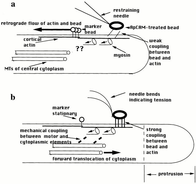
Highly schematic diagram of the forces and mechanical connections inferred from the results of Suter et al. (19). (a) During the latency period after attachment of ApCAM-coated beads, restraint of the bead causes it to stop, with no evidence of tension exerted in the needle. Retrograde actin flow continues uninterrupted, suggesting a weak, slipping interaction between the bead and the underlying actin network. The question marks are intended to illustrate that the results provide no information about the presence or absence of connection(s) between the central cytoplasm and the myosin driving retrograde flow. (b) After a latency period, firm connections develop between the ApCAM bead and the actin, associated with clustering of ApCAM beneath the bead. Restraint of the bead now causes development of tension in the needle and retrograde flow to stop. Because the actin is no longer able to slip relative to the surface, the tension generated by the myosin motors is now accomodated by forward flow of cytoplasm at the leading edge and in the central microtubule-containing cytoplasm. This indicates mechanically resistant connections among the surface, actin, myosin motors, and surrounding cytoplasm. See text for further explanation.
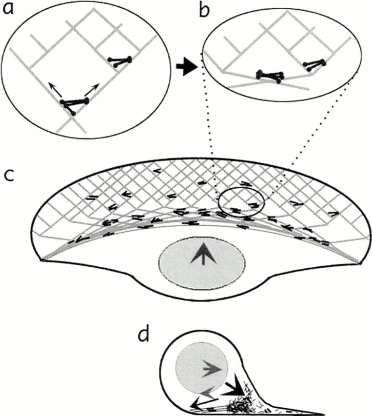
Diagram illustrating the dynamic network model of Svitkina et al. (20) in a locomoting fish keratocyte. (a and b) A network of actin (light grey lines) and myosin (dark bipolar filaments) contracts causing reorganization of the network. Myosin is clustered and actin is brought into alignment as parallel bundles. (c) Seen at the level of the entire cell, this network contraction causes forward translocation of the cell body. (d) In this cross-section, the forward rolling of the cell body is seen as a combination of the forward-directed force and a drag force at the bottom of the cell body/nucleus created by the accumulation of contracted network. Similar to nondrive wheels of a car, the combination of forward force and dragging along the bottom surface causes rotation of a rounded object.
Comment on
-
The Ig superfamily cell adhesion molecule, apCAM, mediates growth cone steering by substrate-cytoskeletal coupling.J Cell Biol. 1998 Apr 6;141(1):227-40. doi: 10.1083/jcb.141.1.227. J Cell Biol. 1998. PMID: 9531561 Free PMC article.
