. 1998 Apr 6;141(1):21-9.
doi: 10.1083/jcb.141.1.21.

Yeast nuclei display prominent centromere clustering that is reduced in nondividing cells and in meiotic prophase

Affiliations

Yeast nuclei display prominent centromere clustering that is reduced in nondividing cells and in meiotic prophase

Q Jin et al. J Cell Biol. .

Abstract

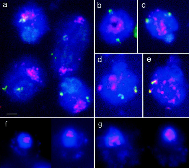
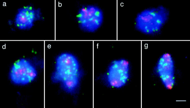
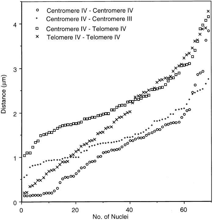
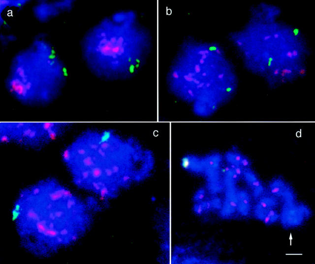
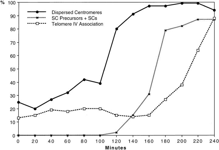
Chromosome arrangement in spread nuclei of the budding yeast, Saccharomyces cerevisiae was studied by fluorescence in situ hybridization with probes to centromeres and telomeric chromosome regions. We found that during interphase centromeres are tightly clustered in a peripheral region of the nucleus, whereas telomeres tend to occupy the area outside the centromeric domain. In vigorously growing cultures, centromere clustering occurred in approximately 90% of cells and it appeared to be maintained throughout interphase. It was reduced when cells were kept under stationary conditions for an extended period. In meiosis, centromere clusters disintegrated before the emergence of the earliest precursors of the synaptonemal complex. Evidence for the contribution of centromere clustering to other aspects of suprachromosomal nuclear order, in particular the vegetative association of homologous chromosomes, is provided, and a possible supporting role in meiotic homology searching is discussed.


Figures

Figure 1
Figure 1
Representative nuclei of SK1 cells from (a, b, f) logarithmic growth and (c–e, g) stationary phase culture. (a–e) Spread nuclei; (f and g) intact nuclei. (Red) Centromeres. Telomeric region of chromosome IVR, green. (Blue) DAPI-stained chromatin. (a) Nuclei show centromere clusters near the periphery. Telomeres are dispersed within the nuclear chromatin. A slight preference for accumulation of telomere signals in the centromere-distant domain of the nucleus was noted (see text). The nucleus right on top is at anaphase with centromeres at the two opposite poles of the elongated nucleus and telomeres IVR in between. (b) Plan view of the centromeric pole of a nucleus showing centromeres arranged in a ring. (c–e) Nuclei displaying aspects of the progressive reduction of centromere clustering in a stationary culture. (f and g) Examples of intact nuclei from three-dimensionally embedded spheroplasts. Cells from a logarithmic culture show centromere clusters (f) and cells from a stationary culture show dispersed centromeres (g). Notice the ring-like arrangement of centromeres in the left nucleus in f which is similar to b. Nuclei are delineated in strong blue, spheroplasts are lightly blue due to DAPI staining of the mitochondrial DNA. Bar, 2 μm.
Figure 2
Figure 2
FISH of centromeric (red) and telomeric (green) regions in spread mitotic nuclei of the haploid bar1 strain. (Blue) DAPI-stained chromatin. (a–c) Interphase nuclei with clustered centromeric regions. Telomeres are apparently randomly distributed in the nucleus in a, whereas in b and c there is some association of telomere signals (see text). (d–g) Mitotic nuclei showing different degrees of separation of centromere clusters (pictures taken at t = 75 min of a mitotic time course), probably representing metaphase and anaphase stages. (d) The centromere cluster is split into two equal-sized patches. (e and f) The clusters consisting of the centromeres of chromatids are separated further and most telomeres are assembled between them. (g) The nucleus is oblong with the centromeres at the most distant poles, and the telomeres are separated to the two halves of the nucleus. Note that the constriction in the middle of the dividing nucleus, that is typical of anaphase/telophase in intact cells (for example see Byers, 1981), is not retained after spheroplasting. Bar, 2 μm.
Figure 3
Figure 3
Pairwise distances between homologous chromosome regions within the centromere cluster (Centromeres IV), nonhomologous chromosome regions within the centromere cluster (Centromeres IV – Centromere-near III), the centromere and the telomere of the long arm of chromosome IV, and between the homologous telomeres IV in the diploid strain SK1. The distances between the centers of FISH signals were measured in 69 nuclei which were differentially stained for these three regions. For nonhomologous signals all four possible distances were pooled for each nucleus. All measured distances were independently plotted in increasing order, i.e., symbols on the same ordinate do not necessarily represent distances in one and the same nucleus. This format of presentation adopted from Weiner and Kleckner (1994) is highly suitable for accentuating minute differences in average distances.
Figure 4
Figure 4
Change in the nuclear architecture upon transfer to sporulation medium. (a) Early in meiosis, centromere (red) clustering is striking and telomeric regions on the long arms of homologous chromosomes IV (green) tend to be located near the nuclear periphery opposite to the centromere cluster. As can be seen from the two examples depicted in a, this polarization may bring about a closer than random association between homologous telomeres, since their distribution is restricted to a relatively small domain of the nucleus. At later time points nuclei with scattered centromeres become predominant. In early nuclei of this type, homologous telomeres IV produce separate signals (b), whereas later the telomeric signals fuse (c) in the course of meiotic pairing. For the frequencies of the various structural aspects at different time points in the meiotic culture see Fig. 5. (d) At pachytene bivalents are condensed. The red FISH signals indicate centromeres of individual bivalents. The green spot marks the end of the synapsed long arms of chromosome IV. The region to the right, which is devoid of centromere signals, is the nucleolus (arrow). Chromatin is stained blue with DAPI. Bar, 2 μm.
Figure 5
Figure 5
Meiotic time course showing the increase of frequency of nuclei with dispersed centromeres (i.e., loss of centromere clustering) and the appearance of SCs and their precursors (short axial elements and short synapsed segments). For each time point (samples taken at 0 to 240 min in sporulation medium in 20 min intervals) 100 nuclei were analyzed by FISH and light microscopy of silver-stained preparations (for example see Loidl et al., 1991) for centromere clustering, homologous associations of telomeres IVR, and presence of SCs or SC-precursors. The increase in the frequency of nuclei without clustered centromeres precedes the first appearance of SC-precursors at ∼120 min. Still later (180 min) the association of homologous chromosomal regions (telomere-near sites on chromosome IVR) above mitotic background levels indicates the onset of synapsis.

References

    1. Adachi Y, Yanagida M. Higher order chromosome structure is affected by cold-sensitive mutations in a Schizosaccharomyces pombe gene crm1 +which encodes a 115-kD protein preferentially localized in the nucleus and its periphery. J Cell Biol. 1989;108:1195–1207. - PMC - PubMed
    1. Aragón-Alcaide L, Reader S, Beven A, Shaw P, Miller T, Moore G. Association of homologous chromosomes during floral development. Curr Biol. 1997;7:905–908. - PubMed
    1. Avivi L, Feldman M. Arrangement of chromosomes in the interphase nucleus of plants. Hum Genet. 1980;55:281–295. - PubMed
    1. Bartholdi MF. Nuclear distribution of centromeres during the cell cycle of human diploid fibroblasts. J Cell Sci. 1991;99:255–263. - PubMed
    1. Bass HW, Marshall WF, Sedat JW, Agard DA, Cande WZ. Telomeres cluster de novo before the initiation of synapsis: a three-dimensional spatial analysis of telomere positions before and during meiotic prophase. JCell Biol. 1997;137:5–18. - PMC - PubMed

Publication types