. 1998 Apr 6;141(1):61-70.
doi: 10.1083/jcb.141.1.61.

Cargo selection by the COPII budding machinery during export from the ER

Affiliations

Cargo selection by the COPII budding machinery during export from the ER

M Aridor et al. J Cell Biol. .

Abstract

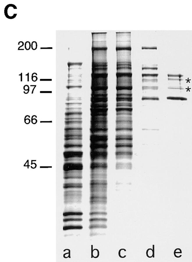
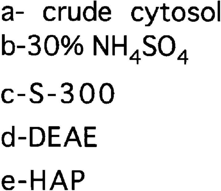
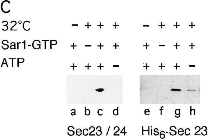
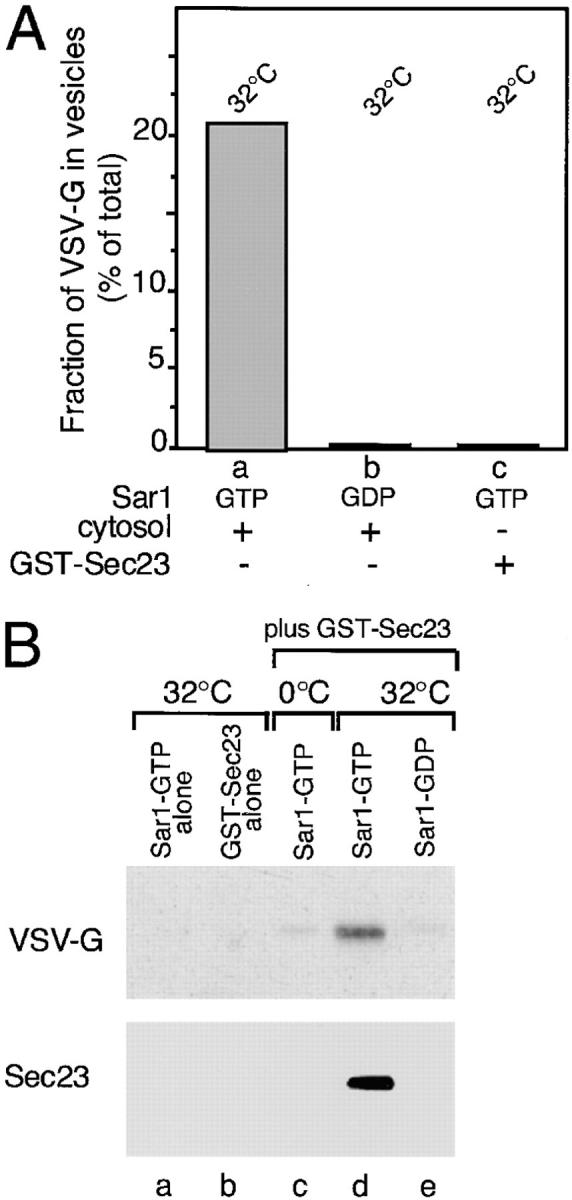
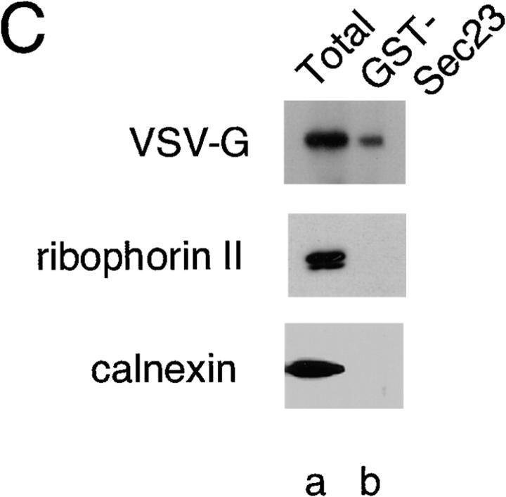
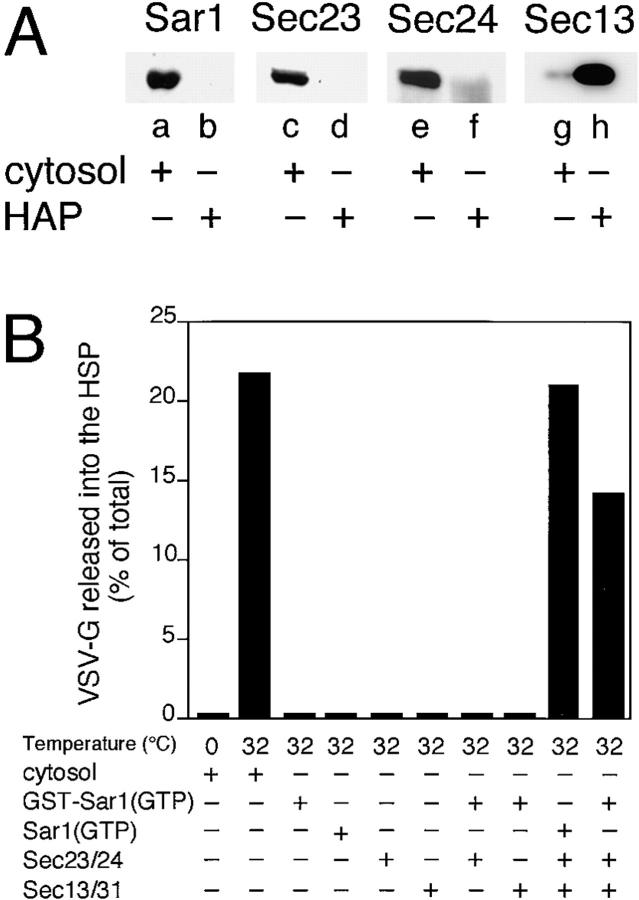
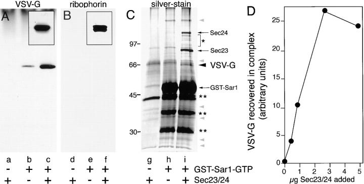
Cargo is selectively exported from the ER in COPII vesicles. To analyze the role of COPII in selective transport from the ER, we have purified components of the mammalian COPII complex from rat liver cytosol and then analyzed their role in cargo selection and ER export. The purified mammalian Sec23-24 complex is composed of an 85-kD (Sec23) protein and a 120-kD (Sec24) protein. Although the Sec23-24 complex or the monomeric Sec23 subunit were found to be the minimal cytosolic components recruited to membranes after the activation of Sar1, the addition of the mammalian Sec13-31 complex is required to complete budding. To define possible protein interactions between cargo and coat components, we recruited either glutathione-S-transferase (GST)-tagged Sar1 or GST- Sec23 to ER microsomes. Subsequently, we solubilized and reisolated the tagged subunits using glutathione-Sepharose beads to probe for interactions with cargo. We find that activated Sar1 in combination with either Sec23 or the Sec23-24 complex is necessary and sufficient to recover with high efficiency the type 1 transmembrane cargo protein vesicular stomatitis virus glycoprotein in a detergent-soluble prebudding protein complex that excludes ER resident proteins. Supplementing these minimal cargo recruitment conditions with the mammalian Sec13-31 complex leads to export of the selected cargo into COPII vesicles. The ability of cargo to interact with a partial COPII coat demonstrates that these proteins initiate cargo sorting on the ER membrane before budding and establishes the role of GTPase-dependent coat recruitment in cargo selection.


Figures

Figure 1
Figure 1
Purification of Sec23–24 from rat liver cytosol. The Sec23–24 complex was purified from rat liver cytosol as described in the Materials and Methods. A and B illustrate typical elution profiles from the DEAE and HAP chromatography steps, respectively. (C), A silver-stained gel of representative pooled fractions from crude cytosol (3.3 μg) (lane a), ammonium sulfate precipitate (3 μg) (lane b), S-300–Sepharose (2.8 μg) (lane c), DEAE (0.7 μg) (lane d), and HAP (0.2 μg) (lane e) columns. The asterisks in e, partial proteolytic breakdown products of Sec24 based on Western blotting. Molecular markers are indicated in the left margin of C (kD).
Figure 1
Figure 1
Purification of Sec23–24 from rat liver cytosol. The Sec23–24 complex was purified from rat liver cytosol as described in the Materials and Methods. A and B illustrate typical elution profiles from the DEAE and HAP chromatography steps, respectively. (C), A silver-stained gel of representative pooled fractions from crude cytosol (3.3 μg) (lane a), ammonium sulfate precipitate (3 μg) (lane b), S-300–Sepharose (2.8 μg) (lane c), DEAE (0.7 μg) (lane d), and HAP (0.2 μg) (lane e) columns. The asterisks in e, partial proteolytic breakdown products of Sec24 based on Western blotting. Molecular markers are indicated in the left margin of C (kD).
Figure 2
Figure 2
Sec23 and the Sec23–24 are functional GAP proteins for the mammalian Sar1 protein. Recombinant Sar1 was incubated in the presence or absence of GST– Sec23 or purified Sec23–24 and then GAP activity was monitored as described in the Materials and Methods.
Figure 3
Figure 3
Sec 23 represents the minimal coat component that is recruited to the membranes by the Sar1 GTPase. (A) Microsomes were incubated with rat liver cytosol for 10 min on ice or at 32°C in the absence or presence of 1 μg of activated Sar1–GTP mutant with or without the ATP regenerating system. In all cases, Sar1–GTP was added together with 100 μM GTP. Membranes were pelleted, washed, and then the amount of Sec23 bound was determined by Western blotting with Sec23-specific antibody as described in the Materials and Methods. (B) Microsomes were incubated with the ATP regenerating system, GTPγS (100 μM), Sar1–GTP, and/or 2 mM GTP as indicated. Membranes were pelleted, washed, and then Western blots were quantitated as described in the Materials and Methods. The amount of Sec23 bound is reported as the percentage of maximal binding observed in incubations containing Sar1–GTP and the ATP regenerating system. (C) Microsomes were incubated with purified Sec23–24 complex or His6-tagged Sec23 with the indicated reagents as described for A. Membranes were pelleted, washed, and then the amount of Sec23 bound was determined by Western blotting. The typical results of three independent experiments are shown.
Figure 3
Figure 3
Sec 23 represents the minimal coat component that is recruited to the membranes by the Sar1 GTPase. (A) Microsomes were incubated with rat liver cytosol for 10 min on ice or at 32°C in the absence or presence of 1 μg of activated Sar1–GTP mutant with or without the ATP regenerating system. In all cases, Sar1–GTP was added together with 100 μM GTP. Membranes were pelleted, washed, and then the amount of Sec23 bound was determined by Western blotting with Sec23-specific antibody as described in the Materials and Methods. (B) Microsomes were incubated with the ATP regenerating system, GTPγS (100 μM), Sar1–GTP, and/or 2 mM GTP as indicated. Membranes were pelleted, washed, and then Western blots were quantitated as described in the Materials and Methods. The amount of Sec23 bound is reported as the percentage of maximal binding observed in incubations containing Sar1–GTP and the ATP regenerating system. (C) Microsomes were incubated with purified Sec23–24 complex or His6-tagged Sec23 with the indicated reagents as described for A. Membranes were pelleted, washed, and then the amount of Sec23 bound was determined by Western blotting. The typical results of three independent experiments are shown.
Figure 4
Figure 4
VSV-G can be detected in a complex with Sec23. (A) Salt-washed microsomes prepared as described in Materials and Methods were incubated at 32°C for 30 min in the presence of rat liver cytosol (a and b) or GST–Sec23 (c) in the presence of the inactive Sar1A (GDP- restricted) mutant (b) (1 μg) or the activated Sar1A (GTP-restricted) (a and c) mutant (1 μg). The amount of VSV-G (% of total) released from the ER in COPII vesicles was determined as described (Rowe et al., 1996). (B) Salt-washed microsomes were incubated on ice (c) or at 32°C (a, b, d, and e) for 30 min without (a) or with GST– Sec23 (b–e) in the absence (b) or presence of either the GTP- restricted (a, c, and d) or GDP-restricted (e) Sar1 mutant. Membranes were pelleted, solubilized, and incubated with GS beads and then the amount of GST–Sec23 or VSV-G recovered on the beads was determined using Western blotting as described in Materials and Methods. (C) Salt-washed microsomes were incubated in the presence of the activated Sar1–GTP mutant and then GST–Sec23 was pelleted, solubilized, and then incubated with GS beads as described in Materials and Methods. The amount of VSV-G (top panel), ribophorin II (middle panel) or calnexin (bottom panel) in the total sample (a) or that eluted from beads (b) was determined by Western blotting. The typical results of three independent experiments are shown.
Figure 4
Figure 4
VSV-G can be detected in a complex with Sec23. (A) Salt-washed microsomes prepared as described in Materials and Methods were incubated at 32°C for 30 min in the presence of rat liver cytosol (a and b) or GST–Sec23 (c) in the presence of the inactive Sar1A (GDP- restricted) mutant (b) (1 μg) or the activated Sar1A (GTP-restricted) (a and c) mutant (1 μg). The amount of VSV-G (% of total) released from the ER in COPII vesicles was determined as described (Rowe et al., 1996). (B) Salt-washed microsomes were incubated on ice (c) or at 32°C (a, b, d, and e) for 30 min without (a) or with GST– Sec23 (b–e) in the absence (b) or presence of either the GTP- restricted (a, c, and d) or GDP-restricted (e) Sar1 mutant. Membranes were pelleted, solubilized, and incubated with GS beads and then the amount of GST–Sec23 or VSV-G recovered on the beads was determined using Western blotting as described in Materials and Methods. (C) Salt-washed microsomes were incubated in the presence of the activated Sar1–GTP mutant and then GST–Sec23 was pelleted, solubilized, and then incubated with GS beads as described in Materials and Methods. The amount of VSV-G (top panel), ribophorin II (middle panel) or calnexin (bottom panel) in the total sample (a) or that eluted from beads (b) was determined by Western blotting. The typical results of three independent experiments are shown.
Figure 5
Figure 5
GST-tagged Sar1–GTP supports vesicle budding from salt-washed microsomal membranes. (A) Salt-washed microsomes prepared as described in Materials and Methods were incubated in a budding reaction with cytosol on ice (a) or for 30 min at 32°C without (b) or with wild-type Sar1 (1.5 μg) (c), Sar1-GTP (4 μg) (d), or a GST-tagged Sar1–GTP (10 μg) (e) as indicated. The amount of VSV-G (% of total) released from the ER into the HSP was determined as described (Rowe et al., 1996). (B) Salt-washed microsomes were incubated in a budding reaction supplemented with a GST-tagged Sar1–GTP (4 μg) for 30 min at 32°C. The HSP of seven budding reactions was collected and then subjected to immunoisolation with magnetic beads alone (a) or beads coupled with a monoclonal antibody to the VSV-G tail (P5D4) as described (Rowe et al., 1996) (b). GST–Sar1–GTP bound to beads was detected by Western blotting using a polyclonal antibody to Sar1. Molecular weight markers are indicated on the left. The typical results of three independent experiments are shown.
Figure 7
Figure 7
Vesicle budding from the ER requires Sar1, Sec23–24, and Sec13–31. (A) The mammalian Sec13–31 complex was partially purified from rat liver cytosol as described in the Materials and Methods. The presence of Sar1, Sec23, Sec24, and Sec13 in crude cytosol (a, c, e, and g) and the HAP pool (b, d, f, and h), respectively, was determined by Western blotting with specific antibody (refer to Materials and Methods). Note the absence of Sar1 or Sec23–24 in the HAP fraction highly enriched in the Sec13–31 complex. (B) ER microsomes were incubated in the presence of ATP, GTP, and purified components for 30 min at 32°C and then the amount of VSV-G released into the HSP was determined as described (Rowe et al., 1996). The reactions were carried out in a 40-μl vol supplemented with either 400 μg of rat liver cytosol, 2 μg of GST–Sar1–GTP, 1 μg of His6–Sar1– GTP, 1 μg of Sec23–24 complex, and 42 μg of the Sec13/31–containing fraction as indicated. The typical results of three independent experiments are shown.
Figure 6
Figure 6
VSV-G can be isolated in a complex with a GST-tagged Sar1–GTP and the mammalian Sec23–24 complex. (A–C) Salt-washed microsomes were incubated at 32°C for 30 min in the presence of Sec23–24 complex (lanes a, d, and g), GST-tagged Sar1–GTP (lanes b, e, and h), or both (lanes c, f, and i). Membranes were pelleted, solubilized, and then incubated with GS beads as described in Materials and Methods. The amount of VSV-G (A) or ribophorin II (B) recovered on beads was determined using Western blotting as described in Materials and Methods. Insets in A and B show control microsomal membranes on the same blot that were probed for either VSV-G (A) or ribophorin II (B). For immunoblot analysis, the equivalent of four budding reactions (supplemented with a total of 5 μg Sec23–24 (A and B) and 8 μg of GST-tagged Sar1–GTP) were combined. (C) For silver stain analysis, the equivalent of six budding reactions supplemented with 12 μg of GST-tagged Sar1–GTP and 4 μg of Sec23–24 were combined. In C, molecular weight markers are indicated to the left of the gel. *, Sec24 breakdown products; **, GST–Sar1–GTP breakdown products. (D) Salt-washed microsomes were incubated at 32°C for 30 min in the standard budding reaction in the presence of the GST–Sar1–GTP and the indicated amounts of Sec23–24. The relative amounts of VSV-G recovered in the GST–Sar1 complex was determined as described in the Materials and Methods.
Figure 8
Figure 8
Immunolocalization of VSV-G in semi-intact cells incubated in the presence of cytosol or purified COP II components. Incubation of semiintact cells in the presence of ATP and the indicated components and localization of VSV-G using immunoelectron microscopy (immunodiffusion technique) was carried out as described previously (Balch et al., 1994). Semiintact cells were incubated in the presence of cytosol (A); the Sar1–GTP (9 μg), Sec23–24 (2.5 μg) and Sec13–31 (200 μg) in a 200-μl reaction vol (B); or Sar1–GTP and Sec23–24 in the absence of Sec13–31 or cytosol (C). In A and B, note the increased density of gold particles in ER-associated buds and ER-derived vesicles or pre-Golgi intermediates (arrowheads) when compared to the markedly reduced density of gold particles on total ER membranes. Arrow in top right panel of B highlights regions of localized concentration of VSV-G on ER membranes adjacent to regions of budding activity. Such structures were generally not observed in the presence of cytosol (A). (C) VSV-G can be readily detected in ER- associated buds and regional patches on the ER surface (arrowheads). Bars, 0.1 μM.
Figure 8
Figure 8
Immunolocalization of VSV-G in semi-intact cells incubated in the presence of cytosol or purified COP II components. Incubation of semiintact cells in the presence of ATP and the indicated components and localization of VSV-G using immunoelectron microscopy (immunodiffusion technique) was carried out as described previously (Balch et al., 1994). Semiintact cells were incubated in the presence of cytosol (A); the Sar1–GTP (9 μg), Sec23–24 (2.5 μg) and Sec13–31 (200 μg) in a 200-μl reaction vol (B); or Sar1–GTP and Sec23–24 in the absence of Sec13–31 or cytosol (C). In A and B, note the increased density of gold particles in ER-associated buds and ER-derived vesicles or pre-Golgi intermediates (arrowheads) when compared to the markedly reduced density of gold particles on total ER membranes. Arrow in top right panel of B highlights regions of localized concentration of VSV-G on ER membranes adjacent to regions of budding activity. Such structures were generally not observed in the presence of cytosol (A). (C) VSV-G can be readily detected in ER- associated buds and regional patches on the ER surface (arrowheads). Bars, 0.1 μM.

References

    1. Aridor M, Balch WE. Principles of selective transport: coat complexes hold the key. Trends Cell Biol. 1996a;6:315–320. - PubMed
    1. Aridor M, Balch WE. Timing is everything. Nature. 1996b;383:220–221. - PubMed
    1. Aridor M, Bannykh SI, Rowe T, Balch WE. Sequential coupling between COPII and COPI vesicle coats in endoplasmic reticulum-to-Golgi transport. J Cell Biol. 1995;131:875–893. - PMC - PubMed
    1. Balch WE, McCaffery JM, Plutner H, Farquhar MG. Vesicular stomatitis virus glycoprotein is sorted and concentrated during export from the endoplasmic reticulum. Cell. 1994;76:841–852. - PubMed
    1. Bannykh SI, Rowe T, Balch WE. Organization of endoplasmic reticulum export complexes. J Cell Biol. 1996;135:19–35. - PMC - PubMed

Publication types

MeSH terms