. 1998 Apr 6;141(1):175-85.
doi: 10.1083/jcb.141.1.175.

Rho guanosine triphosphatase mediates the selective stabilization of microtubules induced by lysophosphatidic acid

Affiliations

Rho guanosine triphosphatase mediates the selective stabilization of microtubules induced by lysophosphatidic acid

T A Cook et al. J Cell Biol. .

Abstract

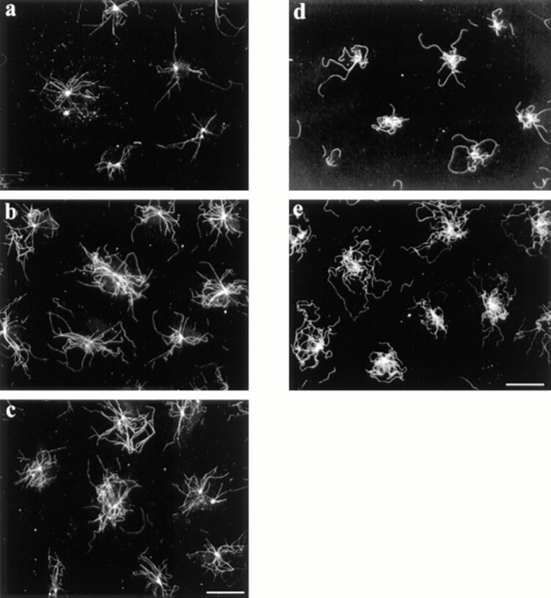
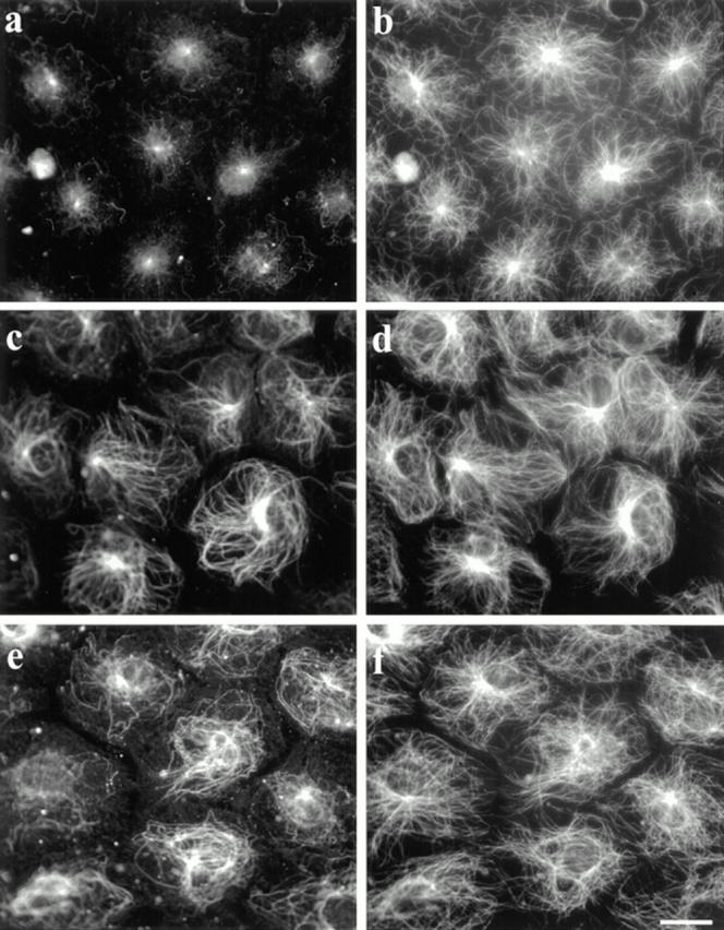
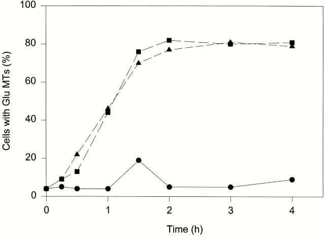
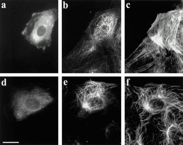
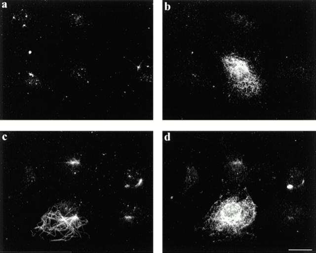
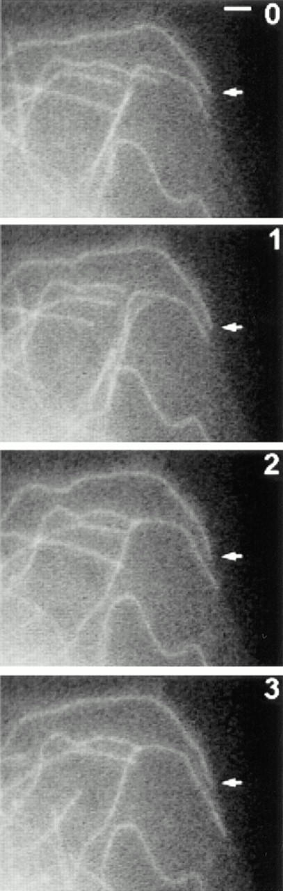
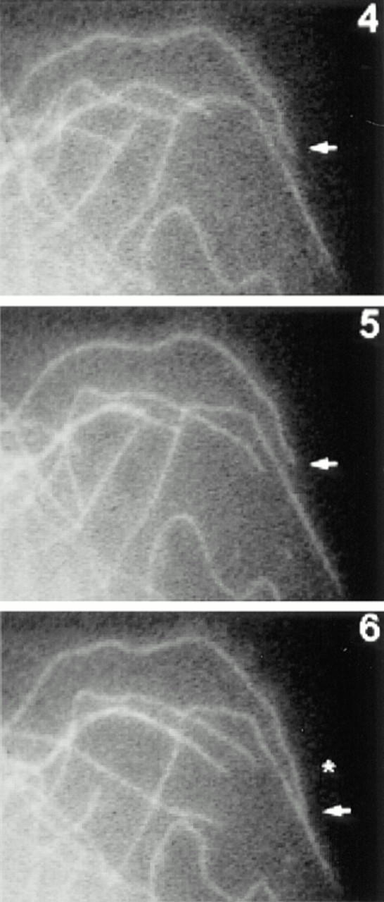
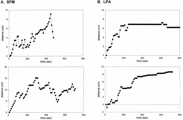
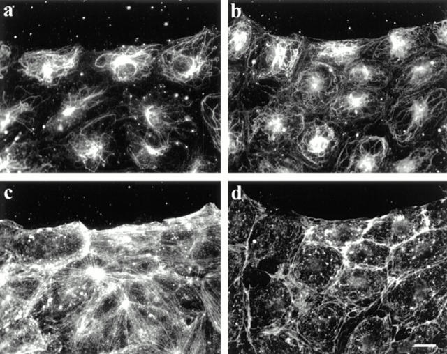
The asymmetric distribution of stable, posttranslationally modified microtubules (MTs) contributes to the polarization of many cell types, yet the factors controlling the formation of these MTs are not known. We have found that lysophosphatidic acid (LPA) is a major serum factor responsible for rapidly generating stable, detyrosinated (Glu) MTs in serum-starved 3T3 cells. Using C3 toxin and val14 rho we showed that rho was both necessary and sufficient for the induction of Glu MTs by LPA and serum. Unlike previously described factors that induce MT stability, rho induced the stabilization of only a subset of the MTs and, in wound-edge cells, these stable MTs were appropriately oriented toward the leading edge of the cell. LPA had little effect on individual parameters of MT dynamics, but did induce long states of pause in a subset of MTs near the edge of the cell. Rho stimulation of MT stability was independent of actin stress fiber formation. These results identify rho as a novel regulator of the MT cytoskeleton that selectively stabilizes MTs during cell polarization by acting as a switch between dynamic and stable states of MTs rather than as a modulator of MT assembly and disassembly.


Figures

Figure 3
Figure 3
LPA stimulates formation of nocodazole- and dilution-resistant MTs. For nocodazole stability, SFM-treated cells were refed SFM alone (a) or SFM containing 1 μM LPA (b) or 0.5% CS (c) for 5 min. Nocodazole was added to a final concentration of 2 μM and cells were incubated for 30 min. Cells were then detergent extracted to remove monomeric tubulin, washed, fixed, and stained for β-tubulin. For dilution resistance, SFM-treated cells were refed SFM alone (d) or SFM containing 1 μM LPA (e) for 5 min. The cells were then washed, detergent extracted, washed again, and incubated in PEM at 37°C for 1 h. Coverslips were fixed and stained with antibodies to β-tubulin. Bar, 20 μm.
Figure 1
Figure 1
LPA induces Glu MT formation in serum-starved 3T3 fibroblasts. SFM-treated cells were refed SFM (a and b), SFM containing 1 μM LPA (c and d), or SFM containing 0.5% CS (e and f) for 2 h. Cells were fixed and stained with antibodies specific for Glu (a, c, and e) or Tyr (b, d, and f) tubulin. Bar, 20 μm.
Figure 2
Figure 2
LPA and CS induce Glu MTs with a similar time course. SFM-treated cells were refed SFM alone (•) or SFM containing 1 μM LPA (▪) or 0.5% CS (▴) and then incubated for the indicated time before fixation and staining for Glu MTs. Scoring was as described in Table I. Results shown are averages of three separate experiments.
Figure 4
Figure 4
C3 toxin inhibits LPA-induced formation of Glu MTs. Botulinum C3 toxin (1 μg/ml) was microinjected into serum-starved cells along with human IgG as a marker. The medium was then replaced with fresh SFM containing 1 μM LPA and cells were incubated for 90 min. After fixation in −20°C methanol, coverslips were quadrupally stained with antibodies to human IgG (a), Glu tubulin (b), actin (c), and Tyr tubulin (d). Bar, 20 μm.
Figure 5
Figure 5
Microinjected val14 rho induces formation of Glu MTs. A wounded monolayer of serum-starved cells was microinjected with val14 rho along with human IgG as a marker and then incubated an additional 90–120 min before fixation and triple immunofluorescence staining (a–c are from one field, d–f are from another). Injected cells were detected with coumarin-conjugated antibodies to human IgG (a and d). Injected cells contain many Glu MTs, whereas uninjected neighboring cells contain few or none (b and e). Val14 rho also induced stress fiber formation, as shown in c with a monoclonal antibody to actin. Tyr MTs were largely unaffected by val14 rho injection (f). Bar, 20 μm.
Figure 6
Figure 6
Microinjected val14 rho induces formation of nocodazole resistant MTs. Serum-starved cells were microinjected with either IFA antibodies (a and b) or IFA antibodies and val14 rho (c and d). Cells were then incubated for 30 min, treated with 2 μM nocodazole for 1 h, and then extracted and fixed. Injected cells were detected with rhodamine-conjugated antibodies to mouse IgG (b and d) and nocodazole-resistant MTs were detected with Glu tubulin antibodies (a and c). Only val14 rho–injected cells contain nocodazole resistant MTs (c). Bar, 20 μm.
Figure 7
Figure 7
Dynamics of MTs in LPA-treated cells. Serum-starved cells were injected with R-tubulin. The next day cells were treated with 1 μM LPA for 1–2 h and then fluorescent images were acquired at 10-s intervals. Shown are R-tubulin–labeled MTs at the edge of a live cell at 1-min intervals. Many MT ends can be seen to undergo changes in length during the recording. Arrow designates a long-paused MT (see text). An asterisk indicates a change in position of the long-paused MT at 6 min. Bar, 2 μm.
Figure 7
Figure 7
Dynamics of MTs in LPA-treated cells. Serum-starved cells were injected with R-tubulin. The next day cells were treated with 1 μM LPA for 1–2 h and then fluorescent images were acquired at 10-s intervals. Shown are R-tubulin–labeled MTs at the edge of a live cell at 1-min intervals. Many MT ends can be seen to undergo changes in length during the recording. Arrow designates a long-paused MT (see text). An asterisk indicates a change in position of the long-paused MT at 6 min. Bar, 2 μm.
Figure 8
Figure 8
Life history plots of MTs in SFM-treated cells and LPA-treated cells. Representative life history plots for typical MTs in control SFM-treated cells (a) and for long-paused MTs in LPA-treated cells (b). Data points indicate the change in position of the end of a single MT at 10-s intervals. MTs in SFM-treated cells undergo frequent transitions between growth and shortening phases, whereas long-paused MTs in LPA-treated cells do not grow or shrink for long periods of time.
Figure 9
Figure 9
Cytochalasin D treatment does not block LPA-induced Glu MT formation. A wounded monolayer of serum-starved cells was treated for 15 min with SFM alone (a and c) or SFM containing 0.5 μM cytochalasin D (b and d). LPA was then diluted into the medium to a final concentration of 0.1 μM, and cells were incubated an additional 90 min before fixation and staining. Cytochalasin D blocked LPA-induced stress fibers (d) but not Glu MTs (b). Bar, 20 μm.

References

    1. Amano M, Mukai H, Ono Y, Chihara K, Matsui T, Hamajima Y, Okawa K, Iwamatsu A, Kaibuchi K. Identification of a putative target for rho as the serine-threonine kinase protein kinase N. Science. 1996;271:648–650. - PubMed
    1. Amano M, Chihara K, Kimura K, Fukata Y, Nakamura N, Matsuura Y, Kaibuchi K. Formation of actin stress fibers and focal adhesions enhanced by rho-kinase. Science. 1997;275:1308–1311. - PubMed
    1. Baas PW, Black MM. Individual microtubules in the axon consist of domains that differ in both composition and stability. J Cell Biol. 1990;111:495–509. - PMC - PubMed
    1. Bershadsky A, Chausobsky A, Becker E, Lyubimova A, Geiger B. Involvement of microtubules in the control of adhesion-dependent signal transduction. Curr Biol. 1996;6:1279–1289. - PubMed
    1. Bramblett GT, Goedert M, Jakes R, Merrick SE, Trojanowski JQ, Lee VM-Y. Abnormal tau phosphorylation at ser396 in Alzheimer's disease recapitulates development and contributes to reduced microtubule binding. Neuron. 1993;10:1089–1099. - PubMed

Publication types

MeSH terms