. 1998 Apr 6;141(1):281-6.
doi: 10.1083/jcb.141.1.281.

The tyrosine kinase p56lck mediates activation of swelling-induced chloride channels in lymphocytes

Affiliations

The tyrosine kinase p56lck mediates activation of swelling-induced chloride channels in lymphocytes

A Lepple-Wienhues et al. J Cell Biol. .

Abstract

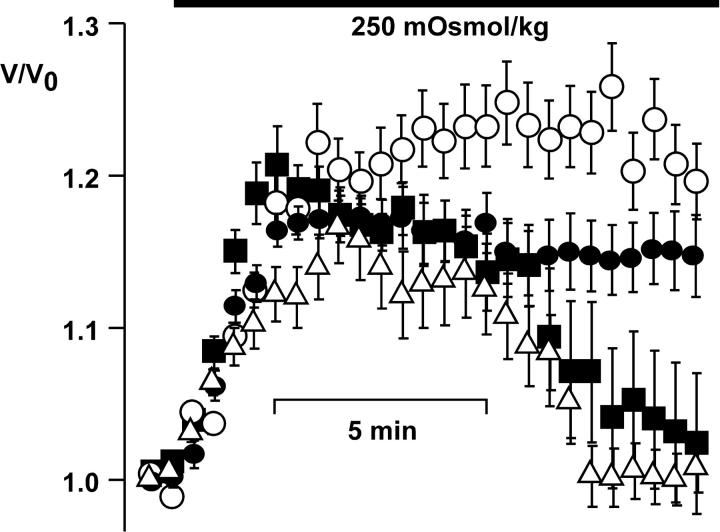
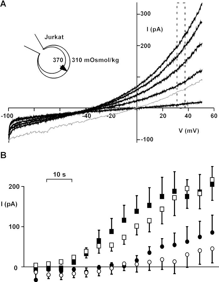
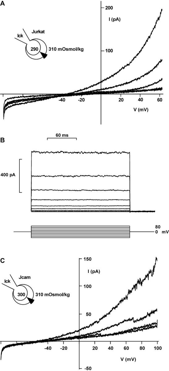
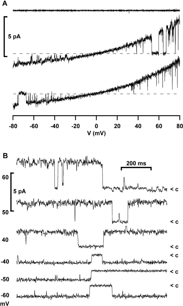
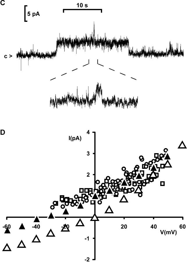
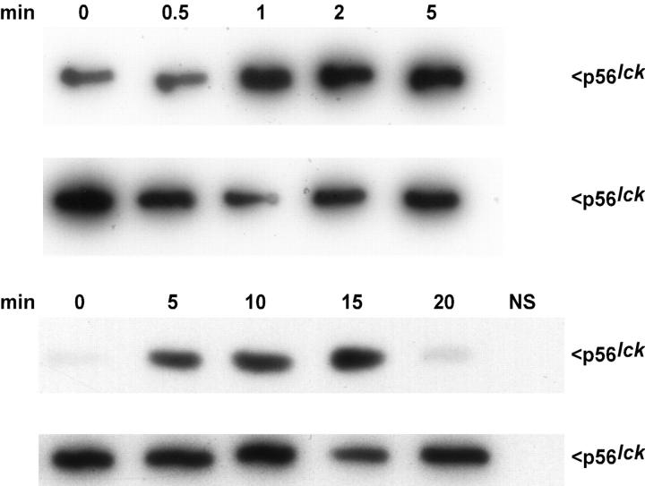
Osmotic cell swelling activates Cl- channels to achieve anion efflux. In this study, we find that both the tyrosine kinase inhibitor herbimycin A and genetic knockout of p56lck, a src-like tyrosine kinase, block regulatory volume decrease (RVD) in a human T cell line. Activation of a swelling-activated chloride current (ICl-swell) by osmotic swelling in whole-cell patch-clamp experiments is blocked by herbimycin A and lavendustin. Osmotic activation of ICl-swell is defective in p56lck-deficient cells. Retransfection of p56lck restores osmotic current activation. Furthermore, tyrosine kinase activity is sufficient for activation of ICl-swell. Addition of purified p56lck to excised patches activates an outwardly rectifying chloride channel with 31 pS unitary conductance. Purified p56lck washed into the cytoplasm activates ICl-swell in native and p56lck-deficient cells even when hypotonic intracellular solutions lead to cell shrinkage. When whole-cell currents are activated either by swelling or by p56lck, slow single-channel gating events can be observed revealing a unitary conductance of 25-28 pS. In accordance with our patch-clamp data, osmotic swelling increases activity of immunoprecipitated p56lck. We conclude that osmotic swelling activates ICl-swell in lymphocytes via the tyrosine kinase p56lck.


Figures

Figure 1
Figure 1
RVD is blocked by herbimycin A. Cells were superfused with hypotonic solution as indicated. After the initial swelling, control cells restored their volume within 13 min (▪; n = 24). No RVD was seen in cells pretreated with herbimycin A (○; n = 13) or in p56lck-deficient JCaM1.6 cells (•; n = 12). RVD was restored in JCaM1.6 cells retransfected with p56lck (▵; n = 30). Error bars show SEM.
Figure 2
Figure 2
Osmotic activation of ICl−swell is defective in p56lck-deficient JCaM1.6 cells. (A) Osmotic swelling activates an outwardly rectifying anion conductance. The reversal potential <−40 mV and the small inward current indicate poor permeability for intracellular glutamate. Traces are shown 1, 28, 42, 56, and 77 s after break-in. The lower trace (thin line) was recorded from a cell with symmetrical Cl concentrations 35 s after break-in, demonstrating intrinsic outward rectification. No leak correction was performed. Instantaneous IV-plots were obtained using 200-ms voltage ramps. (B) Outward currents at 35 mV in Jurkat cells (▪), p56lck-deficient JCaM1.6 cells (•), p56lck-retransfected JCaM1.6/ lck+ cells (□), and Jurkat cells pretreated for 8 h with 10 μM herbimycin A (○; n = 13, 10, 10, and 8, respectively). Error bars show SEM. Leak currents determined at −55 mV were subtracted.
Figure 3
Figure 3
ICl activated by p56lck. (A) ICl in a shrinking Jurkat cell with 10 U/ml purified p56lck in the pipette. Representative traces are shown 1, 121, 218, 284, and 371 s after break-in (no leak subtraction). (B) Lack of inactivation of outward currents induced by purified p56lck. 200-ms pulses beginning at −80 mV, with 20-mV increments, were applied every 5 s. Leak and capacitive currents were subtracted. (C) ICl in a shrinking JCaM1.6 cell with 10 U/ml purified p56lck in the pipette. Traces are shown 307, 373, and 482 s after break-in. The lowest trace shows block of the current with 500 μM DIDS. Note the current steps indicating single-channel transitions. 250-ms voltage ramps, no leak subtraction.
Figure 4
Figure 4
Single channels in excised patches and whole- cell recordings. (A) Activation of an outwardly rectifying Cl channel by purified p56lck (2 U/ml + 10 μM ATP) added to the internal surface of an excised patch (Jurkat T cells; n = 6). Instantaneous IV-plots were obtained using voltage ramps from −80 to 80 mV. The upper trace was taken before, the lower traces 3 min after addition of the enzyme (symmetrical NaCl solution, leak subtracted). (B) Continuous recordings from an excised inside-out patch activated by purified p56lck, showing closure events (<c) of the outwardly rectifying channel at the indicated holding potentials (filter 500 Hz). (C) Single-channel transitions in a continuous whole- cell recording from a JCaM1.6 cell with 10 U/ml p56lck in the pipette (same conditions as in Fig. 3 C). Upward deflection indicates channel opening. Two channels are visible in this trace. The inset shows a channel opening at a 10-fold magnified time scale. Note the slow gating. The holding potential was 80 mV, filter 500 Hz. (D) Current transitions in whole-cell currents resemble outwardly rectifying chloride channels in excised patches. □, Whole-cell current transitions activated by slow swelling of a Jurkat cell with hypertonic intracellular solution (330 mOsmol/kg, Cs+-glutamate); ○, whole-cell current transitions activated by p56lck in a JCaM1.6 cell with slightly hypotonic intracellular solution (300 mOsmol/kg, Cs+-glutamate); ▴, excised inside-out patch activated by purified p56lck with Cs+-glutamate in the bath; ▵, excised patch activated by purified p56lck with NaCl in the bath. The extracellular solution always contained NaCl (310 mOsmol/kg).
Figure 4
Figure 4
Single channels in excised patches and whole- cell recordings. (A) Activation of an outwardly rectifying Cl channel by purified p56lck (2 U/ml + 10 μM ATP) added to the internal surface of an excised patch (Jurkat T cells; n = 6). Instantaneous IV-plots were obtained using voltage ramps from −80 to 80 mV. The upper trace was taken before, the lower traces 3 min after addition of the enzyme (symmetrical NaCl solution, leak subtracted). (B) Continuous recordings from an excised inside-out patch activated by purified p56lck, showing closure events (<c) of the outwardly rectifying channel at the indicated holding potentials (filter 500 Hz). (C) Single-channel transitions in a continuous whole- cell recording from a JCaM1.6 cell with 10 U/ml p56lck in the pipette (same conditions as in Fig. 3 C). Upward deflection indicates channel opening. Two channels are visible in this trace. The inset shows a channel opening at a 10-fold magnified time scale. Note the slow gating. The holding potential was 80 mV, filter 500 Hz. (D) Current transitions in whole-cell currents resemble outwardly rectifying chloride channels in excised patches. □, Whole-cell current transitions activated by slow swelling of a Jurkat cell with hypertonic intracellular solution (330 mOsmol/kg, Cs+-glutamate); ○, whole-cell current transitions activated by p56lck in a JCaM1.6 cell with slightly hypotonic intracellular solution (300 mOsmol/kg, Cs+-glutamate); ▴, excised inside-out patch activated by purified p56lck with Cs+-glutamate in the bath; ▵, excised patch activated by purified p56lck with NaCl in the bath. The extracellular solution always contained NaCl (310 mOsmol/kg).
Figure 5
Figure 5
Cell swelling activates p56lck. The activity of p56lck induced by hypoosmotic cell swelling is shown at two different time scales. Jurkat cells (4 × 10⁶ per sample) were incubated with 250 mOsmol/kg solution for the indicated time, lysed, and then p56lck was immunoprecipitated from postcentrifugation supernatants. Activity of p56lck was determined by autophosphorylation in the presence of [32P]γATP. Samples were separated by 10% SDS-PAGE and analyzed by autoradiography. Nonspecific control immunoprecipitate performed with irrelevant affinity-purified polyclonal rabbit immunoglobulins is lacking kinase activity (lower panel right lane, NS). The second blot in each panel shows an aliquot of immunoprecipitated p56lck immunoblotted with anti-p56lck antibody demonstrating comparable amounts of protein in each lane. The results are representative of four independent experiments.

References

    1. August A, Dupont B. Activation of extracellular signal-regulated protein kinase (ERK/MAP kinase) following CD28 cross-linking: activation in cells lacking p56lck. Tissue Antigens. 1995;46:155–162. - PubMed
    1. Cahalan M.D., and R.S. Lewis. 1988. Role of potassium and chloride channels in volume regulation by T lymphocytes. In Cell Physiology of Blood. R.B. Gunn, and J.C. Parker, editors. Rockefeller University Press, New York. 281–301. - PubMed
    1. Deutsch C, Lee SC. Cell volume regulation in lymphocytes. Renal Physiol Biochem. 1988;3:260–276. - PubMed
    1. Doroshenko P, Neher E. Volume-sensitive chloride conductance in bovine chromaffin cell membrane. J Physiol (Lond) 1992;449:197–218. - PMC - PubMed
    1. Fischer H, Machen TE. The tyrosine kinase p60c-src regulates the fast gate of the cystic fibrosis transmembrane conductance regulator chloride channel. Biophys J. 1996;71:3073–3082. - PMC - PubMed

Publication types

MeSH terms