. 1998 Apr;116(4):1289-98.
doi: 10.1104/pp.116.4.1289.

Biological activity of reducing-end-derivatized oligogalacturonides in tobacco tissue cultures

Affiliations

Biological activity of reducing-end-derivatized oligogalacturonides in tobacco tissue cultures

M D Spiro et al. Plant Physiol. 1998 Apr.

Abstract

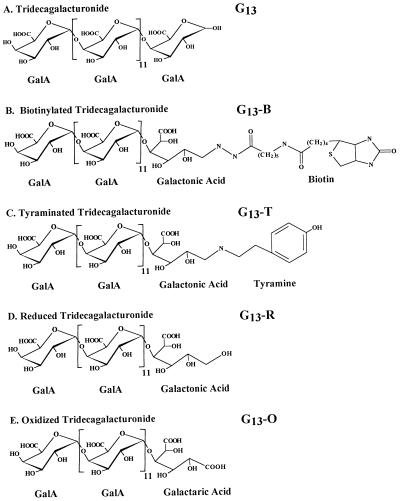
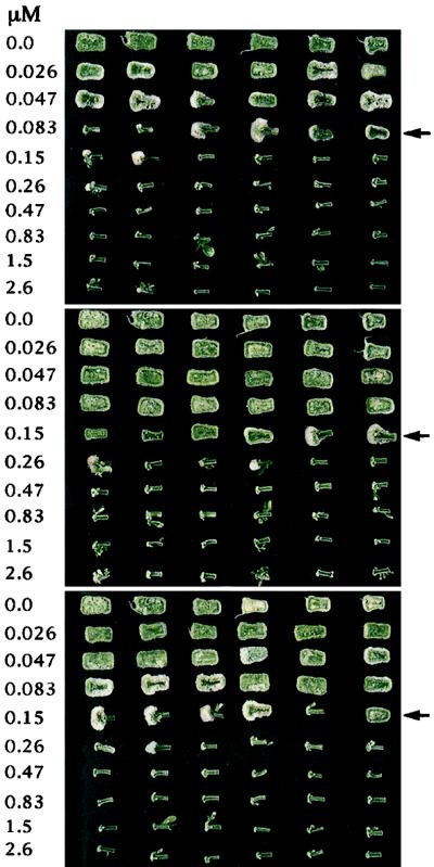
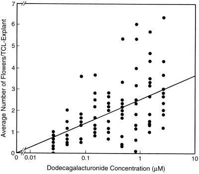
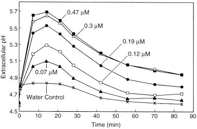
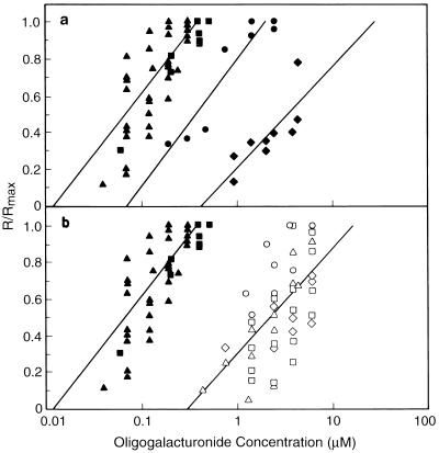
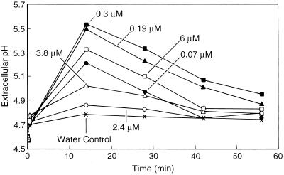
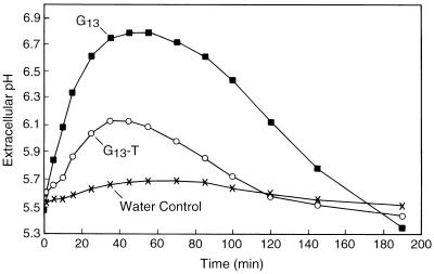
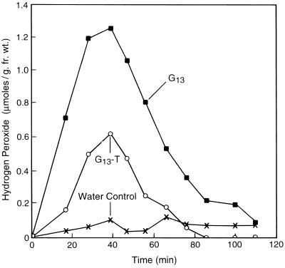
The biological activity of reducing-end-modified oligogalacturonides was quantified in four tobacco (Nicotiana tabacum) tissue culture bioassays. The derivatives used were oligogalacturonides with the C-1 of their reducing end (a) covalently linked to a biotin hydrazide, (b) covalently linked to tyramine, (c) chemically reduced to a primary alcohol, or (d) enzymatically oxidized to a carboxylic acid. These derivatives were tested for their ability to (a) alter morphogenesis of N. tabacum cv Samsun thin cell-layer explants, (b) elicit extracellular alkalinization by suspension-cultured cv Samsun cells, (c) elicit extracellular alkalinization by suspension-cultured N. tabacum cv Xanthi cells, and (d) elicit H2O2 accumulation in the cv Xanthi cells. In all four bioassays, each of the derivatives had reduced biological activity compared with the corresponding underivatized oligogalacturonides, demonstrating that the reducing end is a key element for the recognition of oligogalacturonides in these systems. However, the degree of reduction in biological activity depends on the tissue culture system used and on the nature of the specific reducing-end modification. These results suggest that oligogalacturonides are perceived differently in each tissue culture system.


Figures

Figure 1
Figure 1
Structures of G13 and the four reducing-end derivatives of G13 used in this study.
Figure 2
Figure 2
The TCL explants from a representative tobacco morphogenesis bioassay comparing the activities of G12 (top), G13-B (middle), and G13-T (bottom). The explants were incubated in growth medium containing the indicated oligogalacturonide concentrations, with six replicate TCL explants per concentration. The arrows indicate the concentrations at which oligogalacturonides induce a distinct change in the overall morphology of the TCL explants (the morphogenetic switch concentrations).
Figure 3
Figure 3
Plot of the log of G12 concentration versus the average number of flowers induced per TCL explant. The data from 12 experiments are shown, and each data point represents the average number of flowers produced by the six replicate TCL explants used for each treatment in a given experiment. In the absence of G12, no flowers were formed. The line represents the dose-response curve for G12 as determined by least-squares analysis using log concentration.
Figure 4
Figure 4
Representative time courses of the extracellular alkalinization by suspension-cultured cv Samsun cells treated with the indicated concentrations of G12 at time 0.
Figure 5
Figure 5
Effect of the DP (a) and reducing-end derivatization (b) of oligogalacturonides on their ability to induce extracellular alkalinization by suspension-cultured cv Samsun cells. The data were plotted as the alkalinization response of each sample divided by the alkalinization response induced by a saturating concentration of G12 in that experiment (R/Rmax). The R/Rmax values for the various oligogalacturonides are indicated as follows: G12, ▴; G13, ▪; G15, •; G9, ♦; G13-T, ⋄; G13-B, □; G13-O, ○; and G13-R, ▵. The data for G12 and G13 are shown on both plots to aid in comparison. The lines represent the linear portion of the dose-response curves as determined by least-squares analysis using log concentration. Separate dose-response curves were generated using combined data from G12 and G13 and combined data from the four reducing-end derivatives of G13 (see text).
Figure 6
Figure 6
Representative time courses for extracellular alkalinization by cv Samsun cells induced by the indicated concentrations of G12 (closed symbols) or G13-T (open symbols). More than a 30-fold higher concentration of G13-T, compared with G12, is required to induce an equivalent alkalinization response.
Figure 7
Figure 7
Representative time courses of extracellular alkalinization by cv Xanthi cells elicited by G13 and of G13-T. All treatments were at 8.7 μm.
Figure 8
Figure 8
Representative time courses of H2O2 accumulation in the growth medium of cv Xanthi cells elicited by the addition of G13 and G13-T. These measurements were carried out simultaneously using the same suspension cells as used for the measurement of extracellular alkalinization (Fig. 7). All treatments were at 8.7 μm.

Similar articles

Cited by

References

    1. Apostol I, Heinstein PF, Low PS. Rapid stimulation of an oxidative burst during elicitation of cultured plant cells. Role in defense and signal transduction. Plant Physiol. 1989;90:109–116. - PMC - PubMed
    1. Basse CW, Fath A, Boller T. High affinity binding of a glycopeptide elicitor to tomato cells and microsomal membranes and displacement by specific glycan suppressors. J Biol Chem. 1993;268:14724–14731. - PubMed
    1. Baureithel K, Felix G, Boller T. Specific, high affinity binding of chitin fragments to tomato cells and membranes. Competitive inhibition of binding by derivatives of chitin fragments and a nod factor of Rhizobium. J Biol Chem. 1994;269:17931–17938. - PubMed
    1. Bellincampi D, Salvi G, De Lorenzo G, Cervone F, Marfà V, Eberhard S, Darvill A, Albersheim P. Oligogalacturonides inhibit the formation of roots on tobacco explants. Plant J. 1993;4:207–213.
    1. Bishop PD, Pearce G, Bryant JE, Ryan CA. Isolation and characterization of the proteinase inhibitor-inducing factor from tomato leaves. Identity and activity of poly- and oligogalacturonide fragments. J Biol Chem. 1984;259:13172–13177. - PubMed

Publication types

LinkOut - more resources