

A surface-exposed region of a novel outer membrane protein (P66) of Borrelia spp. is variable in size and sequence
- PMID: 9537355
- PMCID: PMC107070
- DOI: 10.1128/JB.180.7.1618-1623.1998
A surface-exposed region of a novel outer membrane protein (P66) of Borrelia spp. is variable in size and sequence
Abstract
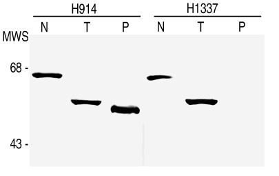
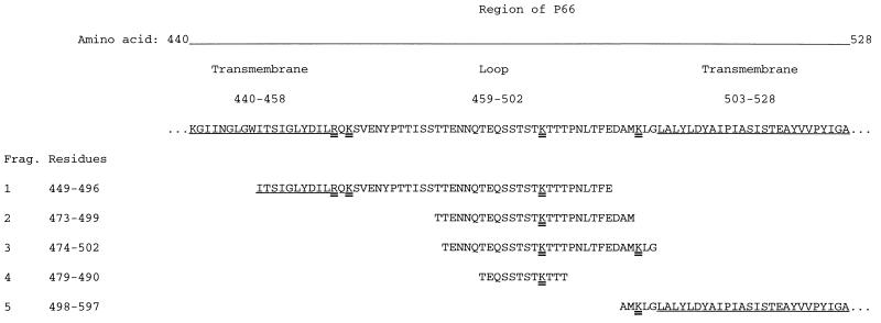
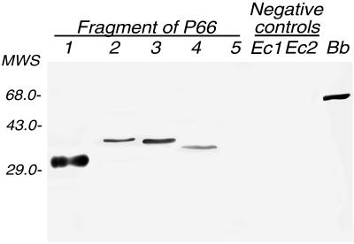
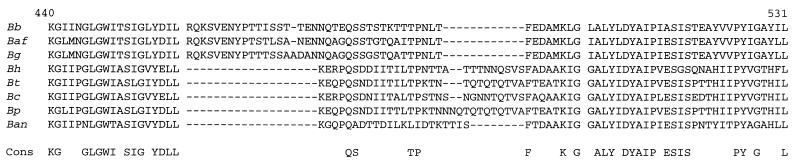
A model of the 66-kDa outer membrane protein (P66) of Lyme disease Borrelia spp. predicts a surface-exposed loop near the C terminus. This region contains an antigen commonly recognized by sera from Lyme disease patients. In the present study, this region of P66 and homologous proteins of other Borrelia spp. were further investigated by using monoclonal antibodies, epitope mapping of P66 of Borrelia burgdorferi, and DNA sequencing. A monoclonal antibody specific for B. burgdorferi bound to the portion of P66 that was accessible to proteolysis in situ. The linear epitope for the antibody was mapped within a variable segment of the surface-exposed region. To further study this protein, the complete gene of Borrelia hermsii for a protein homologous to P66 was cloned. The deduced protein was 589 amino acids in length and 58% identical to P66 of B. burgdorferi. The B. hermsii P66 protein was predicted to have a surface-exposed region in the same location as that of B. burgdorferi's P66 protein. With primers designed on the basis of conserved sequences and PCR, we identified and cloned the same regions of P66 proteins of Borrelia turicatae, Borrelia parkeri, Borrelia coriaceae, and Borrelia anserina. The deduced protein sequences from all species demonstrated two conserved hydrophobic regions flanking a surface-exposed loop. The loop sequences were highly variable between different Borrelia spp. in both sequence and size, varying between 35 and 45 amino acids. Although the actual function of P66 of Borrelia spp. is unknown, the results suggest that its surface-exposed region is subject to selective pressure.
Figures
References
-
- Åsbrink E, Hovmark A, Hederstedt B. The spirochetal etiology of acrodermatitis chronica atrophicans Herxheimer. Acta Dermato-Venereol. 1984;64:506–512. - PubMed
-
- Ausubel F M, Brent R, Kingston R E, Moore D D, Seidman J G, Smith J A, Struhl K, editors. Current protocols in molecular biology. New York, N.Y: Wiley; 1993.
-
- Barbour A. Clonal polymorphisms of surface antigens in a relapsing fever Borrelia spp. In: Jackson G, editor. Pathogenesis of bacterial infection. Heidelberg, Germany: Springer-Verlag; 1985. pp. 235–245.
Publication types
MeSH terms
Substances
Associated data
- Actions
- Actions
- Actions
- Actions
- Actions
Grants and funding
LinkOut - more resources
Full Text Sources
Other Literature Sources
Molecular Biology Databases
Miscellaneous
